Review
. 2023 Apr 26;13(5):747.
doi: 10.3390/biom13050747.

A New Generation of IMiDs as Treatments for Neuroinflammatory and Neurodegenerative Disorders

Affiliations
Review

A New Generation of IMiDs as Treatments for Neuroinflammatory and Neurodegenerative Disorders

Katherine O Kopp et al. Biomolecules. .

Abstract

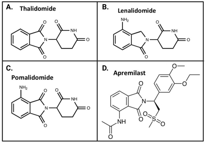
The immunomodulatory imide drug (IMiD) class, which includes the founding drug member thalidomide and later generation drugs, lenalidomide and pomalidomide, has dramatically improved the clinical treatment of specific cancers, such as multiple myeloma, and it combines potent anticancer and anti-inflammatory actions. These actions, in large part, are mediated by IMiD binding to the human protein cereblon that forms a critical component of the E3 ubiquitin ligase complex. This complex ubiquitinates and thereby regulates the levels of multiple endogenous proteins. However, IMiD-cereblon binding modifies cereblon's normal targeted protein degradation towards a new set of neosubstrates that underlies the favorable pharmacological action of classical IMiDs, but also their adverse actions-in particular, their teratogenicity. The ability of classical IMiDs to reduce the synthesis of key proinflammatory cytokines, especially TNF-α levels, makes them potentially valuable to reposition as drugs to mitigate inflammatory-associated conditions and, particularly, neurological disorders driven by an excessive neuroinflammatory element, as occurs in traumatic brain injury, Alzheimer's and Parkinson's diseases, and ischemic stroke. The teratogenic and anticancer actions of classical IMiDs are substantial liabilities for effective drugs in these disorders and can theoretically be dialed out of the drug class. We review a select series of novel IMiDs designed to avoid binding with human cereblon and/or evade degradation of downstream neosubstrates considered to underpin the adverse actions of thalidomide-like drugs. These novel non-classical IMiDs hold potential as new medications for erythema nodosum leprosum (ENL), a painful inflammatory skin condition associated with Hansen's disease for which thalidomide remains widely used, and, in particular, as a new treatment strategy for neurodegenerative disorders in which neuroinflammation is a key component.

Keywords: Alzheimer’s disease; Parkinson’s disease; cereblon; erythema nodosum leprosum; immunomodulatory imide drugs (IMiDs); neurodegeneration; neuroinflammation; pomalidomide; thalidomide; traumatic brain injury.


Conflict of interest statement

N.H.G., D.T., S-C.H., N.V., and D.S.K. are inventors on patents related to novel thalidomide and pomalidomide analogs. They have assigned their rights in entirety to their respective institutions, and hence have no personal rights to these patents. The National Institute on Aging, NIH, has a Cooperative Research and Development Agreement with AevisBio Inc. (Daejeon, Republic of Korea) to support the evaluation of novel IMiDs in neurological disorders. DSK is an employee and shareholder in AevisBio Inc (Daejeon, Republic of Korea). All other authors declare no conflict of interest.

Figures

Figure 1
Figure 1
Cycle of neuroinflammation and neurodegeneration. The accumulation of aggregated pathogenic proteins, often in a neurodegenerative disease state, stimulates excessive generation and release of pro-inflammatory molecules, including cytokines, such as TNF-α, IL-1β, and IL-6. These pro-inflammatory molecules promote neuroinflammation and neurodegeneration. Chronically elevated levels of these cytokines and chemokines promote further degradation of tissue and functional abnormalities associated with neurodegeneration, which leads to more release and a feed-forward loop. Created with BioRender.com (accessed on 17 February 2023).
Figure 2
Figure 2
Microglial activation and neuroinflammation. (A) In a quiescent state, microglia survey their environment for insults and injury in the CNS. Once they detect damage signals, such as DAMPs, cell debris, cytokines, and chemokines, microglia transition into their M1 activated state. M1 microglia are pro-inflammatory and release cytokines and chemokines that serve to activate other cells. In contrast, M2 microglia have anti-inflammatory and phagocytic functions, clearing cell debris. (B) Chronically activated microglia exist in a pro-inflammatory, hyper-reactive primed state and demonstrate enhanced sensitivity to inflammatory stimuli and, thereby, contribute to chronic neuroinflammation and neurodegeneration. Image adapted from Glotfelty et al., 2019 [8].
Figure 3
Figure 3
Positive feedback loop of TNF-α-promoted neuroinflammation. Inflammatory stimuli, often associated with the pathophysiological characteristics of a neurodegenerative disease, are neurotoxic and activate peripheral and glial cells. Activation of peripheral and glial cells increases levels of TNF-α, which binds and activates its receptor TNFR1. TNFR1 signaling stimulates apoptosis and activates NF-κB, a transcription factor that enhances the production of pro-inflammatory cytokines and further activates glial cells, causing microglia and astrocytes to adopt their reactive states. Activated microglia and astrocytes contribute to the neuroinflammatory environment and neuronal death, stimuli which promote additional glial cell activation, recommencing the cycle. Image adapted from Jung et al., 2019 [21].
Figure 4
Figure 4
Molecular mechanisms of IMiD action. (A) An IMiD compound interacts with cereblon (CRBN) via its glutarimide moiety, altering the substrate preferences of the ubiquitin-E3 ligase complex and leading to the degradation of specific target proteins. (B) IMiDs compete with CRBN for binding to the CD147-MCT1 complex, destabilizing the complex and inhibiting its ability to enhance tumor survival. (C) An IMiD compound binds to the 3′ untranslated region (UTR) of TNF-α mRNA, serving to destabilize the mRNA and reduce protein expression of TNF-α. Image adapted from Jung et al., 2021 [27]. Created with BioRender.com.
Figure 5
Figure 5
Chemical structures of US FDA-approved IMiDs: thalidomide (A) and analogs lenalidomide (B), pomalidomide (C), and apremilast (D). Image adapted from Jung et al., 2021 [27].
Figure 6
Figure 6
Cereblon binding and reduced SALL4 degradation of NAP, 3,6′-DP, and 1,6′-DP, as compared to older generation IMiDs. (A) Binding to cereblon was quantified utilizing a cereblon/BRD3 binding FRET assay. When 5 μM pomalidomide (Pom), thalidomide (Thal), lenalidomide (Len), and NAP were tested and compared to a vehicle control (Con), which was set at 0% cereblon inhibition, NAP was not statistically different from the control value, indicating that NAP does not bind cereblon. *** p < 0.001 versus control value. (B) 3,6′-DP and 1,6′-DP bind cereblon with similar potency to pomalidomide (Pom). (C) In H9 hES cells with 20 μM Thal, Pom, and NAP treatments, SALL4 expression was measured and quantified relative to β-actin expression. *** p < 0.001 versus control value; ### p < 0.001 relative to the NAP value by Tukey’s multiple comparisons test. (D) In H9 cells, SALL4 protein expression was measured using Western blots, following 3 μM treatments of vehicle (control), Thal, Pom, 3,6′-DP, and 1,6′-DP. SALL4 expression was normalized to β-actin levels. 3,6′-DP and 1,6′-DP significantly elevated SALL4 levels relative to Pom treatment. * p < 0.05, **** p < 0.0001: treatments versus control (i.e., DMSO/vehicle); ## p < 0.01: treatments versus Pom of same concentration (values: mean ± SEM, n = 4/group). Panels A and C were adapted from Hsueh et al., 2021 [28]; Panels B and D were adapted from Tsai et al., 2022 [108].
Figure 7
Figure 7
Behavioral assay data of Lin and colleagues (2020) reveal cognitive and behavioral benefits of 3,6′-DP. Scores of sham animals with no injury, TBI animals treated with vehicle (control), and TBI animals treated with 0.5 or 0.1 mg/kg body weight pomalidomide (Pom) or 3,6′-DP are shown 24 h post-TBI induction. We quantified body asymmetry using the EBST (A), neurological function using mNSS (B), sensorimotor function using a tactile sensory test (C), and motor coordination using a beam walk (D). Data represent the mean ± S.E.M. (n = 5 in each group). * p < 0.05, ** p < 0.01, *** p < 0.001 versus the Sham group; ## p < 0.01, ### p < 0.001 versus the TBI + Veh group; + p < 0.05, +++ p < 0.001 versus the TBI + Pom (0.5 mg/kg body weight) group. Image adapted from Lin et al., 2020 [105].
Figure 8
Figure 8
Behavioral assay data of Huang and colleagues (2021) demonstrate beneficial effects of 3,6′-DP on locomotion and anxiety (A) and short-term memory (B) 24 h and seven days post CCI TBI induction. (A) The mean distance traveled, expressed as the mean ± SEM, at 24-h and seven-day time points for sham and TBI animals treated with vehicle (Veh), pomalidomide (Pom), or 3,6′-DP. Both Pom and 3,6′-DP ameliorated TBI-induced locomotion deficits and anxiety-related behaviors at the seven-day time point; 3,6′-DP uniquely improved these behaviors at the 24-h time point. (B) The discrimination indices, expressed as the mean ± SEM, at 24-h and seven-day time points for sham and TBI animals treated with Veh, Pom, or 3,6′-DP. 3,6′-DP entirely remediated the TBI-induced short-term memory deficit seven days following injury; Pom only partially restored short-term memory function at the seven-day point. * p < 0.05, ** p < 0.01, *** p < 0.001 versus different time points in the same group; ## p < 0.01, ### p < 0.001 versus TBI + Veh group at the same time point. + p < 0.05 versus TBI + Pom group at the same time point (n = 5 in each group). Statistical differences were analyzed using one-way ANOVA, followed by Tukey’s post hoc comparisons. The image was adapted from Huang et al., 2021 [106].
Figure 9
Figure 9
In the CA1 region of the hippocampus, 3,6′-DP and, to a lesser extent, pomalidomide (Pom) treatments mitigate TBI-induced neuroinflammation/microgliosis, as indicated by reduced levels of activated microglia (Iba1+), astrogliosis, as indicated by reduced levels of reactive astrocytes (GFAP+), and neurodegeneration, as indicated by reduced number of degenerating cells (FJC+). All photomicrographs and cell quantifications were taken seven days post TBI. All treatments were administered at a 0.5 mg/kg body weight dose. (A) Photomicrographs of Iba1-stained CA1 tissue (scale bar: 60 μm). (B) Iba1+ activated microglia quantified in the CA1 region. (C) Photomicrographs of GFAP-stained CA1 tissue (scale bar: 60 μm). (D) GFAP+ reactive astrocytes quantified in the CA1 region. (E) Photomicrographs of FJC-stained CA1 tissue (scale bar: 100 μm). (F) FJC+ degenerating cells quantified in the CA1 region. Data are expressed as mean ± S.E.M. * p < 0.05, ** p < 0.01, *** p < 0.001 versus Sham + Veh group; # p < 0.05, ## p < 0.01, ### p < 0.001 versus TBI + Veh group; ++ p < 0.01 versus TBI + Pom group (n = 5 per group). Statistical differences were analyzed using independent sample t-tests and one-way ANOVA, followed by Tukey’s post hoc comparisons. Image adapted from Huang et al., 2021 [106].
Figure 10
Figure 10
Improvements in stroke-induced infarct volume and body asymmetry with 3,6′-DP, 1,6′-DP and pomalidomide (Pom) treatments. Treatments (equimolar to 20 mg/kg body weight Pom) were administered via intraperitoneal (i.p.) injection following one hour of MCAo and 30 min of reperfusion. (A) Infarct volumes of MCAo rats treated with Pom and its dithionated analogs 3,6′-DP and 1,6′-DP. (B) Body asymmetry as quantified by number of contralateral turns in the EBST. Non-MCAo rats perform roughly equal numbers of ipsi- and contralateral turns; injured rats exhibit a bias toward contralateral turns. Data are expressed as mean + standard error of the mean (S.E.M., n = 4 per group), Dunnett’s t-test * p < 0.05, ** p < 0.01 versus MCAo + vehicle group. Image adapted from Tsai et al., 2022 [108].
Figure 11
Figure 11
Cognitive behavioral markers ameliorated with 3,6′-DP treatment. Treatments were administered at 26.4 mg/kg body weight (pomalidomide (Pom)) or 29.5 mg/kg body weight (3,6′-DP) for four months. We calculated composite behavioral scores to increase the sensitivity between groups (WT control (Cntl), 5xFAD vehicle (Veh), 5xFAD Pom, and 5xFAD 3,6′-DP). ** p < 0.01 5xFAD veh versus WT cntl groups; ** p < 0.01 5xFAD veh versus 5xFAD 3,6′-DP groups by one-way ANOVA followed by Dunnett’s post-hoc tests. Image adapted from Lecca et al., 2022 [109].
Figure 12
Figure 12
3,6′-DP and pomalidomide (Pom) mitigate microglial activation, reactive astrogliosis, synaptic loss, and neurodegeneration. 5xFAD mice were treated with vehicle (Veh), 29.5 mg/kg body weight 3,6′-DP, or 26.4 mg/kg body weight Pom i.p. daily for four months. (A) We quantified Iba1 total area occupied in immunohistochemical assays to measure activated microglia levels in the hippocampus. (B) Photomicrographs of Iba1+ cells (scale bar: 30 μm). (C) We used GFAP immunohistochemical assays to measure astrogliosis, expressing GFAP+ cells as a percentage of the brain area occupied. (D) Photomicrographs of GFAP+ cells (scale bar: 30 μm). (E) We quantified PSD-95+ dendritic spines as a measure of intact synapses in the hippocampus. (F) Representative images of PSD-95+ dendritic spines (green) located on MAP2+ dendrites (scale bar: 3 μm). (G) We quantified degenerating neurons by identifying FJC+ cells in the hippocampus. (H) Representative images of FJC-stained brain section (20×, tile scan over entire area of brain section, scale bar: 1 mm). * p < 0.05, *** p < 0.001, **** p < 0.0001 versus WT control (Cntl) group; * p < 0.05, ** p < 0.01 versus 5xFAD Veh group by one-way ANOVA followed by Dunnett’s post hoc test (n = 4–5 per group). Image adapted from Lecca et al., 2022 [109].
Figure 13
Figure 13
NAP treatment ameliorates TBI-induced behavioral impairments one or two weeks post-TBI. (A) The EBST was used to quantify body asymmetry. NAP treatment (30 mg/kg body weight i.p.) improved this measure one and two weeks post-TBI, although the difference at two weeks did not reach significance. (B) The ART was used to measure sensorimotor function, as TBI mice spend more time removing stickers from their contralateral paw than control mice. NAP treatment mitigated this impairment, reaching a significant difference at the two-week time point. (C) TBI mice demonstrated balance and motor coordination deficits in the BWT. NAP treatment reduced contralateral foot fall abnormalities associated with TBI, with the difference at one week reaching significance. Analysis by two-way ANOVA followed by Bonferroni’s test: *** p < 0.001. Data are expressed as mean ± SEM; n = 9 (TBI + Saline), n = 24 (TBI + NAP). Image adapted from Hsueh et al., 2021 [28].
Figure 14
Figure 14
NAP treatment two weeks post TBI reduced microglial activation and promoted resting morphologies in the ipsilateral (injured) thalamus of TBI mice. (A) We utilized Iba1 immunofluorescence staining to image microglia in four forms: ramified, intermediate, amoeboid, and round. We classified each morphological form using the following criteria: (1) activated: amoeboid and round forms; (2) resting/quiescent: ramified long branching processes with a small cell body; and (3) intermediate transition forms. Scale bar: 5 μm. (B) Numbers of microglia in activated versus resting states in the ipsilateral thalamus. NAP treatment (30 mg/kg body weight i.p.) substantially decreased the number of microglia in active form relative to the TBI + saline group. *** p < 0.001, resting versus active form within TBI + NAP group; ### p < 0.001, active form of microglia in TBI + saline versus TBI + NAP groups. Two-way ANOVA with Bonferroni’s t-test for multiple comparisons. Image adapted from Hsueh et al., 2021 [28].

Similar articles

Cited by

References

    1. Alam Q., Alam M.Z., Mushtaq G., Damanhouri G.A., Rasool M., Kamal M.A., Haque A. Inflammatory Process in Alzheimer’s and Parkinson’s Diseases: Central Role of Cytokines. Curr. Pharm. Des. 2016;22:541–548. doi: 10.2174/1381612822666151125000300. - DOI - PubMed
    1. Bradburn S., Murgatroyd C., Ray N. Neuroinflammation in mild cognitive impairment and Alzheimer’s disease: A meta-analysis. Ageing Res. Rev. 2019;50:1–8. doi: 10.1016/j.arr.2019.01.002. - DOI - PubMed
    1. Dionisio-Santos D.A., Olschowka J.A., O’Banion M.K. Exploiting microglial and peripheral immune cell crosstalk to treat Alzheimer’s disease. J. Neuroinflamm. 2019;16:74. doi: 10.1186/s12974-019-1453-0. - DOI - PMC - PubMed
    1. Troncoso-Escudero P., Parra A., Nassif M., Vidal R.L. Outside in: Unraveling the Role of Neuroinflammation in the Progression of Parkinson’s Disease. Front. Neurol. 2018;9:860. doi: 10.3389/fneur.2018.00860. - DOI - PMC - PubMed
    1. Hirsch E.C., Hunot S. Neuroinflammation in Parkinson’s disease: A target for neuroprotection? Lancet Neurol. 2009;8:382–397. doi: 10.1016/S1474-4422(09)70062-6. - DOI - PubMed

Publication types

MeSH terms