. 2023 May 9:14:1113404.
doi: 10.3389/fneur.2023.1113404. eCollection 2023.

Increased expression of cell adhesion molecules in myofasciitis

Affiliations

Increased expression of cell adhesion molecules in myofasciitis

Xue Ma et al. Front Neurol. .

Abstract

Background: Myofasciitis is a heterogeneous group of diseases pathologically characterized by inflammatory cell infiltration into the fascia. Endothelial activation plays a critical role in the pathogenesis of the inflammatory response. However, the expression of cellular adhesion molecules (CAMs) in myofasciitis has not been investigated.

Methods: Data on clinical features, thigh magnetic resonance imaging, and muscle pathology were collected from five patients with myofasciitis. Immunohistochemical (IHC) staining and Western blot (WB) of the muscle biopsies from patients and healthy controls were performed.

Results: Increased levels of serum pro-inflammatory cytokines, including IL-6, TNF-α, and IL-2R, were detected in four patients. IHC staining and WB indicated significantly increased expression of cell adhesion molecules in blood vessels or inflammatory cells within the perimysium in muscle and fascia tissues of patients with myofasciitis compared to controls.

Conclusion: The up-regulation of CAMs in myofasciitis indicates endothelial activation, which may be potential therapy targets for the treatment of myofasciitis.

Keywords: cell adhesion molecules; dysregulated immune response; endothelial activation; myofasciitis; myopathy.


Conflict of interest statement

The authors declare that the research was conducted in the absence of any commercial or financial relationships that could be made as a potential conflict of interest.

Figures

Figure 1
Figure 1
MRI findings at time of diagnosis in five patients with myofasciitis (A) Increased signal abnormalities were noted in deep fascia of bilateral fascia of vastus medialis muscle and semimembranosus (white arrowheads) on fat suppression T2-weighted thigh MRI in Patient 1. (B) MRI of the lower extremities displayed hyperintensity in superficial fasciae (bright arrowheads) and deep fasciae (black arrowheads) on fat suppression T2-weighted images in Patient 2. (C) Fat suppression T2-weighted thigh MRI in Patient 3 showed thickening and increased signal in superficial (bright arrowheads) and deep (black arrowheads) facias of posterior thigh muscles. (D) Muscular edema and thickening fascia (black arrowhead) of posterior thigh muscles on fat suppression T2-weighted MRI images were obvious in Patient 4. (E) Thickening deep fascia (black arrowheads) had high signal intensity on fat suppression T2-weighted MRI of thigh in Patient 5. (F) MRI of forearm in Patient 5 showed hyperintensity in deep fasciae (black arrowhead).
Figure 2
Figure 2
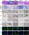
Immunohistochemical illustration of inflammatory infiltrate and immune-mediated factors in biopsied specimen of patients with myofasciitis. (A–E) Hematoxylin and eosin staining showed the numerous inflammatory cells in fascia and perivascular collections of inflammatory cells with minimal muscle damage in these patients. Original magnification: ×200. (F–J) A few eosinophils (black arrows) were observed in Patient 3. Original magnification: ×400. (K–O) A substantial infiltration of CD68+ macrophages were noted in the affected fascia in these patients with myofasciitis. Original magnification: ×200. (P–T) Abundant CD4+ T lymphocytes infiltrated into the fascia in these patients. Scale bar, 200 μm. Original magnification: ×200. (U–Y) Scatter CD8+ T lymphocytes were found in these patients. Original magnification: ×200. (Z–Dd) Scatter staining of CD20+ B lymphocytes was noted in fascia of Patient 2. Rare B lymphocytes were detected in Patient 1, Patient 3, Patient 4, and Patient 5. Original magnification: ×400. (Ee–Ii) The MHC-I on the sarcolemma close to thickened fascia and inflammatory cells was up-regulated in these patients. Original magnification: ×400. (Ji–Nn) MAC was positive with a vascular strong staining pattern in these patients. Original magnification: ×400.
Figure 3
Figure 3
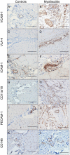
The expression of cell adhesion molecules in controls and patients with myofasciitis. (A,B) The weak expression of VCAM-1 in controls and up-regulated VCAM-1 on blood vessels and a few inflammatory cells in Patient 3. A and B, scale bar, 50 μm. (C,D) No positive staining of VLA-4 in controls and the expression of VLA-4 on scattered inflammatory cells in Patient 2. C and D, scale bar, 100 μm. (E,F) The low-level expression of ICAM-1 on vascular endothelium cells in controls and overexpressed ICAM-1 on vascular endothelial cells and a few inflammatory cells in Patient 2. E and F, scale bar, 50 μm. (G,H) No positive staining of CD11a/CD18 in controls and the expression of CD11a/CD18 on scatter inflammatory cells in Patient 3. G and H, scale bar, 100 μm. (I,J) The expression of PECAM-1 on blood vessels in controls and up-regulated PECAM-1 on blood vessels in Patient 3, especially in actively involved areas. (I,G), scale bar, 100 μm. (K,L) Very weak positive staining of CD146 on vascular endothelium cells in controls and strong intensity of CD146 on blood vessels in Patient 2. (K,L), scale bar, 100 μm.
Figure 4
Figure 4
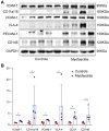
Western blot of cells adhesion molecules in muscle biopsies from controls and patients with myofasciitis. (A) Representative Western blot of ICAM-1, CD11a/CD18, VCAM-1, VLA-4, PECAM-1, and CD146 in muscle biopsies for five controls and four patients, including Patient 1, Patient 2, Patient 3, and Patient 5. (B) Quantitative analysis of ICAM-1, CD11a/CD18, VCAM-1, VLA-4, PECAM-1, and CD146 relative expression. n = 5 for controls, n = 4 for myofasciitis. *p < 0.05. Statistical analyses were performed by Mann–Whitney U test.

Similar articles

References

    1. Pinal-Fernandez I, Selva-O' Callaghan A, Grau JM. Diagnosis and classification of eosinophilic fasciitis. Autoimmun Rev. (2014) 13:379–82. doi: 10.1016/j.autrev.2014.01.019, PMID: - DOI - PubMed
    1. Preuße C, Goebel HH, Held J, Wengert O, Scheibe F, Irlbacher K, et al. . Immune-mediated necrotizing myopathy is characterized by a specific Th1-M1 polarized immune profile. Am J Pathol. (2012) 181:2161–71. doi: 10.1016/j.ajpath.2012.08.033, PMID: - DOI - PubMed
    1. Turkcapar N, Sak SD, Saatci M, Duman M, Olmez U. Vasculitis and expression of vascular cell adhesion molecule-1, intercellular adhesion molecule-1, and E-selectin in salivary glands of patients with Sjögren's syndrome. J Rheumatol. (2005) 32:1063–70. PMID: - PubMed
    1. Nishiya K, Tanimoto N, Hashimoto K, Hashizume M, Tominaga A. Serum and synovial fluid levels of interleukin-5 in a patient with eosinophilic fasciitis. Ann Rheum Dis. (1996) 55:935–6. doi: 10.1136/ard.55.12.935, PMID: - DOI - PMC - PubMed
    1. Wasserman SI, Seibold JR, Medsger TA, Jr, Rodnan GP. Serum eosinophilotactic activity in eosinophilic fasciitis. Arthritis Rheum. (1982) 25:1352–6. doi: 10.1002/art.1780251111, PMID: - DOI - PubMed

LinkOut - more resources