. 2023 May;13(5):230045.
doi: 10.1098/rsob.230045. Epub 2023 May 24.

Correlative super-resolution analysis of cardiac calcium sparks and their molecular origins in health and disease

Affiliations

Correlative super-resolution analysis of cardiac calcium sparks and their molecular origins in health and disease

Miriam E Hurley et al. Open Biol. 2023 May.

Abstract

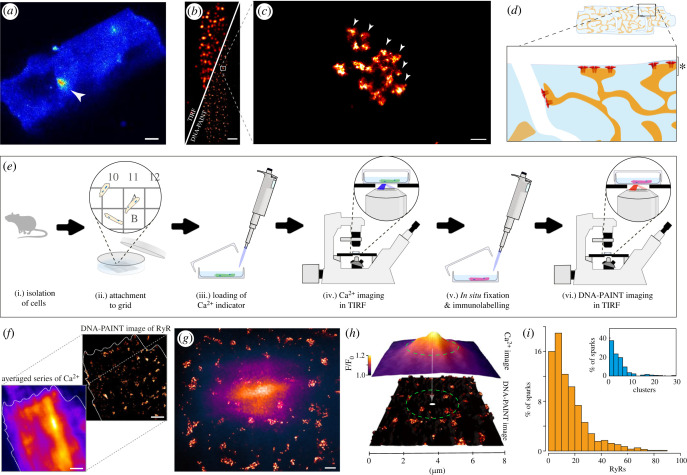
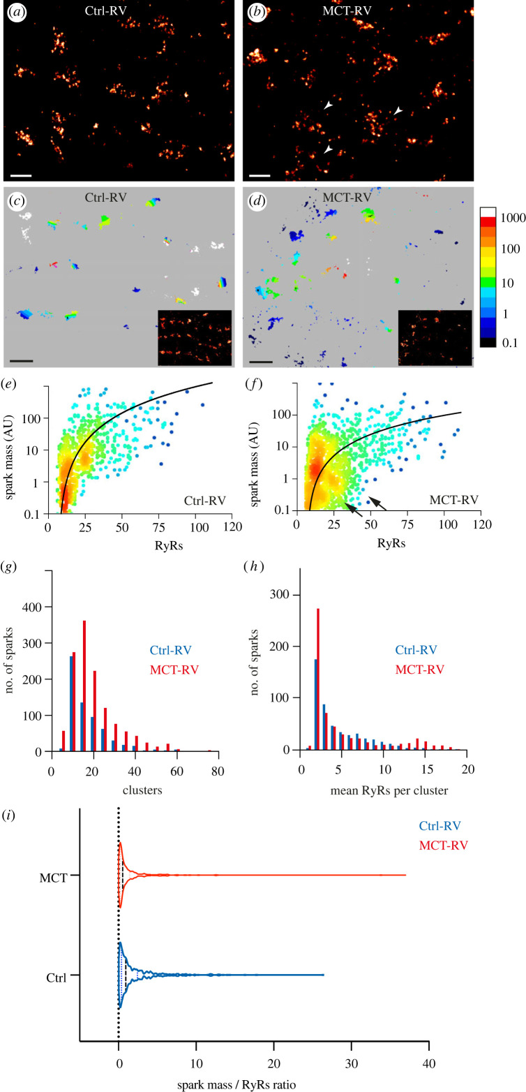
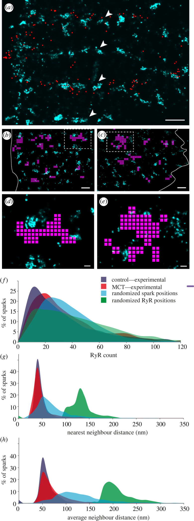
Rapid release of calcium from internal stores via ryanodine receptors (RyRs) is one of the fastest types of cytoplasmic second messenger signalling in excitable cells. In the heart, rapid summation of the elementary events of calcium release, 'calcium sparks', determine the contraction of the myocardium. We adapted a correlative super-resolution microscopy protocol to correlate sub-plasmalemmal spontaneous calcium sparks in rat right ventricular myocytes with the local nanoscale RyR2 positions. This revealed a steep relationship between the integral of a calcium spark and the sum of the local RyR2s. Segmentation of recurring spark sites showed evidence of repeated and triggered saltatory activation of multiple local RyR2 clusters. In myocytes taken from failing right ventricles, RyR2 clusters themselves showed a dissipated morphology and fragmented (smaller) clusters. They also featured greater heterogeneity in both the spark properties and the relationship between the integral of the calcium spark and the local ensemble of RyR2s. While fragmented (smaller) RyR2 clusters were rarely observed directly underlying the larger sparks or the recurring spark sites, local interrogation of the channel-to-channel distances confirmed a clear link between the positions of each calcium spark and the tight, non-random clustering of the local RyR2 in both healthy and failing ventricles.

Keywords: DNA-PAINT; calcium signalling; correlative light microscopy; nanodomains; ryanodine receptor.


Conflict of interest statement

Authors declare no competing interests.

Figures

Figure 1.
Figure 1.
An experimental framework for spatially correlating Ca2+ sparks to the underlying RyR2 channel organization. (a) A TIRF micrograph of a spontaneous Ca2+ spark (white arrowhead) in the sub-surface regions of a rat right ventricular myocyte loaded with Fluo-4 AM and immersed in 5 mM [Ca2+]o. (b) A split view of a similar, fixed rat right ventricular myocyte immuno-stained for RyR2 imaged with standard TIRF microscopy (upper-left) and DNA-PAINT (lower-right), demonstrating the level of resolution improvement in the latter. (c) Magnified view of the region indicated by the box in panel (b) illustrating a single RyR2 cluster. The individual punctate densities of labelling (arrowheads) represented individual RyR2 channels; (d) A schematic illustration of a ventricular cardiomyocyte where the SR forms junctions with both the surface plasmalemma and the t-tubular invaginations. The magnified view illustrates the predominantly sub-surface RyR2 clusters observed with the thin TIRF field (depth indicated by asterisk). (e) Experimental pipeline developed to allow correlative Ca2+ spark TIRF imaging and DNA-PAINT of acutely isolated cardiomyocytes. (f) The image alignment involves the upscaling and registration of a time-averaged, two-dimensional version of the Ca2+ image series to the density-rendered super-resolution DNA-PAINT image. (g) An overlay between a registered Ca2+ spark (purple/orange colour-table) and the local super-resolution image of RyR2 clusters (red hot colour-table). (h) Local sampling within the ‘Ca2+ spark footprint’ (dashed circle) involved the interrogation of RyR2 channel and cluster position within a circular window whose diameter was equal to the average FWHM of the spark. (i) percentage histograms of the total RyR2 puncta counts (main panel) and of the total number of unique clusters (inset) within the ‘spark footprint’. Scale bars: (a) 5 µm; (b) 2 µm; (c) 50 nm; (f) 1 µm; (g) 500 nm.
Figure 2.
Figure 2.
Local interrogation of RyR2 organization in Ctrl and MCT- RV cardiomyocytes. DNA-PAINT super-resolution maps of RyR2 labelling in (a) Ctrl and (b) MCT-RV rat cardiomyocytes. The arrowheads indicate the dissipated morphology of RyR2 clusters in the latter. (c,d) show two-dimensional super-resolution maps of RyR2, colour-coded for local, average Ca2+ spark mass in Ctrl-RV and MCT-RV myocytes, respectively. The insets show the original DNA-PAINT images in the corresponding region (larger versions of the insets shown in electronic supplementary material figure S2); the colour scale represents the average spark mass (in arbitrary units), estimated by xySpark. (e,f) Scattergrams of the Ca2+ spark mass, plotted on log10 scales against the RyR2 locally-counted within each ‘spark footprint’ in Ctrl-RV myocytes (n = 1050 sparks; 15 cells; 6 animals) and MCT-RV myocytes (n = 1529 sparks; 15 cells; 6 animals), the latter featuring sparks with smaller spark mass in regions with larger RyR2 counts. The line fits are y = 0.272x2 – 0.3 and y = 0.022x2 – 0.2 respectively. (g) Overlaid frequency histograms of the number of RyR2 clusters consisting of ≥5 RyR2 within the footprint of sparks recorded in Ctrl-RV (blue; n = 676 sparks; 6 animals) and MCT-RV (red; n = 1326 sparks; 6 animals). (h) Overlaid frequency histograms compare the distributions of mean RyR2 detected per cluster within the sparks recorded from Ctrl-RV and MCT-RV. (i) Shown, is a violin plot of the distribution of the ratio of spark mass to local RyR2 count recorded for each calcium spark in Ctrl-RV (blue) and MCT-RV (red).; medians (0.91 for Ctrl-RV and 0.53 for MCT-RV); mean ± s.d. 2.1 ± 3.1 for Ctrl-RV and 1.8 ± 6.7 for MCT-RV; median shown in dashed-lines and quartiles shown in dotted lines. Scale bars: (a,b) 200 nm; (c,d) 500 nm.
Figure 3.
Figure 3.
Spatial encoding of the spontaneous Ca2+ spark patterns in the RyR2 organization. (a) An overlay of an RyR2 super-resolution image (cyan) of a Ctrl-RV myocyte and a two-dimensional map of the centroids of all the spontaneous Ca2+ sparks (red circles) recorded within a 15 second time window. (b,c) Overlays of the DNA-PAINT super-resolution images of near-surface RyR2 labelling (cyan) and the recurring spark sites (magenta) in Ctrl-RV and MCT-RV myocytes respectively. (d,e) Magnified view of the windows indicated by dashed boxes in panels (b,c) respectively, illustrating that the recurring spark sites typically bridge or tessellate with local groups of RyR2 cluster rather than overlie them. (f) Overlaid percentage histograms of the RyR2 count underneath the spark footprint in Ctrl-RV (blue) and MCT-RV (red) as well as simulations where the detected spark positions were randomized (cyan), and where RyR2 positions were randomized. (g) The equivalent overlays of the percentage histograms of the mean neighbour distance between RyR2 puncta underneath each spark, and (h) the average distance to each of the three nearest neighbours to each RyR2 underneath spark. Scale bars: (a–c) 500 nm; (d,e) 100 nm. See electronic supplementary material, figure S4 for details of the simulation.

Similar articles

Cited by

References

    1. Sun XH, Protasi F, Takahashi M, Takeshima H, Ferguson DG, Franzini-Armstrong C. 1995. Molecular architecture of membranes involved in excitation-contraction coupling of cardiac muscle. J. Cell Biol. 129, 659-671. (10.1083/jcb.129.3.659) - DOI - PMC - PubMed
    1. Ouyang K, Zheng H, Qin X, Zhang C, Yang D, Wang X, Wu C, Zhou Z, Cheng H. 2005. Ca2+ sparks and secretion in dorsal root ganglion neurons. Proc. Natl Acad. Sci. USA 102, 12 259-12 264. (10.1073/pnas.0408494102) - DOI - PMC - PubMed
    1. Pritchard HAT, Griffin CS, Yamasaki E, Thakore P, Lane C, Greenstein AS, Earley S. 2019. Nanoscale coupling of junctophilin-2 and ryanodine receptors regulates vascular smooth muscle cell contractility. Proc. Natl Acad. Sci. USA 116, 21 874-21 881. (10.1073/pnas.1911304116) - DOI - PMC - PubMed
    1. Jayasinghe ID, Munro M, Baddeley D, Launikonis BS, Soeller C. 2014. Observation of the molecular organization of calcium release sites in fast- and slow-twitch skeletal muscle with nanoscale imaging. J. R. Soc. Interface 11, 20140570. (10.1098/rsif.2014.0570) - DOI - PMC - PubMed
    1. Johnson JD, Kuang S, Misler S, Polonsky KS. 2004. Ryanodine receptors in human pancreatic beta cells: localization and effects on insulin secretion. FASEB J. 18, 878-880. (10.1096/fj.03-1280fje) - DOI - PubMed

Publication types

Substances

LinkOut - more resources