. 2023 May 22;14(1):2898.
doi: 10.1038/s41467-023-38514-5.

Vaccinia E5 is a major inhibitor of the DNA sensor cGAS

Affiliations

Vaccinia E5 is a major inhibitor of the DNA sensor cGAS

Ning Yang et al. Nat Commun. .

Abstract

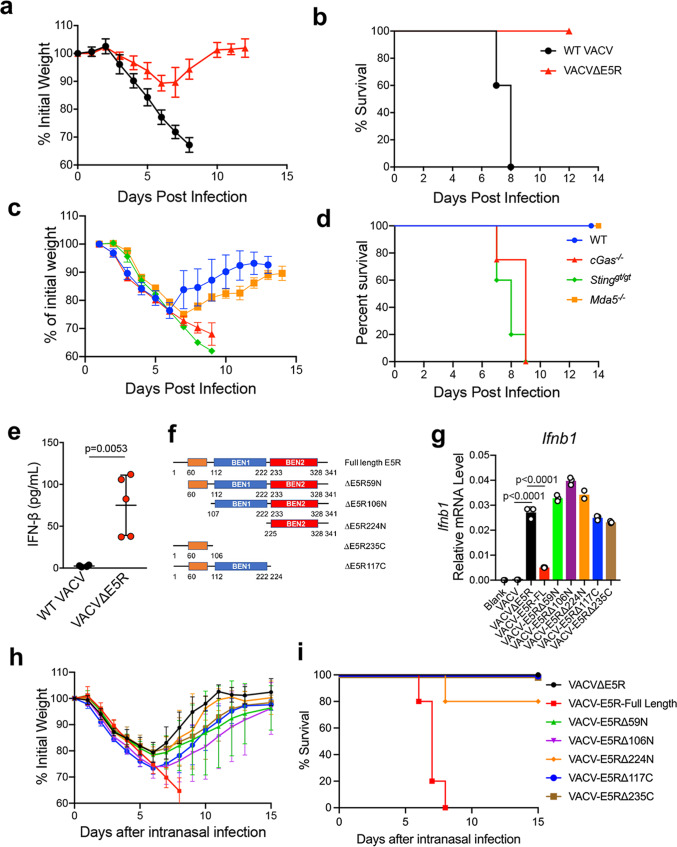
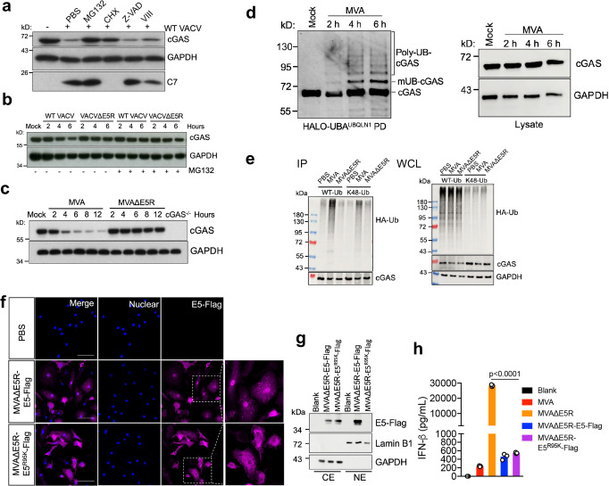
The DNA sensor cyclic GMP-AMP synthase (cGAS) is critical in host antiviral immunity. Vaccinia virus (VACV) is a large cytoplasmic DNA virus that belongs to the poxvirus family. How vaccinia virus antagonizes the cGAS-mediated cytosolic DNA-sensing pathway is not well understood. In this study, we screened 80 vaccinia genes to identify potential viral inhibitors of the cGAS/Stimulator of interferon gene (STING) pathway. We discovered that vaccinia E5 is a virulence factor and a major inhibitor of cGAS. E5 is responsible for abolishing cGAMP production during vaccinia virus (Western Reserve strain) infection of dendritic cells. E5 localizes to the cytoplasm and nucleus of infected cells. Cytosolic E5 triggers ubiquitination of cGAS and proteasome-dependent degradation via interacting with cGAS. Deleting the E5R gene from the Modified vaccinia virus Ankara (MVA) genome strongly induces type I IFN production by dendritic cells (DCs) and promotes DC maturation, and thereby improves antigen-specific T cell responses.


Conflict of interest statement

Memorial Sloan Kettering Cancer Center filed a patent application for the discovery of vaccinia viral inhibitors of the cytosolic DNA-sensing pathway and its use for improving MVA and vaccinia as oncolytic agents and vaccine vectors. The patent has been licensed to IMVAQ Therapeutics. L.D. and N.Y. are co-founders of IMVAQ Therapeutics. The remaining authors declare no competing interests.

Figures

Fig. 1
Fig. 1. Discovery of vaccinia virus E5 as a key inhibitor of the cGAS-dependent type I IFN pathway.
a ELISA analysis of IFN-β levels in the supernatants of BMDCs infected with either WT VACV or MVA at an MOI of 10 for 16 h. Cell lysates were analyzed by immunoblot. n = 3 independent samples. b cGAMP levels in BMDCs infected with either WT VACV or MVA at an MOI of 10. Cells were harvested at 2 and 8 h post-infection. cGAMP levels were measured by LC-MS. c Immunoblot of cGAS, C7, and E3 in BMDCs from WT or cGas−/− mice infected with different vaccinia viruses at MOI of 10 for 16 h. d ELISA analysis of IFN-β levels in BMDCs from WT or cGas−/− mice infected with different vaccinia viruses at MOI of 10 for 16 h. n = 3 independent samples. e ELISA analysis of cGAMP levels in WT BMDCs infected with either WT VACV or VACVΔE5R at MOI of 10 for 16 h. n = 3 independent samples. Two-tailed unpaired Student’s t-test was used for comparisons of two groups in the studies. Data are representative of two independent experiments and presented as mean ± SEM. Source data are provided as a Source Data file.
Fig. 2
Fig. 2. Vaccinia virus E5 is a virulence factor in vivo.
a, b Percentages of initial weight (a) and Kaplan–Meier survival curves (b) of WT C57BL/6J mice (n = 5 in each group) over days post-intranasal infection with either WT VACV or VACV∆E5R at a dose of 2 × 106 pfu per mouse. c, d Percentages of initial weight (c) and Kaplan–Meier survival curves (d) of WT, Mda5−/−, cGas−/−, or Stinggt/gt mice (n = 5 in each group) over days post-intranasal infection with VACV∆E5R at a dose of 2 × 107 pfu per mouse. e ELISA analysis of IFN-β levels in the BALF harvested from WT mice at 48 h post-infection with WT VACV or VACV∆E5R at a dose of 2 × 107 pfu per mouse. n = 5 independent samples. f Schematic diagram of VACV E5R full-length revertant and various VACV E5R truncation mutants. g RT-qPCR analysis of Ifnb levels in WT BMDCs infected with different vaccinia viruses, including VACV, VACV∆E5R, VACV-E5R-FL revertant, and various VACV E5R truncation mutants at MOI of 10 for 6 h. n = 3 independent samples. h, i Percentages of initial weight (h) and Kaplan–Meier survival curves (i) of WT C57BL/6J mice (n = 5 in each group) over days post-intranasal infection with VACV∆E5R, VACV-E5R-full length revertant, and various E5R truncation mutants a dose of 2 × 107 pfu per mouse. Two-tailed unpaired Student’s t-test was used for comparisons of two groups in the studies. Data are presented as mean ± SEM. Source data are provided as a Source Data file.
Fig. 3
Fig. 3. MVA∆E5R strongly induces type I IFN production in a cGAS/STING, IRF3/IRF7-dependent manner.
a RT-qPCR of Ifnb1, Ifna, Ccl4, and Ccl5 gene expression in WT BMDCs infected with either MVA or MVA∆E5R at an MOI of 10 for 6 h. n = 3 independent samples. b ELISA analyses of IFN-β or IFN-α levels in the supernatants of WT BMDCs infected with MVA, MVA∆E5R, Heat-iMVA, or Heat-iMVA∆E5R at an MOI of 10 for 16 h. n = 3 independent samples. c RT-qPCR analysis of E3 gene expression in WT BMDCs infected with different vaccinia viruses at MOI of 10 for 16 h. n = 3 independent samples. d ELISA analyses of IFN-α levels in the supernatants of WT BMDCs infected with MVA or MVA∆E5R at an MOI of 10 for 16 h. n = 3 independent samples. e ELISA analyses of cGAMP levels in WT BMDCs infected with MVA or MVA∆E5R at an MOI of 10 for 16 h. n = 3 independent samples. f ELISA analyses of IFN-β levels in the BMDC from WT, cGas−/−, Stinggt/gt, Irf3−/−, and Irf7−/− mice infected with MVA or MVA∆E5R at an MOI of 10 for 16 h. n = 3 independent samples. ***p < 0.001. Two-tailed unpaired Student’s t-test was used for comparisons of two groups in the studies. Data are presented as mean ± SEM. Source data are provided as a Source Data file.
Fig. 4
Fig. 4. WT VACV or MVA infection induces proteasome-dependent degradation of cGAS.
a Immunoblot of cGAS in MEFs infected with WT VACV at an MOI of 10 for 6 h. Cells were pretreated with cycloheximide (CHX, 25 µg/mL), proteasome inhibitor MG132 (25 µM), pan-caspase inhibitor Z-VAD (50 µM), AKT1/2 inhibitor VIII (10 µM) for 30 min and then infected with WT VACV in the presence of each individual drug. Cells were collected at 6 h post-infection. b, c Immunoblot of cGAS in BMDCs infected with the indicated virus at an MOI of 10. Cells were pre-treated with or without MG132 (25 µM) for 30 min before being infected with the virus. Cells were collected at indicated time post-infection. d BMDCs were infected with MVA at an MOI 10. At indicated time points, cells were collected, and total ubiquitinated proteins were pulled down by Halo-4xUBAUBQLN1 beads. Ubiquitinated cGAS were detected by the anti-cGAS antibody. mUB-cGAS monoubiquitinated cGAS, Poly-UB-cGAS polyubiquitinated cGAS, PD pull-down. e HEK293T cells were co-transfected with V5–cGAS and HA-Ub (WT or K48 only)-expressing plasmids. Twenty-four hours later, cells were infected with MVA or MVAΔE5R at MOI 10 for 6 h in the presence of MG132 (25 µM). cGAS was pulled down by an anti-V5 antibody, and ubiquitinated cGAS was determined by an anti-HA antibody. IP immunoprecipitation. WCL whole cell lysates. f Representative confocal images showing E5-Flag location in BMDCs after MVAΔE5R-E5-Flag or MVAΔE5R-E5R95K-Flag infection at an MOI 10 for 6 h. E5-Flag was stained by an anti-Flag antibody. Scale bar, 50 μm. g Immunoblot of E5-Flag in cytoplasmic and nuclear extracts from BMDCs infected with MVAΔE5R-E5-Flag or MVAΔE5R-E5R95K-Flag at MOI of 10 for 6 h. CE cytoplasmic extract, NE nuclear extract. h ELISA analyses of IFN-β levels in supernatants of BMDCs infected with MVA, MVA∆E5R, MVAΔE5R-E5-Flag or MVAΔE5R- E5R95K-Flag at an MOI of 10 for 16 h. n = 3 independent samples. Two-tailed unpaired Student’s t-test was used for comparisons of two groups in the studies. Data are representative of two (f) or three (ae) independent experiments and presented as mean ± SEM. Source data are provided as a Source Data file.
Fig. 5
Fig. 5. Vaccinia virus E5 interacts with cGAS and promotes cGAS ubiquitination.
a HEK293T cells were co-transfected with V5–cGAS and pcDNA-E5R-Flag or pcDNA3.1 plasmids. Twenty-four hours later, cells were infected with MVAΔE5R at an MOI of 10 for 6 h. V5–cGAS protein levels were determined by an anti-V5 antibody. C7 protein was measured to indicate MVAΔE5R infection. b MEF cells were infected with retrovirus expressing E5R-Flag or mcherry. Forty-eight hours later, cGAS protein levels were determined by an anti-cGAS antibody. c HEK293T cells were transfected with an V5–cGAS and pcDNA-E5R-Flag or pcDNA3.1 plasmids. Twenty-four hours later, cell lysates were treated with or without Benzonase (250 U/mL). V5–cGAS was pulled down by an anti-V5 antibody, and E5-Flag was determined by an anti-Flag antibody. d BMDCs from WT or cGas−/− mice were infected with MVAΔE5R-E5R-Flag at an MOI of 10 for 6 h. Cell lysates were treated with or without Benzonase (250 U/mL). cGAS was pulled down by an anti-cGAS antibody, and E5-Flag was determined by an anti-Flag antibody. e Representative confocal images showing E5-Flag and cGAS locations in BMDCs after MVAΔE5R-E5-Flag infection at an MOI 10 for 6 h. E5-Flag was stained by an anti-Flag antibody. Endogenous cGAS was stained by an anti-cGAS antibody. Scale bar, 15 μm. f HEK293T cells were transfected with a V5–cGAS and pcDNA-E5R-Flag or pcDNA3.1 plasmids. Twenty-four hours later, cells were collected after being treated with MG132 (25 µM) for 6 h, and total ubiquitinated proteins were pulled down by Halo-4xUBAUBQLN1 beads. Ubiquitinated cGAS were detected by the anti-cGAS antibody. mUB-cGAS monoubiquitinated cGAS, Poly-UB-cGAS polyubiquitinated cGAS, PD pull-down. g Working model of vaccinia E5 antagonizes cytoplasmic cGAS. Data are representative of two (e) or three (ad) independent experiments and presented as mean ± SEM. Source data are provided as a Source Data file.
Fig. 6
Fig. 6. MVA∆E5R promotes dendritic cell maturation and antigen-specific CD8+ T cells activation as a vaccine vector.
ac Representative flow cytometry dot plots (a, b) or analysis (c) of CD86 and CD40 expression in WT or cGas−/− BMDC infected with MVA-OVA or MVAΔE5R-OVA at MOI 10 for 16 h. n = 3 independent samples. d Heatmap showing relative expression of selected immune-related genes in WT or cGas−/− BMDC infection with MVA or MVA∆E5R. These include genes involved in antigen presentation, DC activation, IFN and proinflammatory cytokines and chemokines, and innate immune sensors. e Antigen-specific T cell responses after vaccination with MVA-OVA or MVA∆E5R-OVA. C57BL/6J mice were vaccinated with MVA-OVA or MVAΔE5R-OVA via skin scarification (SS) or intradermal injection (ID). One week later, spleens and draining lymph nodes (dLNs) were harvested, and SIINFEKL-specific CD8+ T cells in splenocytes and OVA tetramer-specific CD8+ or Th1 CD4+ T cells in lymph nodes were determined by FACS. n = 5 independent samples. Two-tailed unpaired Student’s t-test was used for comparisons of two groups in the studies. Data are presented as mean ± SEM. Source data are provided as a Source Data file.

Similar articles

Cited by

References

    1. Li XD, et al. Pivotal roles of cGAS-cGAMP signaling in antiviral defense and immune adjuvant effects. Science. 2013;341:1390–1394. doi: 10.1126/science.1244040. - DOI - PMC - PubMed
    1. Schoggins JW, et al. Pan-viral specificity of IFN-induced genes reveals new roles for cGAS in innate immunity. Nature. 2014;505:691–695. doi: 10.1038/nature12862. - DOI - PMC - PubMed
    1. Wu J, et al. Cyclic GMP-AMP is an endogenous second messenger in innate immune signaling by cytosolic DNA. Science. 2013;339:826–830. doi: 10.1126/science.1229963. - DOI - PMC - PubMed
    1. Ablasser, A. & Chen, Z. J. cGAS in action: expanding roles in immunity and inflammation. Science10.1126/science.aat8657 (2019). - PubMed
    1. Wu JJ, et al. Inhibition of cGAS DNA sensing by a herpesvirus virion protein. Cell Host Microbe. 2015;18:333–344. doi: 10.1016/j.chom.2015.07.015. - DOI - PMC - PubMed

Publication types