. 2023 Aug 19;119(10):1915-1927.
doi: 10.1093/cvr/cvad078.

Inhibition of the extracellular enzyme A disintegrin and metalloprotease with thrombospondin motif 4 prevents cardiac fibrosis and dysfunction

Affiliations

Inhibition of the extracellular enzyme A disintegrin and metalloprotease with thrombospondin motif 4 prevents cardiac fibrosis and dysfunction

Maria Vistnes et al. Cardiovasc Res. .

Abstract

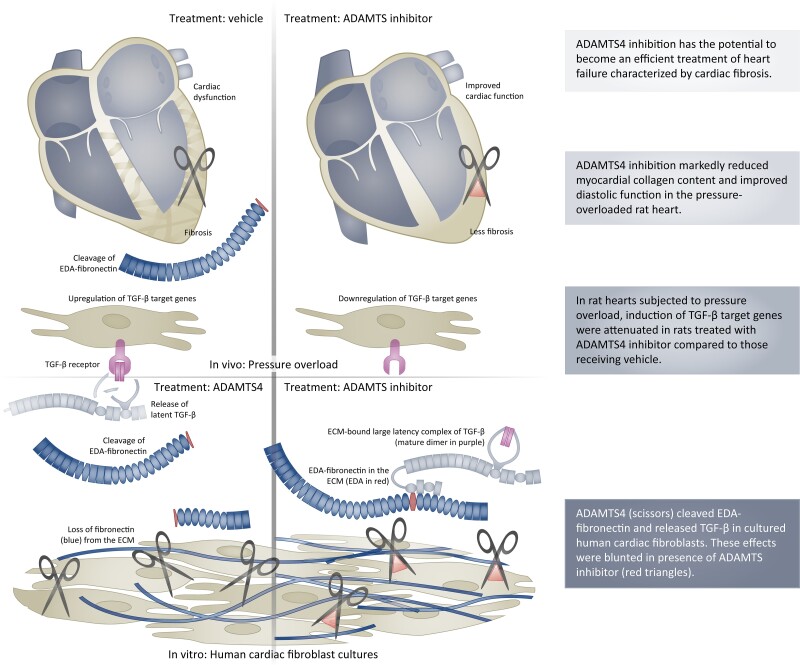
Aims: Heart failure is a condition with high mortality rates, and there is a lack of therapies that directly target maladaptive changes in the extracellular matrix (ECM), such as fibrosis. We investigated whether the ECM enzyme known as A disintegrin and metalloprotease with thrombospondin motif (ADAMTS) 4 might serve as a therapeutic target in treatment of heart failure and cardiac fibrosis.

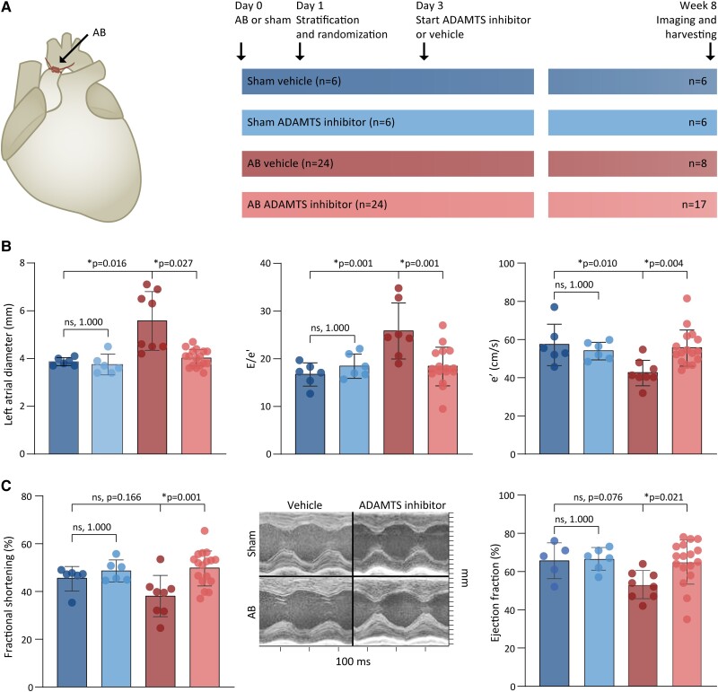
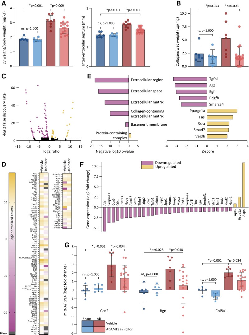
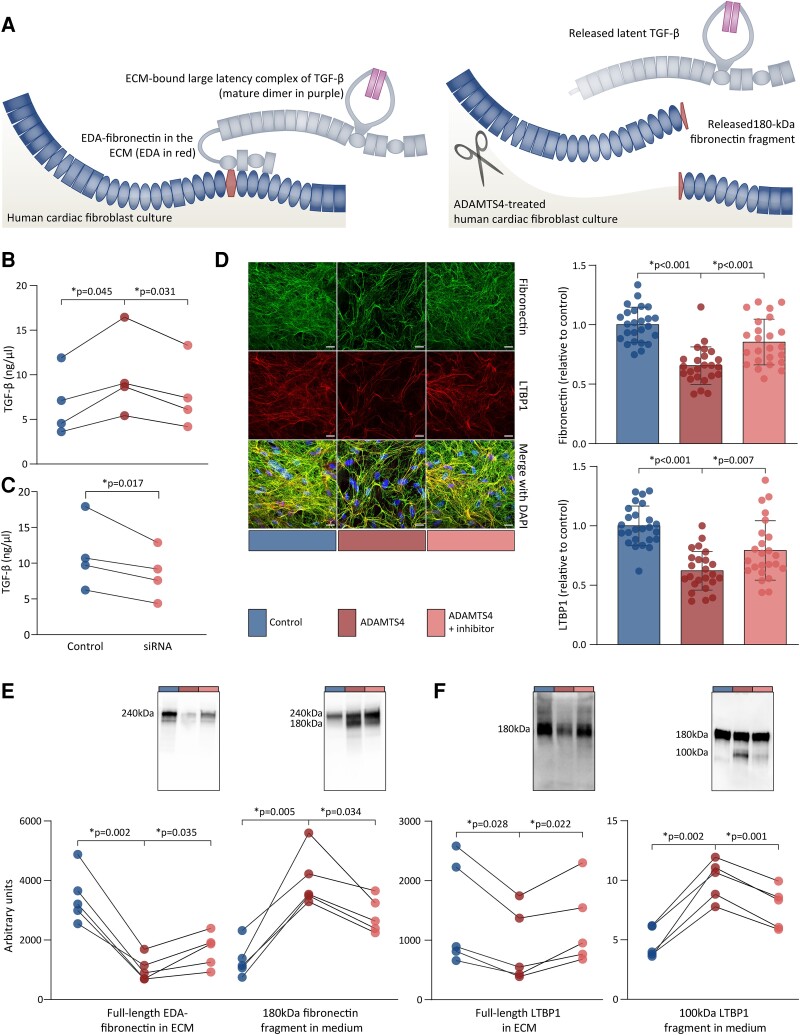
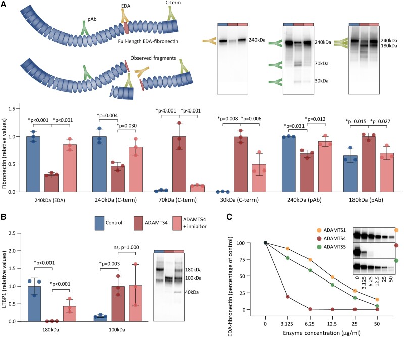
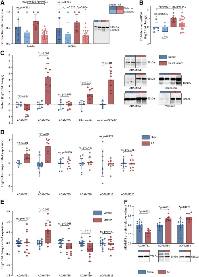
Methods and results: The effects of pharmacological ADAMTS4 inhibition on cardiac function and fibrosis were examined in rats exposed to cardiac pressure overload. Disease mechanisms affected by the treatment were identified based on changes in the myocardial transcriptome. Following aortic banding, rats receiving an ADAMTS inhibitor, with high inhibitory capacity for ADAMTS4, showed substantially better cardiac function than vehicle-treated rats, including ∼30% reduction in E/e' and left atrial diameter, indicating an improvement in diastolic function. ADAMTS inhibition also resulted in a marked reduction in myocardial collagen content and a down-regulation of transforming growth factor (TGF)-β target genes. The mechanism for the beneficial effects of ADAMTS inhibition was further studied in cultured human cardiac fibroblasts producing mature ECM. ADAMTS4 caused a 50% increase in the TGF-β levels in the medium. Simultaneously, ADAMTS4 elicited a not previously known cleavage of TGF-β-binding proteins, i.e. latent-binding protein of TGF-β and extra domain A-fibronectin. These effects were abolished by the ADAMTS inhibitor. In failing human hearts, we observed a marked increase in ADAMTS4 expression and cleavage activity.

Conclusion: Inhibition of ADAMTS4 improves cardiac function and reduces collagen accumulation in rats with cardiac pressure overload, possibly through a not previously known cleavage of molecules that control TGF-β availability. Targeting ADAMTS4 may serve as a novel strategy in heart failure treatment, in particular, in heart failure with fibrosis and diastolic dysfunction.

Keywords: ADAMTS enzymes; Cardiac fibrosis; Extracellular matrix; Heart failure; New therapy.


Conflict of interest statement

Conflict of interest: A patent application filed by the University of Oslo covering the use of ADAMTS4 inhibition in cardiac remodelling and heart failure (WO2015004209A1) is pending in Europe (EP3756667A1) and granted in the United States (US10744155B2). The inventors named in this patent are among the authors (M.V., G.C., J.M.A., I.S., I.G.L., and C.R.C.). The ADAMTS inhibitor used in this study was supplied free of charge by AstraZeneca under a material transfer agreement. A licence agreement with the pharmaceutical company Paradigm Biopharma for the development of pentosane polysulfate (PPS) in the treatment of cardiac remodelling and heart failure was signed between the University of Oslo and Paradigm in 2017, and a patent for the use of PPS in heart failure has been filed by Paradigm Biopharma (PCT/AU2022/051301) where M.V. and G.C. are among the inventors. M.V. has performed consulting services for Paradigm to support this development. The PPS was not used in this study. M.V. has participated in advisory board for Pharmacosmos (iron supplementation in heart failure).

Figures

Graphical Abstract
Graphical Abstract
ADAMTS4 inhibition prevents cardiac fibrosis by reducing the release of ECM-bound TGF-β due to EDA-fibronectin cleavage. TGF, transforming growth factor; ADAMTS4, a disintegrin and metalloprotease with thrombospondin motif; EDA, extra domain A; ECM, extracellular matrix.
Figure 1
Figure 1
Study design and effects of ADAMTS4 inhibition on cardiac function in pressure-overloaded rat hearts. (A) Study design for the testing of ADAMTS inhibitor in AB rats by four groups: (i) sham rats that received vehicle (dark blue); (ii) sham rats that received ADAMTS inhibitor (light blue); (iii) AB rats that received vehicle (red); and (iv) AB rats that received ADAMTS inhibitor (pink). (B) Diastolic function assessed by echocardiography; left atrial diameter (left), E/e′ ratio (middle), and diastolic tissue velocity at mitral annulus, i.e. e′ (right). (C) Systolic function evaluated in terms of fractional shortening measured by echocardiography (left) and ejection fraction measured by magnetic resonance imaging (right). Representative images for M-mode mid-ventricular recordings (middle). Bars represent mean ± 1 SD. Groups were compared by one-way ANOVA with planned comparisons followed by Bonferroni correction for the following comparisons: sham vehicle vs. sham ADAMTS inhibitor, sham vehicle vs. AB vehicle, and AB vehicle vs. AB ADAMTS inhibitor. P < 0.05 are considered significant and marked with *. AB, aortic banding; ADAMTS, a disintegrin and metalloprotease with thrombospondin motif.
Figure 2
Figure 2
Effects of ADAMTS inhibition on remodelling and disease pathways. (A) LV weight to body weight measured at necropsy (left) and interventricular wall thickness measured by echocardiography (right) 8 weeks after AB or sham surgery (sham vehicle n = 6, sham ADAMTS inhibitor n = 6, AB vehicle n = 8, AB ADAMTS inhibitor n = 17). (B) Fibrosis in LV as determined by the total collagen content as a proportion of wet weight quantified by HPLC. (C) Volcano plot showing the expression of genes with a false discovery rate less than 0.15 (black dots), up-regulated (yellow dots), and down-regulated genes (purple dots). Dotted line indicates a false discovery rate of 0.15. (D) Heatmap showing log2-transformed normalized counts of DEGs that were down-regulated (left) and up-regulated (right) in AB rats treated with vehicle and ADAMTS inhibitor. Genes with LOC and AARB prefixes are omitted. (E) Enrichment of DEGs in cellular compartments assessed by overrepresentation test of genes that were down-regulated (purple) or up-regulated (yellow) in AB rats treated with ADAMTS inhibitor compared with those treated with vehicle (left). Upstream regulators identified in IPA ranked by their Z-score (right). (F) Gene expression of DEGs that are identified as TGF-β target genes by IPA. (G) mRNA expression of selected TGF-β-inducible genes in myocardial samples determined by RT–qPCR (sham vehicle n = 5, sham ADAMTS inhibitor n = 6, AB vehicle n = 7, AB ADAMTS inhibitor n = 17). Bars represent mean ± 1 SD. Groups were compared by one-way ANOVA with planned comparisons followed by Bonferroni correction for the following comparisons: sham vehicle vs. sham ADAMTS4 inhibitor, sham vehicle vs. AB vehicle, and AB vehicle vs. AB ADAMTS inhibitor. P < 0.05 were considered significant and marked with *. LV, left ventricle; AB, aortic banding; ADAMTS4, a disintegrin and metalloprotease with thrombospondin motif; DEGs, differentially expressed genes; TGF, transforming growth factor; HPLC, high performance liquid chromatography; IPA, Ingenuity pathways analysis; RT–qPCR, real-time quantitative polymerase chain reaction.
Figure 3
Figure 3
Disruption of TGF-β and its ECM-anchoring proteins by ADAMTS4 in human cardiac fibroblast cultures. Human adult (B) or foetal (C–E) cardiac fibroblasts were cultured for 7 days and treated with DMSO or ADAMTS4 with or without ADAMTS inhibitor for 24 h. (A) Illustration shows how EDA-fibronectin (blue and red) anchors the large latency complex of TGF-β (grey and purple) to the ECM (left). In response to ADAMTS4 (scissor), fibronectin fragments and latent TGF-β are released from the ECM (right). (B) Total TGF-β levels in heat-activated media as quantified by tMLC from four independent experiments. (C) Total TGF-β levels in heat-activated media as quantified by tMLC from four independent experiments using siRNA to knock down ADAMTS4. (D) Representative images of immunofluorescence staining of fibronectin and LTBP1 in the ECM. Area of fibronectin and LTBP1 staining relative to cell number (DAPI) were quantified and normalized to DMSO controls. Data represent quantifications of confocal Z-stacks (n = 8–9 per experiment) from three independent experiments and the graph bars represent mean ± 1 SD. Scale bars represent 20 μm. (E) Immunoblots show full-length EDA-fibronectin (240 kDa) and fragments (180 kDa) in the ECM and medium, respectively. (F) LTBP1 full-length (180 kDa) and fragments (100 kDa) in ECM and medium, respectively. Immunoreactive bands were quantified and normalized to total protein levels. Comparisons between groups were assessed by one-way ANOVA for correlated samples (B, D, and E) or planned comparisons (C) with Bonferroni correction for multiple testing of controls vs. ADAMTS4 and ADAMTS4 vs. ADAMTS inhibitor. Data points from the same experiment are connected by lines. P < 0.05 were considered significant and are marked with *. TGF, transforming growth factor; DMSO, dimethyl sulfoxide; tMLC, transfected mink lung epithelial cells; DAPI, 4′,6-diamidino-2-phenylindole; LTBP, latent TGF-β-binding protein; EDA, extra domain A; ECM, extracellular matrix; ADAMTS4, a disintegrin and metalloprotease with thrombospondin motif.
Figure 4
Figure 4
ADAMTS4-mediated cleavage of EDA-fibronectin and LTBP1. Human cardiac fibroblasts were cultured for 7 days, before lysates were treated with DMSO or ADAMTS4 with or without ADAMTS inhibitor. (A) Fibronectin cleavage assessed by antibodies specific for EDA and C-terminus (C-term), in addition to a polyclonal antibody (pAb). Relative values to control (full-length protein) or to ADAMTS4-treated lysates (fragments) are shown. Representative immunblots shown. Illustration shows the fibronectin fragments that were detected by the three antibodies. Cleavage sites were as indicated by the size of the fragments and antibody epitopes. (B) Representative immunoblot and quantification showing LTBP1 cleavage by ADAMTS4. (C) Concentration response of fibronectin cleavage by ADAMTS4 (red) in comparison with ADAMTS1 (yellow) and -5 (green), determined by the amount of full-length EDA-fibronectin detected by EDA-specific antibody in response to increasing enzyme concentrations. Immunoreactive bands were normalized to total protein levels. Data represent quantifications from three independent experiments, and the graph bars represent mean ± 1 SD (A and B). Comparisons between groups assessed by one-way ANOVA with Bonferroni correction for the following comparisons: control vs. ADAMTS4; and ADAMTS4 vs. ADAMTS inhibitor. For 70 kDa C-terminal fibronectin fragment and LTBP1, log2-transformed values are used. P < 0.05 were considered significant and are marked with *. DMSO, dimethyl sulfoxide; EDA, extra domain A; LTBP, latent TGF-β-binding protein; ADAMTS4, a disintegrin and metalloprotease with thrombospondin motif.
Figure 5
Figure 5
ADAMTS4 activity is increased in the myocardium of rats and patients with cardiac dysfunction. (A) The myocardial amount of full-length fibronectin (240 kDa) and its cleavage fragment (180 kDa) as determined by immunoblots in sham vehicle (n = 6), sham ADAMTS inhibitor (n = 6), AB vehicle (n = 7), and AB ADAMTS inhibitor (n = 17). Representative blots shown. (B) mRNA levels of EDA-fibronectin in myocardial samples from AB rats determined by RT–qPCR in sham vehicle (n = 5), sham ADAMTS inhibitor (n = 6), AB vehicle (n = 7), and AB ADAMTS inhibitor (n = 17). (C) The levels of active ADAMTS1, -4, and -5 proteins, fibronectin 180 kDa fragments, and versican DPEAAE fragments in myocardial samples from explanted human failing hearts (red) compared with healthy donor hearts (blue). Representative blots shown for ADAMTS4 (left), EDA-fibronectin (middle), and versican DPEAAE fragments (right). (D) mRNA levels of ADAMTS1, -4, -5, -9, -15, and -20 in myocardial samples from rats 6 weeks after AB (n = 8) or sham (n = 8). (E) Log2-transformed mRNA levels of ADAMTS1, -4, -5, -9, and -15 in adult human cardiac fibroblasts exposed to stretch (n = 11) compared to control conditions (n = 11). ADAMTS20 levels were not detectable. (F) The levels of ADAMTS1, -4, and -5 protein in myocardial lysates from rats 6 weeks after AB (n = 8) or sham (n = 8) relative to sham group. Bars represent mean ± 1 SD. Groups were compared by one-way ANOVA with planned comparisons followed by Bonferroni correction for the following comparisons: sham vehicle vs. sham ADAMTS inhibitor, sham vehicle vs. AB vehicle, and AB vehicle vs. AB ADAMTS inhibitor. Groups were compared by Student’s t-test for the following comparisons: sham vs. AB, control vs. stretched cells, and donor vs. heart failure patients. P < 0.05 were considered significant and marked with *. AB, aortic banding; EDA, extradomain A; ADAMTS4, a disintegrin and metalloprotease with thrombospondin motif; RT–qPCR, real-time quantitative polymerase chain reaction; DCM, dilated cardiomyopathy.

Similar articles

Cited by

References

    1. Heidenreich PA, Albert NM, Allen LA, Bluemke DA, Butler J, Fonarow GC, Ikonomidis JS, Khavjou O, Konstam MA, Maddox TM, Nichol G, Pham M, Piña IL, Trogdon JG. Forecasting the impact of heart failure in the United States. Circ Heart Fail 2013;6:606–619. - PMC - PubMed
    1. Taylor CJ, Ordóñez-Mena JM, Roalfe AK, Lay-Flurrie S, Jones NR, Marshall T, Hobbs FDR. Trends in survival after a diagnosis of heart failure in the United Kingdom 2000–2017: population based cohort study. BMJ 2019;364:l223. - PMC - PubMed
    1. Virani SS, Alonso A, Aparicio HJ, Benjamin EJ, Bittencourt MS, Callaway CW, Carson AP, Chamberlain AM, Cheng S, Delling FN, Elkind MSV, Evenson KR, Ferguson JF, Gupta DK, Khan SS, Kissela BM, Knutson KL, Lee CD, Lewis TT, Liu J, Loop MS, Lutsey PL, Ma J, Mackey J, Martin SS, Matchar DB, Mussolino ME, Navaneethan SD, Perak AM, Roth GA, Samad Z, Satou GM, Schroeder EB, Shah SH, Shay CM, Stokes A, VanWagner LB, Wang N-Y, Tsao CW. Heart disease and stroke statistics—2021 update. Circulation 2021;143:e254–e743. - PubMed
    1. Sweeney M, Corden B, Cook SA. Targeting cardiac fibrosis in heart failure with preserved ejection fraction: mirage or miracle? EMBO Mol Med 2020;12:e10865. - PMC - PubMed
    1. Weidemann F, Herrmann S, Störk S, Niemann M, Frantz S, Lange V, Beer M, Gattenlöhner S, Voelker W, Ertl G, Strotmann JM. Impact of myocardial fibrosis in patients with symptomatic severe aortic stenosis. Circulation 2009;120:577–584. - PubMed

Publication types