. 2023 Apr 25;9(1):44.
doi: 10.1038/s41421-023-00531-5.

The asymmetrical ESR1 signaling in muscle progenitor cells determines the progression of adolescent idiopathic scoliosis

Affiliations

The asymmetrical ESR1 signaling in muscle progenitor cells determines the progression of adolescent idiopathic scoliosis

Xiexiang Shao et al. Cell Discov. .

Abstract

Adolescent Idiopathic Scoliosis (AIS) is a common pediatric skeletal disease highly occurred in females. The pathogenesis of AIS has not been fully elucidated. Here, we reveal that ESR1 (Estrogen Receptor 1) expression declines in muscle stem/progenitor cells at the concave side of AIS patients. Furthermore, ESR1 is required for muscle stem/progenitor cell differentiation and disrupted ESR1 signaling leads to differentiation defects. The imbalance of ESR1 signaling in the para-spinal muscles induces scoliosis in mice, while reactivation of ESR1 signaling at the concave side by an FDA approved drug Raloxifene alleviates the curve progression. This work reveals that the asymmetric inactivation of ESR1 signaling is one of the causes of AIS. Reactivation of ESR1 signaling in para-spinal muscle by Raloxifene at the concave side could be a new strategy to treat AIS.


Conflict of interest statement

The authors declare no competing interests.

Figures
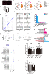
Fig. 1
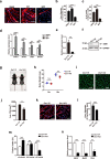
Fig. 1. ESR1 expression decreases in muscle stem/progenitor cells at the concave side of AIS patients.
a Representative immunofluorescent staining of bilateral para-spinal muscle sections from an AIS patient. Cryosections were obtained from the convex and concave sides of AIS patients. The cryosections were stained with anti-Laminin antibody. Red indicated laminin; Blue indicated DAPI staining of nuclei. The merged images were shown. Scale bars: 100 μm. b Statistical analysis of the cross-sectional area (CSA) of myofibers. At least 350 fibers were analyzed for each sample. Error bars indicated standard deviation (n = 6). **P < 0.01. c Muscle tissues harvested from the concave and convex side of AIS patients were subjected for scRNA-seq. Samples from two patients were used. Total 16,930 cells were analyzed. The UMAP plots of main cell types for muscles originated from concave and convex side were shown. FAPs indicated fibro/adipogenic progenitors; ECs indicated epithelia cells; SMCs indicated smooth muscle cells; MuSCs/progenitors indicated muscle stem/progenitor cells. d Dot plot of highly expressed genes in each cell type. The size of dots indicated the percentage of cells expressing the designated genes. The color of dots represented gene expression level. e Violin plot of highly expressed genes in each cell type. f Percentage of each cell type at the concave and convex side. g UMAP plots of genes marked muscle stem/progenitor cells. h Pathway enrichment analysis of differentially expressed genes in muscle stem/progenitor cells located at the concave and convex side of AIS patients. Red bars indicated the enriched terms up-regulated in muscle stem/progenitor cells located at the concave side. Blue bars indicated the enriched terms down-regulated in muscle stem/progenitor cells located at the concave side. The enriched terms related to myogenesis were highlighted by the red solid frames and the terms related to estrogen signaling were highlighted by the red dashed frames. i Dot graph of ESR1 and ESR2 expression in each cell types identified in muscle located at the concave and convex side of AIS patients, respectively. j RT-qPCR analysis of ESR1 and ESR2 gene expression levels in muscle stem/progenitor cells isolated from para-spinal muscles derived from non-scoliosis (NS, n = 5), congenital scoliosis (CS, n = 8) and adolescent idiopathic scoliosis (AIS, n = 25) patients. Error bars indicated standard deviation. **P < 0.01; ns indicated no significant changes. k ELISA analysis of ESR1 level in muscle stem/progenitor cells isolated from para-spinal muscles derived from non-scoliosis (NS, n = 5), congenital scoliosis (CS, n = 8) and adolescent idiopathic scoliosis (AIS, n = 25) patients. Error bars indicated standard deviation. **P < 0.01; ns indicated no significant changes.
Fig. 2
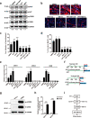
Fig. 2. ESR1 is required for muscle stem/progenitor cell differentiation.
a Representative immunofluorescent staining of differentiating human muscle stem/progenitor cells treated with DMSO, ESR1 agonist PPT, or ESR1 antagonist MPP for six days. Red indicated MyHC; Blue indicated DAPI staining of nuclei. The merged images were shown. Scale bars: 100 μm. b Statistical analysis of average diameters of myotubes. Error bars indicated standard deviation (n = 5). ***P < 0.001. c Statistical analysis of differentiation efficiency. Error bars indicated standard deviation (n = 5). ***P < 0.001. d Expression levels of differentiation markers. Total RNA was extracted from differentiating cells treated by DMSO, ESR1 agonist PPT, or ESR1 antagonist MPP for six days followed by RT-qPCR analysis. Error bars indicated standard deviation (n = 3). ***P < 0.001, **P < 0.01. e Gene expression level of Esr1 in Esr1 KO muscle stem cells. The primary muscle stem cells were isolated from Pax7-CreERT2; Esr1f/f mice after tamoxifen induction. Esr1 expression level was next analyzed by RT-qPCR. Error bars indicated standard deviation (n = 3). **P < 0.01. f Immunoblotting of Esr1 in Esr1 KO muscle stem cells. The primary muscle stem cells were isolated from Pax7-CreERT2; Esr1f/f mice after tamoxifen induction. GAPDH was served as control. g Pictures of 8-weeek-old Esr1f/f and Esr1 KO female mice. Scale bar: 1 cm. h Statistical analysis of the body weight of Esr1f/f and Esr1 KO mice. Mice were treated with vehicle (Esr1f/f) or tamoxifen (Esr1 KO) every other day from 2 weeks old to 3 weeks old. Five female mice were weighted at 3 weeks old and 8 weeks old in each group. Error bars indicated standard deviation (n = 5). **P < 0.01; ns indicated no significant changes. i Representative immunofluorescent staining of cryosection of para-spinal muscle derived from Esr1f/f and Esr1 KO mice, respectively. Green indicated laminin; Blue indicated DAPI staining of nuclei. The merged images were shown. Scale bars: 100 μm. j Statistical analysis of cross-sectional area of para-spinal muscle derived from Esr1f/f and Esr1 KO mice, respectively. At least 500 fibers were analyzed for each sample. Error bars indicated standard deviation (n = 4). **P < 0.01. k Representative immunofluorescent staining of myotubes differentiated from muscle stem cells isolated from Esr1f/f and Esr1 KO mice, respectively. Red indicated MyHC; Blue indicated DAPI staining of nuclei. The merged images were shown. Scale bars: 100 μm. l Statistical analysis of the average diameters of myotubes differentiated from Esr1f/f or Esr1 KO muscle stem cells. Error bars indicated standard deviation (n = 5). ***P < 0.001. m Fusion index of myotubes differentiated from Esr1f/f or Esr1 KO muscle stem cells. Error bars indicated standard deviation (n = 5). ***P < 0.001; ns indicated no significant changes. Fusion index of myotubes differentiated from Esr1f/f or Esr1 KO muscle stem cells. Error bars indicated standard deviation (n = 5). ***P < 0.001; ns indicated no significant changes. n Expression levels of differentiation markers in myotubes differentiated from Esr1f/f or Esr1 KO muscle stem cells. Error bars indicated standard deviation (n = 3). **P < 0.01; ***P < 0.001.
Fig. 3
Fig. 3. ESR1 activates AKT signaling in human muscle stem/progenitor cells.
a Representative immunoblotting of phosphorylated AKT, total AKT, phosphorylated CREB, total CREB, and GAPDH of human muscle stem/progenitor cells with different treatments. Upon initiation of differentiation, DMSO, estradiol, ESR1 agonist PPT, or ESR1 antagonist MPP was added in culture, respectively. Cells were treated for 2 h and harvested for analysis. Total proteins were extracted and subjected for immunoblotting. The quantification of the band intensity was listed below each band. b Representative immunofluorescent staining of MyHC with differentiated human muscle stem/progenitor cells. Human muscle stem/progenitor cells were differentiated for 6 days and treated with DMSO, PPT, SC79, AKTi-1/2, 666-15, PPT combined with AKTi-1/2, and PPT combined with 666-15, respectively. PPT, ESR1 agonist; MPP, ESR1 antagonist; SC79, AKT signaling agonist; AKTi-1/2, AKT signaling inhibitor; 666-15, CREB inhibitor. Red indicated MyHC; blue indicated DAPI staining of nuclei. The merged images were shown. Scale bars: 100 μm. c Statistical analysis of the average diameter of myotubes by small molecule treatment. Error bars indicated standard deviation (n = 5). **P < 0.01; ***P < 0.001. d Statistical analysis of the differentiation efficiency. Error bars indicated standard deviation (n = 5). *P < 0.05; ***P < 0.001. e Expression levels of MYH1, MYH3, and CKM. Human muscle stem/progenitor cells were induced to differentiate for 6 days while treating with various types of small molecules targeting ESR1-AKT-CREB signaling. Total RNA was extracted from each sample and subjected for RT-qPCR analysis. Error bars indicated standard deviation (n = 3). **P < 0.01; ***P < 0.001. f The scheme of luciferase construct. The red triangles indicated predicted CREB binding site (cAMP response element). TSS indicated the transcription starting site. The potential promoter region of MYH1 (–1958 bp to TSS) or MYH3 (–1536 bp to TSS) was cloned into the MYH-Luciferase plasmid. g Expression of ectopic CREB and phosphorylated CREB in luciferase assays. The 293T cells were transfected by plasmid encoding CREB and harvested 2 days after transfection. Total proteins were extracted and subjected for immunoblotting of CREB and phosphorylated CREB. GAPDH served as the internal control. h Statistical analysis of luciferase essays. Error bars indicated standard deviation (n = 3). ***P < 0.001. i ESR1 signaling regulated muscle stem cell differentiation. PPT, ESR1 agonist; SC79, AKT signaling agonist; AKTi-1/2, AKT signaling inhibitor; 666-15, CREB inhibitor.
Fig. 4
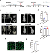
Fig. 4. Asymmetric inactivation of ESR1 in para-spinal muscle leads to scoliosis.
a Scheme of unilateral MPP treatment. Bipedal mice of 3 weeks old were subjected for unilateral intramuscular injection of MPP at the left side of the para-spinal muscle, while DMSO was injected to the opposite side. Continuous injections were performed twice every week for 3 weeks. The spinal alignment was evaluated 5 weeks after the first MPP injection. b Micro-CT images for spinal alignment and thoracic cage symmetry evaluation in bilateral DMSO and unilateral MPP group 5 weeks after the first MPP injection. c In vivo X-ray images of spinal alignment in bilateral DMSO and unilateral MPP group 5 weeks after the first MPP injection. d Statistical analysis of Cobb angle in coronal plane for bilateral DMSO group (n = 7) and unilateral MPP group (n = 8) 5 weeks after the first injection. Error bars indicated standard deviation. ***P < 0.001. e Statistical analysis of kyphosis in sagittal plane for bilateral DMSO group (n = 7) and unilateral MPP group (n = 8) 5 weeks after the first injection. Error bars indicated standard deviation. *P < 0.05. f Representative immunofluorescent staining of para-spinal muscle sections from unilateral MPP group. Cryosections were obtained from the para-spinal muscles isolated from the DMSO injection side and MPP injection side. The cryosections were stained with anti-Laminin antibody. Green indicated laminin; Blue indicated DAPI staining of nuclei. The merged images were shown. Scale bars: 100 μm. g Statistical analysis of the CSA of myofibers from unilateral MPP group. At least 400 fibers were analyzed for each sample. Error bars indicated standard deviation (n = 5). *P < 0.05.
Fig. 5
Fig. 5. Raloxifene activates ESR1-AKT-CREB-MYH signaling and rescues the differentiation defects of concave muscle stem/progenitor cells.
a Representative immunoblotting of AKT, phosphorylated AKT, CREB, and phosphorylated CREB of human muscle stem/progenitor cells. Upon initiation of differentiation, DMSO or Raloxifene was added in culture for 2 h. Then total proteins were extracted and subjected for immunoblotting. GAPDH served as an internal control. The quantification of the band intensity was listed below each band. b Representative immunofluorescent staining of human muscle stem/progenitor cells treated with Raloxifene. Human muscle stem/progenitor cells were isolated from either the convex or concave side of AIS patients. The cells isolated from the concave side (treated by DMSO or Raloxifene) and convex side (treated by estradiol) were differentiated for 6 days and subjected for MyHC immunofluorescent staining. Red indicated MyHC; blue indicated DAPI staining of nuclei. The merged images were shown. Scale bars: 100 μm. c The statistical analysis of the average diameter of myotubes differentiated from bilateral muscle stem/progenitor cells in AIS with different treatments. Error bars indicated standard deviation (n = 5). ***P < 0.001; ns indicated no significant changes. d The statistical analysis of the differentiation efficiency after different treatments. Error bars indicated standard deviation (n = 5). ***P < 0.001; ns indicated no significant changes. e The mRNA level of MYH1, MYH3, and CKM in myotubes with different treatments. Human muscle stem/progenitor cells isolated from the concave side (treated by DMSO or Raloxifene) and convex side (treated by estradiol) were differentiated for 6 days. Total RNA was extracted and subjected for RT-qPCR analysis. Error bars indicated standard deviation (n = 3). **P < 0.01, ***P < 0.001; ns indicated no significant changes.
Fig. 6
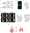
Fig. 6. Raloxifene alleviates the progression of scoliosis.
a Scheme of Raloxifene treatment in AIS animal model. Scoliosis mouse model was generated as aforementioned. After the occurrence of scoliosis, intramuscular Raloxifene injections were performed at the concave para-spinal muscle once every 3.5 days for 2 weeks (from the 5th week to the 7th week). The spinal alignment and para-spinal muscle size were then analyzed at 9th week. b Representative immunofluorescent staining of Raloxifene-treated or DMSO-treated concave para-spinal muscle 2 weeks after final injection. Cryosections were obtained from para-spinal muscle at the concave side with either DMSO mock treatment or Raloxifene treatment and subjected for laminin immunofluorescent staining. Green indicated laminin; blue indicated DAPI staining of nuclei. The merged images were shown. Scale bars: 100 μm. c Statistical analysis of the CSA of myofibers in Raloxifene treated or DMSO treated concave para-spinal muscle. At least 500 fibers were analyzed for each sample. Error bars indicated standard deviation (n = 5). **P < 0.01. d In vivo X-ray images of spinal alignment before and after Raloxifene treatment. e Statistical analysis of Cobb angle in coronal plane after DMSO mock treatment (n = 8) and Raloxifene treatment (n = 8), respectively. 5th week indicated before treatment; 9th week indicated after treatment. Error bars indicated standard deviation (n = 8). ns indicated no significant changes; ***P < 0.001. f Statistical analysis of kyphosis in sagittal plane after DMSO mock treatment (n = 8) and Raloxifene treatment (n = 8), respectively. 5th week indicated before treatment; 9th week indicated after treatment. Error bars indicated standard deviation (n = 8). ns indicated no significant changes; ***P < 0.001. g Asymmetric ESR1 signaling increased the susceptibility to AIS. Restoration of ESR1 signaling by Raloxifene (indicated by R in yellow triangles) alleviated the progression of scoliosis.

Similar articles

Cited by

References

    1. Sarwark JF, Castelein RM, Maqsood A, Aubin CE. The biomechanics of induction in adolescent idiopathic scoliosis: theoretical factors. J. Bone Joint Surg. Am. 2019;101:e22. doi: 10.2106/JBJS.18.00846. - DOI - PubMed
    1. Cheng JC, et al. Adolescent idiopathic scoliosis. Nat. Rev. Dis. Prim. 2015;1:15030. doi: 10.1038/nrdp.2015.30. - DOI - PubMed
    1. Kim W, et al. Clinical evaluation, imaging, and management of adolescent idiopathic and adult degenerative scoliosis. Curr. Probl. Diagn. Radiol. 2018;48:402–414. doi: 10.1067/j.cpradiol.2018.08.006. - DOI - PubMed
    1. Dunn J, et al. Screening for adolescent idiopathic scoliosis: evidence report and systematic review for the US preventive services task force. JAMA. 2018;319:173–187. doi: 10.1001/jama.2017.11669. - DOI - PubMed
    1. Tambe AD, Panikkar SJ, Millner PA, Tsirikos AI. Current concepts in the surgical management of adolescent idiopathic scoliosis. Bone Joint J. 2018;100-B:415–424. doi: 10.1302/0301-620X.100B4.BJJ-2017-0846.R2. - DOI - PubMed