. 2023 May 4;24(9):8242.
doi: 10.3390/ijms24098242.

Effects of Therapeutic Platelet-Rich Plasma on Overactive Bladder via Modulating Hyaluronan Synthesis in Ovariectomized Rat

Affiliations

Effects of Therapeutic Platelet-Rich Plasma on Overactive Bladder via Modulating Hyaluronan Synthesis in Ovariectomized Rat

Jian-He Lu et al. Int J Mol Sci. .

Abstract

Postmenopausal women who have ovary hormone deficiency (OHD) may experience urological dysfunctions, such as overactive bladder (OAB) symptoms. This study used a female Sprague Dawley rat model that underwent bilateral ovariectomy (OVX) to simulate post-menopause in humans. The rats were treated with platelet-rich plasma (PRP) or platelet-poor plasma (PPP) after 12 months of OVX to investigate the therapeutic effects of PRP on OHD-induced OAB. The OVX-treated rats exhibited a decrease in the expression of urothelial barrier-associated proteins, altered hyaluronic acid (hyaluronan; HA) production, and exacerbated bladder pathological damage and interstitial fibrosis through NFƘB/COX-2 signaling pathways, which may contribute to OAB. In contrast, PRP instillation for four weeks regulated the inflammatory fibrotic biosynthesis, promoted cell proliferation and matrix synthesis of stroma, enhanced mucosal regeneration, and improved urothelial mucosa to alleviate OHD-induced bladder hyperactivity. PRP could release growth factors to promote angiogenic potential for bladder repair through laminin/integrin-α6 and VEGF/VEGF receptor signaling pathways in the pathogenesis of OHD-induced OAB. Furthermore, PRP enhanced the expression of HA receptors and hyaluronan synthases (HAS), reduced hyaluronidases (HYALs), modulated the fibroblast-myofibroblast transition, and increased angiogenesis and matrix synthesis via the PI3K/AKT/m-TOR pathway, resulting in bladder remodeling and regeneration.

Keywords: hyaluronan; ovariectomy; overactive bladder; platelet rich plasma.


Conflict of interest statement

The authors declare no conflict of interest.

Figures
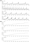
Figure 1
Figure 1
Platelet-rich plasma (PRP) treatment improved voiding behavior and ameliorated bladder overactivity in the ovary hormone deficiency (OHD)-induced overactive bladder (OAB) rat model. Cystometrography parameters (A) and tracing analysis of 24 h voiding behavior (B) were shown. The OVX group significantly increased micturition frequency, pressure, voiding contractions and non-voiding contractions (asterisk), whereas PRP treatment significantly improved bladder voiding pattern and capacity. n = 6 in each group.
Figure 2
Figure 2
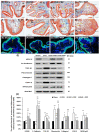
Therapeutic effect of PRP improved OHD-induced pathological alteration by Masson’s trichrome staining, immunostaining and Western blots. (AD, A’D’): Bladder pathological features of the sham group (A,A’), the OVX group (B,B’), the OVX + PRP group (C,C’) and the OVX + PPP group (D,D’). Masson’s trichrome stain showed red-stained smooth muscle and green-stained collagen. In the sham group (A,A’), there were three to five layers of the UL (black arrows), only sparse collagen (blue arrows) and few mononuclear cells (yellow arrows) distributed in the SL (lamina propria). In the OVX group (B,B’), the morphology of bladder was characterized by a thinner layer (black arrows), much mononuclear cells (yellow arrows), collagen accumulation and increased interstitial fibrosis (blue arrows). In contrast, the OVX + PRP group (C,C’) and the OVX + PPP group (D,D’) improved OHD-induced bladder damages by increasing the thicker layer of urothelium (black arrows) and reducing interstitial fibrosis (blue arrows) compared with the OVX group. Additionally, there were many gathered red blood cells (purple arrows) beneath UL and mononuclear cells (yellow arrows) in the SL of the OVX + PRP group. Furthermore, there was a vacuolation in UL (purple arrows) in the OVX + PPP group (D,D’). Scale bar = 100 μm. Original agnification, ×200 (AD). Magnification, × 400 (A’D’). (EH): The distribution of adhesion protein E-Cadherin was expressed by immunostaining. In the sham group (E), the E-Cadherin staining was found in intercellular junctions of urothelium. On the contrary, there was less E-Cadherin staining expression in the thin UL of the OVX group (F), but the immunostaining in the OVX + PRP group (G) and the OVX + PPP group (H) was enhanced in UL. In particular, there were some exfoliated epithelial cells in the lumen of the OVX + PPP group. (I,J): The expressions of urothelial structure (E-Cadherin and UPKIII) and bladder inflammation (TGF-ß1, COX-2 and NFƘB-p65), interstitial fibrosis (fibronectin and collagen I) proteins were analyzed by Western blots. The inflammatory and fibrosis markers were noticeably decreased in the OVX + PRP group and the OVX + PPP group compared to the OVX group. Results were normalized as the sham group (the control group) = 100%. Note: UL, urothelial layer; SL, suburothelial layer; UPKIII, uroplakin III; TGF-ß1, transforming growth factor ß1. Data were expressed as means ± SD for n = 6, * p < 0.05; ** p < 0.01 versus the sham group; †† p < 0.01 versus the OVX group; # p < 0.05; ## p < 0.01 versus the OVX + PRP group.
Figure 3
Figure 3
Effects of PRP instillation strengthened proliferation and tight junction reconstruction. The expressions of urothelial proliferating markers (Ki67 and CK14) and tight junction markers (Claudin-4 and ZO-1) were evaluated by immunostaining (AH) and Western blotting (I,J). (AD): The Ki67 staining (yellow arrows) had less distribution in the bladder tissues of the sham group (A) and the OVX group (B). On the other hand, the immunostaining was markedly expressed in the UL and SL in the OVX + PRP group (C) and the OVX + PPP group (D). The yellow arrows indicated the Ki67-positive cells. (EH): Double-labeled analysis of Claudin-4 (fluorescein isothiocyanate; green, upper panels) and CK14 (rhodamine; red, lower panels) (yellow arrows) was widely distributed in the UL of the sham group (E). The OVX group (F) showed that double labeling was restricted to the thin and disrupted UL. However, the staining of the OVX + PRP group (G) and the OVX + PPP group (H) were obviously expressed in the UL compared to the OVX group (F). Nuclear DNA was labeled with DAPI (blue). The yellow arrows indicated double staining of Claudin-4 and CK14 in urothelium. (AD). (I,J): Quantifications of the percentage of Ki67, CK14, Claudin-4 and ZO-1 were examined by Western blotting. Western blotting analysis showed that PRP instillation increased the expression of urothelial proliferating markers (Ki67 and CK14) and tight junction markers (Claudin-4 and ZO-1), which improved proliferation and tight junction reconstruction. Note: UL, urothelial layer; SL, suburothelial layer; CK, cytokeratin. Original magnification, × 400 (AH). Scale bars = 100 mm. Results were normalized as the sham group = 100%. Data were expressed as means ± SD for n = 6, * p < 0.05; ** p < 0.01 versus the sham group; p < 0.05; †† p < 0.01 versus the OVX group; # p < 0.05 versus the OVX + PRP group.
Figure 4
Figure 4
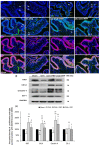
PRP instillation modulated bladder angiogenic remodeling and altered bladder interstitial cells. The angiogenesis related proteins [α-SMA, VEGF, VEGF-R1, VEGF-R2 (VEGF receptor), laminin and integrin-α6 (laminin receptor)] and interstitial cell markers (vimentin, C-Kit and PDGFR) were analyzed by immunostaining (AL) and Western blotting (MP). (AD): In the sham group (A), the α-SMA immunostaining (yellow arrows) was widely distributed in the smooth muscle of microvessels beneath urothelial basal layer and vessels in the suburothelial layer (SL) and muscular layer (ML), including small artery, small vein, arteriole and venule. In the OVX group (B), the staining was reduced in SL compared to the sham group. However, the staining levels in the OVX + PRP group (C) and the OVX + PPP group (D) were significantly increased beneath the urothelial basal layer and in the SL (yellow arrows) compared to the OVX group. Particularly, there are many gathered α-SMA positive-myofibroblasts and microvessels beneath urothelial basal layer, lamina propria and ML in the OVX + PRP group. (EH): Similar immunostaining results with α-SMA were obtained for the laminin expression. The laminin staining was significantly increased beneath the urothelial basal layer, in myofibroblasts and microvessels of SL (yellow arrows) and microvessels of ML (pink arrows) in the OVX + PRP group (G) and the OVX + PPP group (H) compared to the sham group (E) and the OVX group (F). (IL): In the sham group (I), the c-Kit immunostaining was distributed in bladder SL and ML. The C-Kit immunostaining (yellow arrows) in the OVX group (J) was abundantly expressed in the SL and ML compared to the sham group (I). However, the expressions of markers were obviously reduced in the OVX +PRP group (K) and the OVX +PPP group (L) compared to the OVX group. Nucleus was stained by DAPI (blue). Scale bars = 100 mm. (M,N): Quantifications of the percentage for angiogenesis associated proteins was quantified by Western blots. (O,P): Quantifications of the percentage for interstitial cell associated proteins were shown. Results were normalized as the sham group = 100%. The protein level in the OVX group was much lower than the sham group. However, the expressions of angiogenesis-associated markers were significantly increased in the OVX + PRP group compared to the OVX group, indicating that PRP improved bladder angiogenic remodeling. Note: UL, urothelial layer; SL, suburothelial layer; ML, muscular layer; α-SMA, alpha-smooth muscle actin; VEGF, vascular endothelial growth factor; PDGFR, platelet-derived growth factor receptors. Data were expressed as means ± SD for n = 6, * p < 0.05; ** p < 0.01 versus the sham group; p < 0.05; †† p < 0.01 versus the OVX group; # p < 0.05; ## p < 0.01 versus the OVX + PRP group.
Figure 5
Figure 5
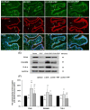
Effects of PRP instillation enhanced the HA receptor expression. The expressions of HA receptors in the bladder were analyzed by immunostaining (AD) and Western blotting (E,F). (AD): In the sham group (A), CD44 expression (green) co-stained with E-Cadherin (red) was mainly distributed in the basal layer of UL (yellow arrows), and slightly co-labeled in stroma cells and vessels of SL (white arrows). (B): In the OVX-treated group, CD44 protein was mainly expressed throughout the ulcerated and disrupted urothelium (yellow arrows) and the stromal cells as well as vessels (white arrows) of the SL. (C,D): In the OVX + PRP group and the OVX + PPP group, CD44 positive cells were predominantly expressed in the basal layer of hyperplasia urothelium (yellow arrows) and were slightly stained in vessels of the SL (white arrows). (E,F): The protein levels of HA receptors (CD44, RHAMM and TLR-4) were investigated in the different groups by Western blotting analysis and normalized to the sham group. The yellow arrows indicated double staining of RHAMM and E-Cadherin in urothelium, and white arrows indicated double staining in stroma cells. Note: UL, urothelial layer; SL, suburothelial layer; HA, hyaluronic acid; RHAMM, receptor for HA-mediated motility; TLR-4, Toll-like receptor-4; GAPDH, glyceraldehyde-3-phosphate dehydrogenase. Data were expressed as means ± SD for n = 6, * p < 0.05; ** p < 0.01 versus the sham group; †† p < 0.01 versus the OVX group; ## p < 0.01 versus the OVX + PRP group.
Figure 6
Figure 6
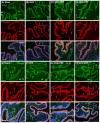
Immunofluorescence analysis was conducted to assess the expression of HA receptors (TLR-4 and RHAMM) in the bladder following treatments. (AD): The double-labeling of TLR-4 (green) and E-Cadherin (red) was performed in the bladder. In the sham group (A), the double-labeling was widely distributed in the urothelial basal layer (yellow arrows) as well as microvessels and vessels (white arrows) in the SL and ML. In the OVX group (B), the co-staining of UL and SL was reduced compared to the sham group. However, the co-staining levels of the OVX + PRP group (C) and the OVX + PPP group (D) were increased throughout the bladder urothelium, including apical, intermediate and basal layers, compared to the OVX group. Particularly, the level of the OVX + PRP group in the SL and ML was much stronger than the OVX + PPP group. (EH): The double-labeling of RHAMM (green) and E-Cadherin (red) was shown in the bladder of the different groups. Similar results were obtained for RHAMM expression coinciding with TLR-4. Yellow arrows indicated the double staining of RHAMM and E-Cadherin in urothelium, while the white arrows indicated the double staining in stroma cells and vessels. Note: UL, urothelial layer; SL, suburothelial layer; ML, muscular layer; RHAMM, receptor for HA-mediated motility; TLR-4, Toll-like receptor-4. Scale bars = 100 mm. Original magnification, ×400 (AH).
Figure 7
Figure 7
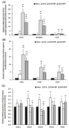
Real-time quantitative PCR of the mRNA levels of HA receptors (CD44, TLR-4 and RHAMM); HA synthase (HAS1 to HAS3) and hyaluronidase [HYAL1 to HYAL4 and sperm adhesion molecule (PH20)] involved in HA synthesis and degradation of bladder tissue. (A) The mRNA levels of HA receptors (CD44, RHAMM, and TLR-4), and (B) HA synthase (HAS2 and HAS3) were significantly declined in the OVX group compared with the sham group, but the levels were abundantly expressed in the OVX + PRP group and the OVX + PPP group compared with the sham group and the OVX group. In addition, the mRNA expressions of HYALs (HYAL2, HYAL3, HYAL4 and PH20) were significantly expressed in the OVX group compared with the sham group. Moreover, the expressions of HYAL2 and HYAL3 were significantly reduced in the PRP group compared to the OVX group (C). Data were expressed as means ± SD. n = 6 in each group (AC). Note: RHAMM, receptor for HA-mediated motility; TLR-4, Toll-like receptor-4; HYAL, hyaluronidase; PH20, sperm adhesion molecule. * p < 0.05; ** p < 0.01 versus the sham group; p < 0.05; †† p < 0.01 versus the OVX group; ## p < 0.01 versus the OVX + PRP group.
Figure 8
Figure 8
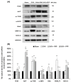
Proposed potential mechanism of PRP instillation that promoted cell proliferation and angiogenesis through PI3K/AKT/m-TOR pathway contribution to the pathogenesis of OHD-induced OAB. (A,B): The levels of signaling related proteins in the bladder, including PI3K, AKT, m-TOR, e-NOS, RAS, ERK1/2, and SOX-9, were quantified by Western blots. In the OVX group, the expressions of PI3K, e-NOS and SOX-9 proteins were significantly declined, but the levels of AKT, m-TOR, RAS and ERK1/2 were significantly promoted as compared with the sham group. Additionally, the expressions of all the above proteins were obviously increased in the OVX + PRP group (except m-TOR, RAS and ERK1/2) as compared to the OVX group. Note: e-NOS, endothelial nitric oxide synthase; ERK, extracellular signal-regulated kinase; GAPDH, glyceraldehyde-3-phosphate dehydrogenase; m-TOR, mammalian target of rapamycin; PI3K, phosphatidylinositol 3-kinase; SOX-9, SRY-box- 9. Data were expressed as means ± SD for n = 6, * p < 0.05; ** p < 0.01 versus the sham group; †† p < 0.01 versus the OVX group; # p < 0.05; ## p < 0.01 versus the OVX + PRP group.
Figure 9
Figure 9
A proposed diagram for the therapeutic effect of PRP improved bladder overactivity induced by OHD in rat model. Accordingly, the OVX group exacerbated bladder pathological damage and interstitial fibrosis through the NFƘB/COX-2 and the RAS/ERK1/2 signaling pathways. In contrast, PRP instillation for 4 weeks regulated the inflammatory fibrotic biosynthesis, promoted cell proliferation, matrix synthesis and enhanced mucosal regeneration through the HA/PI3K/AKT/SOX-9 signaling pathway to ameliorate OHD-induced bladder dysfunction. Moreover, PRP could promote angiogenic potential through the VEGF/VEGF-R and the PI3K/AKT/e-NOS signaling pathways in the pathogenesis of OHD for bladder repair. Note: bFGF, basic fibroblast growth factor; CD44, cluster of differentiation 44; EGF, epidermal growth factor; EMT, epithelial-mesenchymal transition; ERK, extracellular signal-regulated kinase; HA, hyaluronan; HAS, HA synthases (HAS 1-3); HYAL, hyaluronidases (HYAL1-4 and PH20); MAPK, mitogen-activated protein kinase; m-TOR, mammalian target of rapamycin; NO, Nitric oxide; OAB, overactive bladder; PDGF, platelet-derived growth factor; PI3K, phosphatidylinositol 3-kinase; RHAMM, receptor for HA-mediated motility; TLR-4, Toll-like receptor-4; TGF-β, transforming growth factor-β; VEGF, vascular endothelial growth factor.

Similar articles

Cited by

References

    1. Hunskaar S., Burgio K., Diokno A., Herzog A.R., Hjälmås K., Lapitan M.C. Epidemiology and natural history of urinary incontinence in women. Urology. 2003;62:16–23. doi: 10.1016/S0090-4295(03)00755-6. - DOI - PubMed
    1. Kuo H.-C. Videourodynamic precision diagnosis and treatment of lower urinary tract symptoms in women. Urol. Sci. 2021;32:94–101. doi: 10.4103/UROS.UROS_46_21. - DOI
    1. Przydacz M., Golabek T., Dudek P., Lipinski M., Chlosta P. Prevalence and bother of lower urinary tract symptoms and overactive bladder in Poland, an Eastern European Study. Sci. Rep. 2020;10:19819. doi: 10.1038/s41598-020-76846-0. - DOI - PMC - PubMed
    1. Haylen B.T., de Ridder D., Freeman R.M., Swift S.E., Berghmans B., Lee J., Monga A., Petri E., Rizk D.E., Sand P.K., et al. An International Urogynecological Association (IUGA)/International Continence Society (ICS) joint report on the terminology for female pelvic floor dysfunction. Neurourol. Urodyn. 2010;29:4–20. doi: 10.1002/nau.20798. - DOI - PubMed
    1. Iosif C.S., Bekassy Z. Prevalence of genito-urinary symptoms in the late menopause. Acta Obstet. Gynecol. Scand. 1984;63:257–260. doi: 10.3109/00016348409155509. - DOI - PubMed

Substances