. 2023 Jun 13;147(24):1823-1842.
doi: 10.1161/CIRCULATIONAHA.122.061131. Epub 2023 May 9.

Polycomb Group Protein CBX7 Represses Cardiomyocyte Proliferation Through Modulation of the TARDBP/RBM38 Axis

Affiliations

Polycomb Group Protein CBX7 Represses Cardiomyocyte Proliferation Through Modulation of the TARDBP/RBM38 Axis

Kyu-Won Cho et al. Circulation. .

Abstract

Background: Shortly after birth, cardiomyocytes exit the cell cycle and cease proliferation. At present, the regulatory mechanisms for this loss of proliferative capacity are poorly understood. CBX7 (chromobox 7), a polycomb group (PcG) protein, regulates the cell cycle, but its role in cardiomyocyte proliferation is unknown.

Methods: We profiled CBX7 expression in the mouse hearts through quantitative real-time polymerase chain reaction, Western blotting, and immunohistochemistry. We overexpressed CBX7 in neonatal mouse cardiomyocytes through adenoviral transduction. We knocked down CBX7 by using constitutive and inducible conditional knockout mice (Tnnt2-Cre;Cbx7fl/+ and Myh6-MCM;Cbx7fl/fl, respectively). We measured cardiomyocyte proliferation by immunostaining of proliferation markers such as Ki67, phospho-histone 3, and cyclin B1. To examine the role of CBX7 in cardiac regeneration, we used neonatal cardiac apical resection and adult myocardial infarction models. We examined the mechanism of CBX7-mediated repression of cardiomyocyte proliferation through coimmunoprecipitation, mass spectrometry, and other molecular techniques.

Results: We explored Cbx7 expression in the heart and found that mRNA expression abruptly increased after birth and was sustained throughout adulthood. Overexpression of CBX7 through adenoviral transduction reduced proliferation of neonatal cardiomyocytes and promoted their multinucleation. On the other hand, genetic inactivation of Cbx7 increased proliferation of cardiomyocytes and impeded cardiac maturation during postnatal heart growth. Genetic ablation of Cbx7 promoted regeneration of neonatal and adult injured hearts. Mechanistically, CBX7 interacted with TARDBP (TAR DNA-binding protein 43) and positively regulated its downstream target, RBM38 (RNA Binding Motif Protein 38), in a TARDBP-dependent manner. Overexpression of RBM38 inhibited the proliferation of CBX7-depleted neonatal cardiomyocytes.

Conclusions: Our results demonstrate that CBX7 directs the cell cycle exit of cardiomyocytes during the postnatal period by regulating its downstream targets TARDBP and RBM38. This is the first study to demonstrate the role of CBX7 in regulation of cardiomyocyte proliferation, and CBX7 could be an important target for cardiac regeneration.

Keywords: CBX7 protein; cell cycle; cell proliferation; guided tissue regeneration; human; myocytes, cardiac; polycomb-group proteins.


Conflict of interest statement

Disclosures None.

Figures

Figure 1.
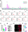
Figure 1.. Expression profiling of Polycomb group genes in mouse hearts and CMs.
A. mRNA expression of PcG genes in the mouse heart. Expression of 18 PcG genes was examined in fetal (E17.5), neonatal (P0), and adult (5 months) hearts by qRT-PCR and the fold change was calculated by normalizing to the fetal heart values. *P < 0.05, **P < 0.01, ***P < 0.001. Mann-Whitney tests were performed comparing fetal vs. neonatal hearts, neonatal vs. adult hearts, and fetal vs. adult hearts. N = 4, each with technical triplicates. B. mRNA levels of the indicated genes in the mouse hearts measured by qRT-PCR at different developmental stages from E10.5 to 60 days after birth. N = 22, each with technical triplicates. C. mRNA levels of Cbx7 in mouse hearts showing perinatal upregulation. **P < 0.01, ***P < 0.001. One-way ANOVA test with Tukey HSD. N = 8, each with technical triplicates. D. Western blot for CBX7 using isolated adult mouse CMs. HEK-293 cells infected with Ad-CBX7 were used as a positive control. E. Double immunostaining of neonatal mouse CMs transduced with Ad-CBX7 for ACTN2 and Ki67 and cultured in the presence of growth factors including IGF-1 and FGF-1 for three days. DAPI (blue). F. Percentages of Ki67+ CMs and multinucleated CMs. *P < 0.05, **P < 0.01, ***P < 0.001. Standard unpaired Student’s t test. More than 500 cells in each group were examined.
Figure 2.
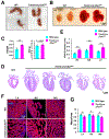
Figure 2.. Neonatal lethality, cardiomegaly, thickening of ventricular walls, and impeded myocardial compaction by cardiomyocyte-specific inhibition of CBX7.
A. Representative photographs of neonatal (P0) wild-type and Tnnt2-Cre;Cbx7fl/+ mice. B. Representative pictures of neonatal (P0) mouse hearts from wild-type and Tnnt2-Cre;Cbx7fl/+ mice. C. The heart to body weight ratio (left) and the body weight (right). ***P < 0.001. Standard unpaired Student’s t test. N = 6 for the wild-type and N = 10 for the mutant, each with technical triplicates. D. H&E stained images of the wild-type and Tnnt2-Cre;Cbx7fl/+ hearts in a four chamber view. E. Wall thickness of RV, IVS, and LV in wild-type and Tnnt2-Cre;Cbx7fl/+. *P < 0.05, **P < 0.01, ***P < 0.001. Standard unpaired Student’s t test. N = 5, each with technical triplicates. F. Representative confocal microscopic images of the LV, RV, and IVS of control (upper panel) and Tnnt2-Cre;Cbx7fl/+ (lower panel) mice at P0 stained for ACTN2 and DAPI. G. Quantification of mass per area. *P < 0.05, **P < 0.01, ***P < 0.001. Standard unpaired Student’s t test. N = 6, each with technical triplicates.
Figure 3.
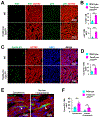
Figure 3.. Increased CM proliferation in the neonatal heart by targeted inhibition of CBX7.
A. Representative confocal microscopic images of neonatal (P0) hearts from wild-type and Tnnt2-Cre;Cbx7fl/+ mice stained for ACTN2, Ki67, and pH3. B. Quantification of Ki67+ or pH3+ CMs out of total CMs. ***P < 0.001. Standard unpaired Student’s t test. N = 5. More than 5000 cells in each group were examined. HPF, High-power field (X10). C-F. Results from immunostaining of neonatal (P0) hearts from wild-type and Tnnt2-Cre;Cbx7fl/+ mice with cyclin B1 and ACTN2. DAPI (blue). Representative confocal microscopic images stained for cyclin B1 and ACTN2 at low magnification (C) and their quantification (D). ***P < 0.001, Standard unpaired Student’s t test. N = 6. More than 5000 cells in each group were examined. Representative confocal microscopic images showing two different subcellular localizations of cyclin B1 in CMs (E) and their quantification (F). **P < 0.01, ***P < 0.001. Standard unpaired Student’s t test. N = 6. 300 cyclin B1+ CMs in 5 different fields were examined.
Figure 4.
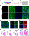
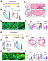
Figure 4.. Increased CM proliferation in the postnatal heart by targeted inhibition of CBX7.
A. A schematic showing the genotype of inducible conditional knockout mice (Myh6-MCM;Cbx7fl/fl). Exon 2 of Cbx7 gene is genetically deleted in CMs upon tamoxifen treatment. B. The experimental timeline for postnatal deletion of Cbx7 in CMs. Vehicle (Control) or tamoxifen (Cbx7 iCKO) were subcutaneously administered to neonatal mice at P0-P2 and the hearts were harvested at P7 or 3 months (mo). C. Validation of CBX7 deletion via western blotting with hearts collected from Myh6-MCM;Cbx7fl/fl mice at 3 mo. N = 3, each with technical triplicates. D. Representative confocal microscopic images of P7 hearts from Myh6-MCM;Cbx7fl/fl mice immunostained for Ki67, ACTN2. DAPI (blue). E. A representative orthogonal projection image of tamoxifen-treated P7 hearts from Myh6-MCM;Cbx7fl/fl mice in panel D. F. Representative confocal microscopic images of P7 hearts from Myh6-MCM;Cbx7fl/fl mice (tamoxifen treated) immunostained for pH3, ACTN2. DAPI (blue). G. Representative confocal microscopic images of P7 hearts from Myh6-MCM;Cbx7fl/fl mice immunostained for cyclin B1, ACTN2. DAPI (blue). H-J. Quantification of CMs positive for Ki67 (H), pH3 (I), and cyclin B1 (J) in P7 hearts from Myh6-MCM;Cbx7fl/fl mice. *P < 0.05, **P < 0.01. Standard unpaired Student’s t test. N = 6. More than 5000 cells in each group were examined. K. Representative H & E stained images of 3-month-old hearts from Myh6-MCM;Cbx7fl/fl mice.
Figure 5.
Figure 5.. Augmented regeneration of neonatal and adult hearts by targeted inhibition of CBX7.
A. The experimental timeline for cardiac apical resection model. Vehicle (Control) or tamoxifen (Cbx7 iCKO) were administered to neonatal mice at P0, which underwent cardiac apical resection surgery at P7, and the hearts were harvested at P28. B. Echocardiographic analyses at P28. LVEF, left ventricular ejection fraction. *P < 0.05. Standard unpaired Student’s t test. N = 8. C. Quantitative analyses of fibrosis area at P28. *P < 0.05. Standard unpaired Student’s t test. Standard unpaired Student’s t test. N = 8. D. Representative 4-chamber view images of Masson’s trichrome-stained hearts at P28. Apex was enlarged on the right. E. Representative confocal microscopic images of P28 hearts from Myh6-MCM;Cbx7fl/fl mice (tamoxifen treated) immunostained for Ki67, pH3, cyclin B1, and ACTN2. DAPI (blue). Arrows indicate CMs positive for either Ki67, pH3, or cyclin B1. F. Quantification of CMs positive for Ki67, pH3, and cyclin B1 in P7 hearts from Myh6-MCM;Cbx7fl/fl mice. *P < 0.05. Standard unpaired Student’s t test. N = 5. G. The experimental timeline for the myocardial infarction (MI) model. Vehicle (Control) or tamoxifen (Cbx7 iCKO) were administered to adult mice (5-months-old). Four weeks later, mice underwent MI surgery. Echocardiographic analyses were performed at W1, W2, and W4. Hearts were harvested at W4 for histological analyses. H. Echocardiographic analyses at W1, W2, and W4. *P < 0.05. Standard unpaired Student’s t test. N = 7-10. I. Quantitative analyses of fibrosis area at W4. **P < 0.01. Standard unpaired Student’s t test. N = 7-10. J. Representative cross-sectional images of Masson’s trichrome-stained hearts at W4. K. Representative confocal microscopic images of W4 hearts from Myh6-MCM;Cbx7fl/fl mice (tamoxifen treated) immunostained for Ki67, pH3, cyclin B1, and ACTN2. DAPI (blue). Arrows indicate CMs positive for either Ki67, pH3, or cyclin B1. L. Quantification of CMs positive for Ki67, pH3, and cyclin B1 in W4 hearts from Myh6-MCM;Cbx7fl/fl mice. *P < 0.05. Standard unpaired Student’s t test. N = 5.
Figure 6.
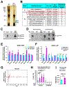
Figure 6.. Interaction of CBX7 and TARDBP and its impact on expression of mitosis-related genes.
A. Immunoprecipitation of CBX7 binding partners. Mouse CBX7 was overexpressed in mouse embryonic fibroblasts (MEFs) via Ad-CBX7. The cytoplasmic protein fraction was isolated and immunoprecipitated using either IgG (control) or anti-CBX7 antibody. Immunoprecipitated samples were subjected to SDS-PAGE followed by silver staining. Indicated bands (#1 and #2) represent candidates for CBX7 binding partners. B. Identified peptides for CBX7 binding partners. Band slices for #1 and #2 underwent MALDI-TOF followed by mass spectrometry. Identified proteins with more than 6 exclusive spectrum counts were listed. Probability for all the listed proteins is higher than 95%. C-D. Immunoprecipitation for TARDBP. The cytoplasmic protein fraction was isolated from MEFs treated with Ad-CBX7 (C) or 3-month-old adult mouse hearts (D) and was immunoprecipitated using either IgG (control) or anti-CBX7 antibody. Immunoprecipitated samples were subjected to SDS-PAGE followed by western blotting with an anti-TARDBP antibody. Input was used as a positive control. Three independent experiments, each with technical triplicates. E. mRNA expression of TARDBP target genes in CBX7-overexpressing CMs. Neonatal mouse CMs were treated with Ad-Mock or Ad-CBX7 for three days and underwent qRT-PCR. Gene expression levels were normalized to Gapdh. *P < 0.05, ***P < 0.001. Standard unpaired Student’s t test. Three independent experiments, each with technical triplicates. GOF: gain of function. F. mRNA expression of TARDBP target genes in Cbx7-haplodeficient (Tnnt2-Cre;Cbx7fl/+) mouse hearts in comparison with neonatal (P0) hearts. Gene expression measured by qRT-PCR and normalized to Gapdh. *P < 0.05, **P < 0.01, ***P < 0.001. Standard unpaired Student’s t test. Three independent experiments, each with technical triplicates. LOF: loss of function G. mRNA expression of Rbm38 gene in the mouse hearts from E10.5 to 486 days after birth. qRT-PCR results. N = 31, each with technical triplicates. H. Percentages of Ki67+ CMs upon Cbx7 knockout and Rbm38 overexpression. CMs were isolated from neonatal (P1) Myh6-MCM;Cbx7fl/fl mice and treated with Ad-Mock or Ad-RBM38 in the presence of 4-OHT at different concentrations. Cells were cultured for 5 days in the absence of growth factors including IGF-1 and FGF-1. *P < 0.05, ***P < 0.001. One-way ANOVA test with Tukey HSD. Three independent experiments. More than 500 cells in each group were examined. I. mRNA expression of Cdkn1a gene in neonatal CMs treated with Ad-Mock or Ad-CBX7. Neonatal (P0) mouse CMs were treated with adenoviral particles for 3 days and subjected to qRT-PCR. ***P < 0.001. Standard unpaired Student’s t test. The representative data for three independent experiments, each with six technical replicates.

Comment in

Similar articles

Cited by

References

    1. Braunwald E The war against heart failure: the Lancet lecture. Lancet. 2015;385:812–24. - PubMed
    1. Velagaleti RS, Pencina MJ, Murabito JM, Wang TJ, Parikh NI, D’Agostino RB, Levy D, Kannel WB and Vasan RS. Long-term trends in the incidence of heart failure after myocardial infarction. Circulation. 2008;118:2057–62. - PMC - PubMed
    1. Ezekowitz JA, Kaul P, Bakal JA, Armstrong PW, Welsh RC and McAlister FA. Declining in-hospital mortality and increasing heart failure incidence in elderly patients with first myocardial infarction. J Am Coll Cardiol. 2009;53:13–20. - PubMed
    1. Senyo SE, Steinhauser ML, Pizzimenti CL, Yang VK, Cai L, Wang M, Wu TD, Guerquin-Kern JL, Lechene CP and Lee RT. Mammalian heart renewal by pre-existing cardiomyocytes. Nature. 2013;493:433–6. - PMC - PubMed
    1. Jopling C, Sleep E, Raya M, Marti M, Raya A and Izpisua Belmonte JC. Zebrafish heart regeneration occurs by cardiomyocyte dedifferentiation and proliferation. Nature. 2010;464:606–9. - PMC - PubMed

Publication types