[Preprint]. 2023 Apr 17:2023.04.17.537203.
doi: 10.1101/2023.04.17.537203.

Human cytomegalovirus attenuates AKT activity by destabilizing insulin receptor substrate proteins

Affiliations

Human cytomegalovirus attenuates AKT activity by destabilizing insulin receptor substrate proteins

Anthony J Domma et al. bioRxiv. .

Update in

Abstract

The phosphoinositide 3-kinase (PI3K)/AKT pathway plays crucial roles in cell viability and protein synthesis and is frequently co-opted by viruses to support their replication. Although many viruses maintain high levels of AKT activity during infection, other viruses, such as vesicular stomatitis virus and human cytomegalovirus (HCMV), cause AKT to accumulate in an inactive state. To efficiently replicate, HCMV requires FoxO transcription factors to localize to the infected cell nucleus (Zhang et. al. mBio 2022), a process directly antagonized by AKT. Therefore, we sought to investigate how HCMV inactivates AKT to achieve this. Subcellular fractionation and live cell imaging studies indicated that AKT failed to recruit to membranes upon serum-stimulation of infected cells. However, UV-inactivated virions were unable to render AKT non-responsive to serum, indicating a requirement for de novo viral gene expression. Interestingly, we were able to identify that UL38 (pUL38), a viral activator of mTORC1, is required to diminish AKT responsiveness to serum. mTORC1 contributes to insulin resistance by causing proteasomal degradation of insulin receptor substrate (IRS) proteins, such as IRS1, which are necessary for the recruitment of PI3K to growth factor receptors. In cells infected with a recombinant HCMV disrupted for UL38 , AKT responsiveness to serum is retained and IRS1 is not degraded. Furthermore, ectopic expression of UL38 in uninfected cells induces IRS1 degradation, inactivating AKT. These effects of UL38 were reversed by the mTORC1 inhibitor, rapamycin. Collectively, our results demonstrate that HCMV relies upon a cell-intrinsic negative feedback loop to render AKT inactive during productive infection.


Figures
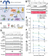
Figure 1:
Figure 1:. HCMV prevents AKT activation in response to serum.
(A): Stimulation of growth factor receptor tyrosine kinases (RTK) causes the recruitment of the scaffolding adaptor insulin receptor substrate 1 (IRS1) to the receptor complex. IRS1 is then phosphorylated, which provides a docking site for recruitment of class IA phosphoinositol 3-kinases (PI3K). PI3K phosphorylates the lipid substrate phosphoinositol 4,5 diphosphate (PIP2) to generate phosphoinositol 3,4,5 triphosphate (PIP3). The pleckstrin homology (PH) domain of AKT binds to PIP3, which drives its localization to membranes where mTORC2 and phosphoinositol-dependent kinase 1 (PDK1) are available to phosphorylate AKT at Ser473 and Thr308, respectively, causing full activation its kinase activity. Once activated, AKT phosphorylates many substrates, such as the proline-rich AKT substrate of 40 kDa (PRAS40) at Thr246, Forkhead box class O (FoxO) family of transcription factors, and the tuberous sclerosis complex 2 protein (TSC2). Phosphorylation of TSC2 leads to activation of mTORC1, which in turn phosphorylates and activates S6 kinase (S6K). S6K phosphorylates S6 to increase protein synthesis. [Schematic generated in BioRender.] (B): Serum-starved fibroblasts were stimulated with insulin or 5% newborn calf serum for the indicated times (min) prior to lysis. Protein extracts were then resolved by SDS-PAGE, transferred to nitrocellulose membranes for Western blot analysis using a phospho-specific antibody to detect AKT phosphorylated at Ser473 compared to AKT (pan) antibody. (C-G): Fibroblasts were serum starved overnight and either mock infected or infected with HCMV strain Towne at multiplicity of infection (MOI=2 TCID50/cell) for the indicated times, h post-infection (hpi). Cells were then either treated (or mock-treated) with serum for 10 min prior to lysis for Western blot analysis. (E-G): Fluorescent signals from dye labeled secondary antibodies were quantified and phospho-specific antibody detection of pAKT Thr308, pAKT Ser473 and PRAS40 Thr246 was normalized to AKT (pan) or PRAS40, respectively. The arithmetic mean was calculated and graphed. Error bars indicate standard error of the mean (SEM) from three independent biological replicates. Where indicated, Ponceau staining was used visualize protein loading and confirm efficient transfer.
Figure 2:
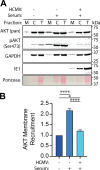
Figure 2:. Membrane recruitment of AKT is defective in HCMV-infected cells.
(A-B) Serum-starved fibroblasts were either mock-infected or infected with HCMV strain Towne (MOI=2 TCID50/cell) for 12 hours and either mock-treated or treated with 5% NCS (serum) for 10 minutes. Samples were mechanically lysed and differentially centrifuged to separate the membrane (M) fraction from the cytoplasmic (C) fraction and subjugated to Western blot analysis of indicated proteins alongside total (T) lysate. Ponceau staining was used as a loading control. (B) The AKT (pan) bands were quantified and the membrane band was normalized to the total lysate band. Quantification was then normalized to -Serum, the arithmetic mean was calculated and values were graphed. Error bars=SEM. n=4.
Figure 3:
Figure 3:. PH-domain fails to recruit to the membrane of HCMV-infected cells.
(A) HeLa cells were transfected with a plasmid expressing GFP fused to the PH domain of AKT and infected with HCMV. In the absence of serum, PI3K signaling is shut off and no PIP3 is generated, so the PH domain of AKT remains in the cytoplasm. However, with the addition of serum, PI3K is activated and PIP3 is generated allowing PH domain binding to PIP3 and membrane recruitment. R25C mutants of the PH domain, even in the presence of serum, fail to bind to PIP3. Figure was created using BioRender. (B) HeLa cells were infected in the presence of 10% fetal bovine serum (FBS) with HCMV strain AD169r131 (MOI=2 TCID50/cell) and lysates were collected at the indicated hours post-infection (hpi). A Western blot analysis was performed probing for indicated proteins. Ponceau staining was used for loading control. n=1. (C-D) HeLa cells were transfected with an expression plasmid harboring the PH-domain of AKT fused to GFP or an R25C mutant of the AKT PH-domain fused to GFP as the transgene. Cells were then either infected or mock-infected with HCMV strain AD169r131 (MOI=2 TCID50/cell) for 12 hours. Cells were then serum-starved for 2 hours and either treated or mock treated with 10% FBS for 10 minutes. (D) Pixel density at the membrane was normalized to cytoplasmic pixel density and graphed. Tukey’s multiple comparisons statistical test was used to compare each condition to each other and labeled with asterisks (ns=P>0.05, *=P<0.05, **=P<0.01, ***=P<0.001, ****P<0.0001). n=3. Error bars=SEM.
Figure 4:
Figure 4:. PDK1 and mTORC2 remain active during HCMV-mediated AKT inactivaiton.
(A) PDK1 and mTORC2 phosphorylate AKT at Thr308 and Ser473 respectively following AKT membrane recruitment. Fibroblasts were transduced with a lentivirus harboring a “tet-on” promoter controlling expression of an AKT1 gene whose PH-domain has been replaced with a myristoylation (myr) signal and genetically fused to an hemagglutinin (HA) tag. Following expression, myrAKT is embedded in the membrane simulating constitutive “membrane recruitment” and is phosphorylated as such by PDK1 and mTORC2. This experiment addresses if the activators downstream of AKT, PDK1 and mTORC2, are active during HCMV infection. Figure was created using BioRender. (B) Induction of myrAKT occured after overnight treatment with doxycycline followed by infection or mock infection with HCMV strain TOWNE (MOI=2 TCID50/cell) for indicated hours post infection (hpi). A Western blot was performed probing for the indicated proteins using Ponceau stain as a readout for loading. In pAKT and Total AKT blots, both endogenously expressed (Endo) AKT and myrAKT (Myr) migrated to different molecular weights due to deletion of PH-domain in myrAKT gene. (C) pAKT (Thr308) signal and (D) pAKT (Ser473) signal were quantified, normalized to AKT (pan) and subsequently normalized to 0 hpi signal at respective molecular weight. The arithmetic mean was calculated and graphed. Error bars=SEM. n=3.
Figure 5:
Figure 5:. UV-treated virus fails to inactivate AKT.
(A) HCMV strain Towne was mock-treated or treated with 125mJ of UV light for indicated seconds (sec) and allowed to infect fibroblasts (MOI=2 TCID50/cell) for 12 hours. A Western blot was performed staining for IE1 with Ponceau stain used as a readout for loading. n=1. (B-C) Fibroblasts were infected with mock-treated or UV-treated HCMV strain Towne (MOI=2 TCID50/cell) in the presence of serum and lysates were taken at the indicated hours post-infection (hpi). A Western blot was performed probing for the indicated proteins using Ponceau stain as a readout for loading. (C) Bands were quantified at 24 hpi, normalized to signal at 1 hpi, and the arithmetic mean was found and graphed. Sidak statistical test were used to compare each condition to the control and labeled with asterisks (ns=P>0.05, *=P<0.05, **=P<0.01, ***=P<0.001, ****P<0.0001). Error bars=SEM. n=3. (D) Serum-starved fibroblasts were either mock-infected or infected with UV-treated or untreated HCMV strain Towne (MOI=2 TCID50/cell) for 12 hours and either mock-treated or treated with 5% NCS (serum) for 10 minutes. Samples were mechanically lysed and differentially centrifuged to separate the membrane (M) fraction from the cytoplasmic (C) fraction and subjugated to Western blot analysis of indicated proteins alongside total (T) lysate.
Figure 6:
Figure 6:. HCMV causes degradation of insulin receptor substrate 1.
(A) Model for AKT inactivation in HCMV infected cells. HCMV UL38 mediated activation of mTORC1 leads to mTORC1 phosphorylation of IRS1, concomitantly inducing IRS1 degradation. This destabilization of IRS1 prevents PI3K recruitment to growth factor receptors (not shown), preventing AKT membrane recruitment and activation. Cartoon was generated using BioRender. (B-C) Fibroblasts were mock-infected or infected with HCMV strain Towne (MOI=2 TCID50/cell) in the presence of serum and lysates were taken at the indicated hours post-infection (hpi). A Western blot analysis detecting the indicated proteins was performed and (C) bands were quantified and phospho-specific quantification was normalized to total protein and arithmetic mean was found and graphed. The dotted line is the phosphorylation of mock-infected controls at 1 hpi, to which all infected time points are normalized to. Error bars=SEM. n=11. (D-E) Similar to Figure 1C, fibroblasts were serum starved overnight and either mock infected or infected with HCMV strain Towne multiplicity of infection (MOI=2 TCID50/cell). At 3 hpi, cells were mock-treated or treated with Rapamycin. At 24 hpi, cells were then either treated or mock-treated with serum for 10 minutes, and lysates were taken for Western blot analysis detection of indicated proteins. Ponceau staining was used for loading control. (E) Bands of the HCMV-infected cells were quantified, and phospho-specific quantification was normalized to total protein then normalized to ‘No Rapamycin’ control and arithmetic mean was found and graphed. Sidak statistical test were used to compare each condition to the control and labeled with asterisks (ns=P>0.05, *=P<0.05, **=P<0.01, ***=P<0.001, ****P<0.0001). Error bars=SEM. n=3.
Figure 7:
Figure 7:. UL38 is sufficient to inactivate AKT.
(A-C) Fibroblasts transduced with a lentivirus harboring a “Tet-on” HA-UL38 expression cassette were either induced for UL38 expression using 100 ng/mL doxycycline (Dox, +) or carrier-alone (−) in the presence of serum. Lysates were taken at indicated times post-treatment (h post treatment, hpt) and analyzed by Western blotting for the indicated proteins and phospho-epitopes. Ponceau stain was used to monitor protein loading and to ensure efficient transfer to nitrocellulose membranes. (B-C) Cells were either Dox induced or mock-induced using carrier alone, and treated or not with 50 nM rapamycin (Rapa) prior to lysis, as indicated. (C) At 24 hpt, the signal intensity from far-red flurophore conjugated antibodies detecting phospho-specific bands or total IRS1 protein were quantified and normalized to those from detection of AKT (pan). Results were graphed relative to a baseline signal of 1.0 determined for untreated control cells (dotted line). The arithmetic mean was calculated and graphed. Tukey’s multiple comparisons statistical test was used to compare each condition to each other and labeled with asterisks (ns=P>0.05, *=P<0.05, **=P<0.01, ***=P<0.001, ****P<0.0001). Error bars=SEM. n=3.
Figure 8:
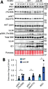
Figure 8:. UL38 inactivates AKT during HCMV infection.
(A-B) Fibroblasts were mock-infected or infected with wild-type (WT) HCMV strain AD169 or an AD169 whose UL38 gene locus has been genetically knocked out for UL38 (ΔUL38) (MOI=2 TCID50/cell). At 1, 6, 12, and 24 h post-infection (hpi), lysates were gathered and a Western blot analysis was carried out to detect the indicated proteins and phospho-epitopes. Ponceau S stain was used to indicate overall protein loading and to ensure efficient transfer to nitrocellulose membranes. (B) Signals from detection of the indicated phospho-specific bands and IRS1 were quantified for 24 hpi samples and normalized to signal from detection of AKT (pan). The arithmetic mean was calculated and graphed. Sidak statistical test were used to compare each condition to the control and labeled with asterisks (ns=P>0.05, *=P<0.05, **=P<0.01, ***=P<0.001, ****P<0.0001). Error bars=SEM. n=3.

Similar articles

References

    1. Chuluunbaatar U, Roller R, Feldman ME, Brown S, Shokat KM, Mohr I. Constitutive mTORC1 activation by a herpesvirus Akt surrogate stimulates mRNA translation and viral replication. Genes Dev. 2010;24: 2627–2639. - PMC - PubMed
    1. Soares JAP, Leite FGG, Andrade LG, Torres AA, De Sousa LP, Barcelos LS, et al. Activation of the PI3K/Akt pathway early during vaccinia and cowpox virus infections is required for both host survival and viral replication. J Virol. 2009;83: 6883–6899. - PMC - PubMed
    1. Xiang K, Wang B. Role of the PI3K-AKT-mTOR pathway in hepatitis B virus infection and replication. Mol Med Rep. 2018;17: 4713–4719. - PubMed
    1. Dunn EF, Connor JH. Dominant inhibition of Akt/protein kinase B signaling by the matrix protein of a negative-strand RNA virus. J Virol. 2011;85: 422–431. - PMC - PubMed
    1. Carsillo M, Kim D, Niewiesk S. Role of AKT kinase in measles virus replication. J Virol. 2010;84: 2180–2183. - PMC - PubMed

Publication types