. 2022 Jan;10(1):10.18103/mra.v10i1.2624.
doi: 10.18103/mra.v10i1.2624. Epub 2022 Jan 27.

Identification of amacrine neurons with a glycinergic and GABAergic phenotype in the mouse retina

Affiliations

Identification of amacrine neurons with a glycinergic and GABAergic phenotype in the mouse retina

Jorge Alberto Pérez-León et al. Med Res Arch. 2022 Jan.

Abstract

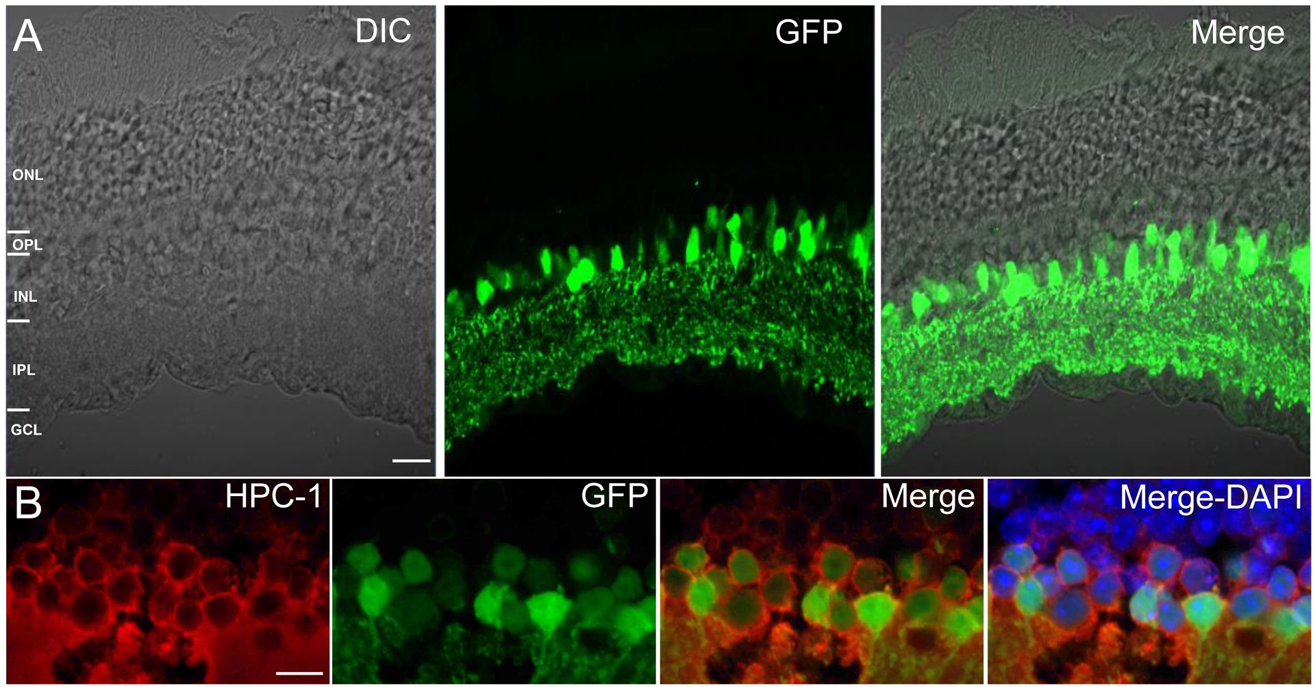
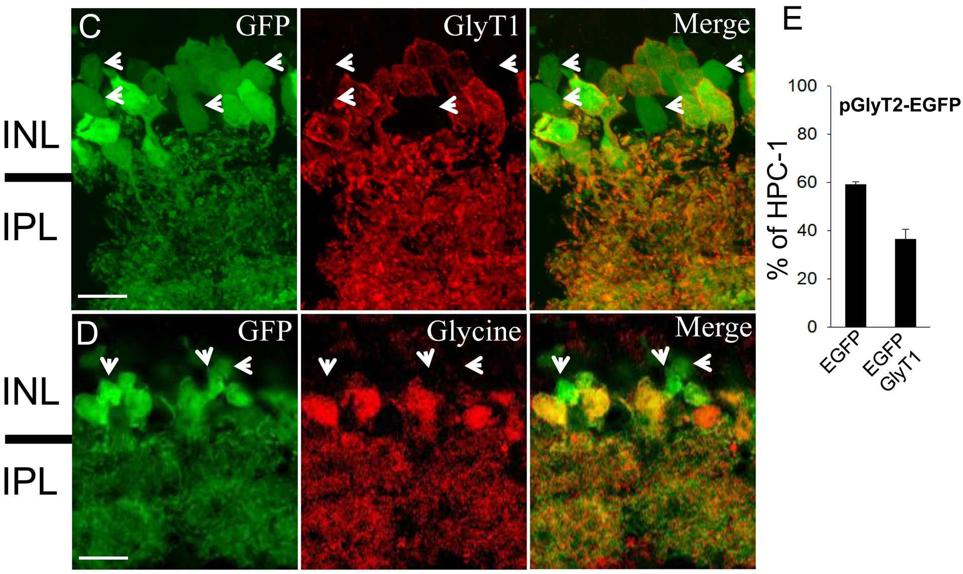
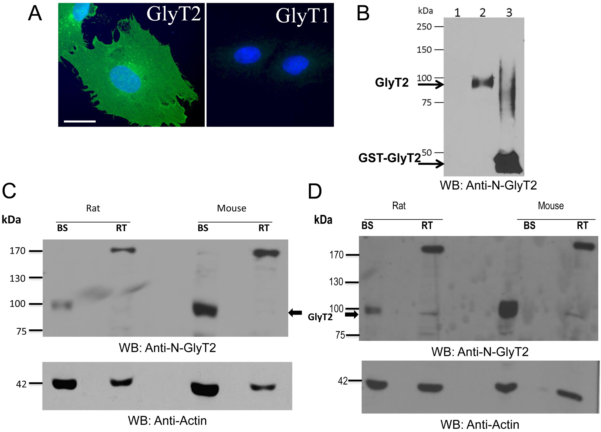
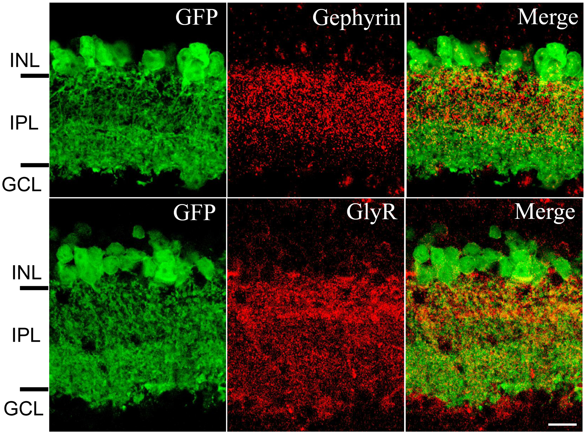
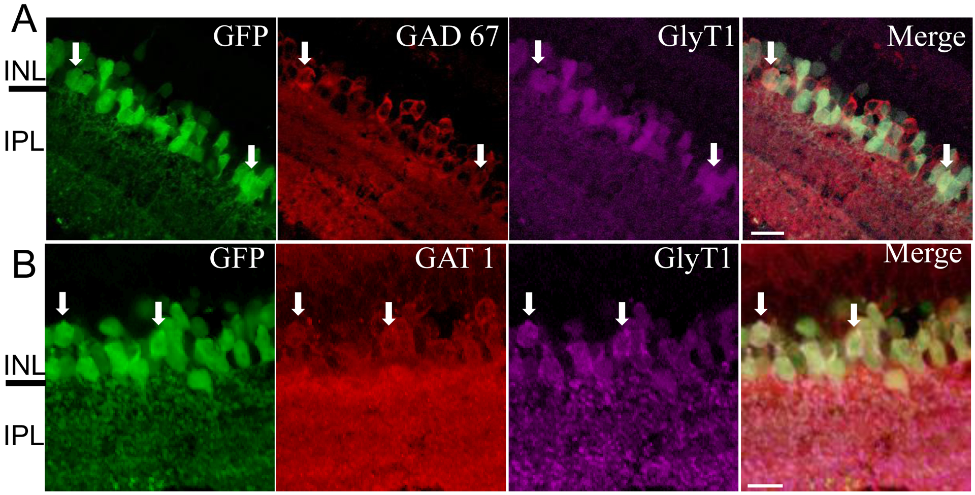
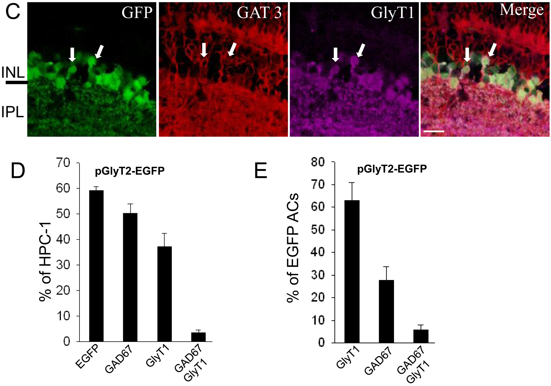
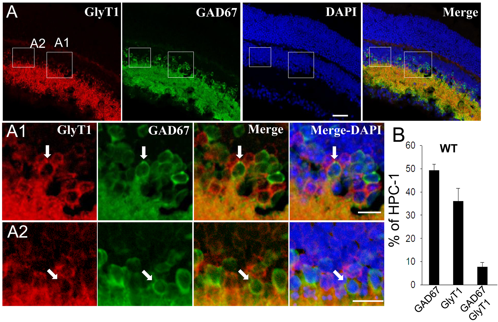
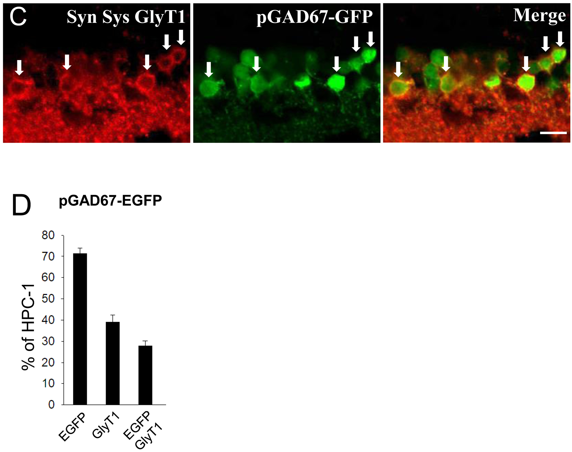
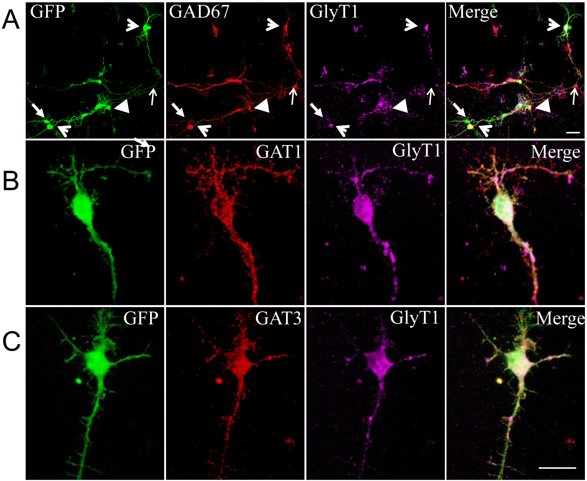
The amacrine neurons in the mammalian retina comprise a large variety of cell types with distinct properties and functions that serve to integrate and modulate signals presented to output neurons. The majority of them use either glycine or GABA as inhibitory neurotransmitters and express the glycine transporter 1 (GlyT1) or glutamic acid decarboxylase (GAD67) and GABA transporters (GAT1 and GAT3), as a glycinergic or GABAergic marker respectively. We report here a novel subpopulation of amacrine neurons expressing both, GABAergic and glycinergic markers, in retinas from wild-type C57BL/6J mice and two transgenic lines. In retinal sections from the transgenic line expressing eGFP under the control of the glycine transporter 2, eGFP expression was exclusively found in cell bodies and dendrites of inhibitory amacrine neurons, identified for their immunoreactivity to syntaxin 1A. All of the glycinergic and a large portion of the GABAergic amacrine neurons contained eGFP; of these, 8-10% of GlyT1 positive neurons were also labeled either with GAD67, GAT1 or GAT3. These findings were confirmed in retinas from a wild-type and a mouse line expressing eGFP under the GAD67 promoter and two different anti-GlyT1 antibodies, showing the presence of a subpopulation with a dual phenotype. Moreover, eGFP-positive dendrites on both mouse lines were found juxtaposed to GlyR subunits and the scaffold protein gephyrin in several areas of the inner plexiform layer, demonstrating the glycinergic character of these neurons. This dual phenotype was also demonstrated in primary retina cultures, in which isolated neurons were positive for GlyT1 and GAD67 or GAT1/3. Altogether, these data provide compelling evidence of a subpopulation of dual inhibitory, glycinergic/GABAergic amacrine neurons. The co-release of both neurotransmitters may serve to strengthen the inhibition on ganglion cells under synaptic hyperexcitability.

Keywords: GABA; Glycine Transporter 1; amacrine neuron; glycine receptor; glycinergic.


Figures

Figure 1.
Figure 1.. Characterization of inhibitory amacrine neurons in the retina of the pGlyT2-EGFP transgenic mouse.
A) Vertical sections from the central retina of the pGlyT2-eGFP mouse were subjected to immunostaining and analyzed by confocal microscopy. Z-stack images were acquired and an optical section from the middle of the stack selected for presentation. Left panel: bright field showing the retinal layers; middle panel, eGFP expression; right panel, merge image. Scale bar, 20 μm. B) Vertical section stained with anti-Syntaxin 1 (HPC-1) antibodies and DAPI followed by CY-3 labeled secondary antibodies. C) Same as B but sections labeled with rabbit antibodies against GlyT1 or D) anti-glycine. Scale bar, 10 μm. Abbreviations: INL, inner nuclear layer; IPL, inner plexiform layer; GCL, ganglion cell layer. E) Quantitation of EGFP-positive or EGFP/GlyT1 neurons. Cell counts were obtained from a minimum of 100 HPC-1 positive cells per retina, for a total of three retinas from three different mice. The plots represent the percent of HPC-1 positive cells +/− SE. Open arrowhead in C and D indicate eGFP labeled cells devoid of GlyT1or Glycine immunoreactivity, respectively.
Figure 2.
Figure 2.. GlyT2 protein detected in the rat and mouse retina.
A) The human GlyT2 or the GlyT1b were expressed in PAE cells and immunostained with purified rabbit anti-GlyT2 antibodies against the N-terminus of GlyT2 (Anti-N-GlyT2) followed by Alexa-488 labeled secondary antibodies and DAPI, Scale bar, 10 μm. B) Cell expressing the human GlyT2 were lysed and subjected to western blot with anti-N-GlyT2 antibodies. Lane 1, parental cells; lane 2, PAE cells expressing GlyT2; lane 3, 100 ng of the fusion protein GST-N-GlyT2. C) Brain stem and retinas were homogenized in RIPA buffer and cleared by centrifugation. Cleared lysates were quantitated and subjected to 8% SDS-PAGE and western blotting with a rabbit GlyT2 and actin antibodies. D) Same as “A” but the tissue was lysed in buffer containing 1 mM Dithiothreitol. The blots are representative of at least three experiments.
Figure 3.
Figure 3.. Localization of eGFP-positive neurons with post-synaptic markers in retinas of the pGlyT2-EGFP transgenic mouse.
Vertical section of the central mouse retina were labeled with antibodies against the protein gephyrin (upper panel) or the GlyR subunits using the mAb4a antibody that recognizes all the receptor subunits, followed by staining with CY-3 labeled secondary antibodies. Confocal Z-stack images were acquired and an optical section from the middle of the stack selected and used for presentation. Scale bar, 10 μm.
Figure 4.
Figure 4.. The mouse retina contains a subpopulation of dual GABAergic and glycinergic amacrine neurons.
A) Vertical sections from the central retina of the pGlyT2-eGFP mouse were immunolabeled with antibodies recognizing the GABA synthetizing enzyme GAD67 and rabbit GlyT1 antibodies or synaptic systems or followed by the corresponding secondary CY-3 and CY-5 labeled antibodies. Z-stack images were acquired through the corresponding channels and an optical section from the middle of the stack selected and used for this figure panels. B) Sections were stained as in A but using the rabbit GABA transporter anti-GAT1 and rat anti-GlyT1 antibodies or C) GAT3 and rat GlyT1. Scale bar 10 μm. Abbreviations: INL, inner nuclear layer; IPL, inner plexiform layer. Open arrowhead in A-C indicate eGFP labeled cells containing GlyT1 and GAD67, or GAT1 or GAT3. D) A minimum of 100 HPC-1 positive cells per retina from three different eyes were used to quantify the number of GAD67 or GlyT1 positive cells and those containing both markers. The plots represent the percent of GAD67 or/and GlyT1 positive cells +/− SE. E) Same as D but normalized by 100 EGFP positive ACs.
Figure 5.
Figure 5.. Co-expression of GAD67 and GlyT1 in amacrine neurons from wild-type retinas and the transgenic mouse expressing eGFP under GAD67 promoter.
A) Vertical sections from the central retina of C57BL/6J mice were subjected to immunostaining with GlyT1 and GAD67 antibodies followed by incubation with CY-3 and Alexa-488 labeled secondary antibodies, respectively. Scale bar, 50 μm. A1 and A2) Magnification of the areas enclosed in panel A, cells labeled for GAD67 and GlyT1 are marked by an arrow. B) Vertical sections were stained with HPC-1, GlyT1 and GAD67 and the resulting images used for quantitation as described in 4D. C) Vertical sections from the central retina of the transgenic line pGAD67-EGFP were labeled with GlyT1 antibody from Synaptic Systems followed by incubation with CY-3 labeled secondary antibodies. Nuclei were stained with DAPI. Arrows indicate co-expression of GlyT1 and eGFP in amacrine neurons. Scale bar, 10 μm for B and C. D) Sections from the pGAD67-EGFP were subjected to quantitation analysis, as described in 4D. Scale bar, 10 μm.
Figure 6.
Figure 6.. GABAergic and glycinergic markers co-expression in cultured cells isolated from pGlyT2-eGFP transgenic mouse.
A) Primary dissociated retinal cultures were fixed with paraformaldehyde and incubated with antibodies recognizing GAD67 and GlyT1 followed by staining with the corresponding CY-3 and CY-5 labeled antibodies, respectively. The inhibitory neurons were heterogeneous and contained eGFP and GlyT1 (long arrows), or eGFP, GAD67 and GlyT1 (open arrowheads), or GAD67 (thin open arrows). B) Cells were subjected to immunostaining with antibodies against GAT1 and GlyT1 or C) against GAT3 and GlyT1 as described under Experimental Procedures. Scale bars, 10 μm.

Similar articles

Cited by

References

    1. Betz H, and Laube B (2006) Glycine receptors: recent insights into their structural organization and functional diversity. J Neurochem 97, 1600–1610 - PubMed
    1. Dutertre S, Becker CM, and Betz H (2012) Inhibitory glycine receptors: an update. J Biol Chem 287, 40216–40223 - PMC - PubMed
    1. Zhang C, and McCall MA (2012) Receptor targets of amacrine cells. Vis Neurosci 29, 11–29 - PMC - PubMed
    1. Johnson JW, and Ascher P (1987) Glycine potentiates the NMDA response in cultured mouse brain neurons. Nature 325, 529–531 - PubMed
    1. Pourcho RG, and Owczarzak MT (1991) Connectivity of glycine immunoreactive amacrine cells in the cat retina. J Comp Neurol 307, 549–561 - PubMed

LinkOut - more resources