. 2023 Apr 6;15(4):926.
doi: 10.3390/v15040926.

An Endogenous Retrovirus Vaccine Encoding an Envelope with a Mutated Immunosuppressive Domain in Combination with Anti-PD1 Treatment Eradicates Established Tumours in Mice

Affiliations

An Endogenous Retrovirus Vaccine Encoding an Envelope with a Mutated Immunosuppressive Domain in Combination with Anti-PD1 Treatment Eradicates Established Tumours in Mice

Joana Daradoumis et al. Viruses. .

Abstract

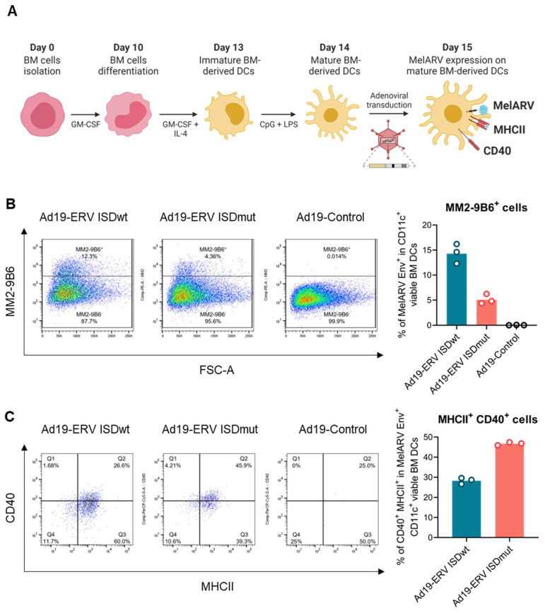
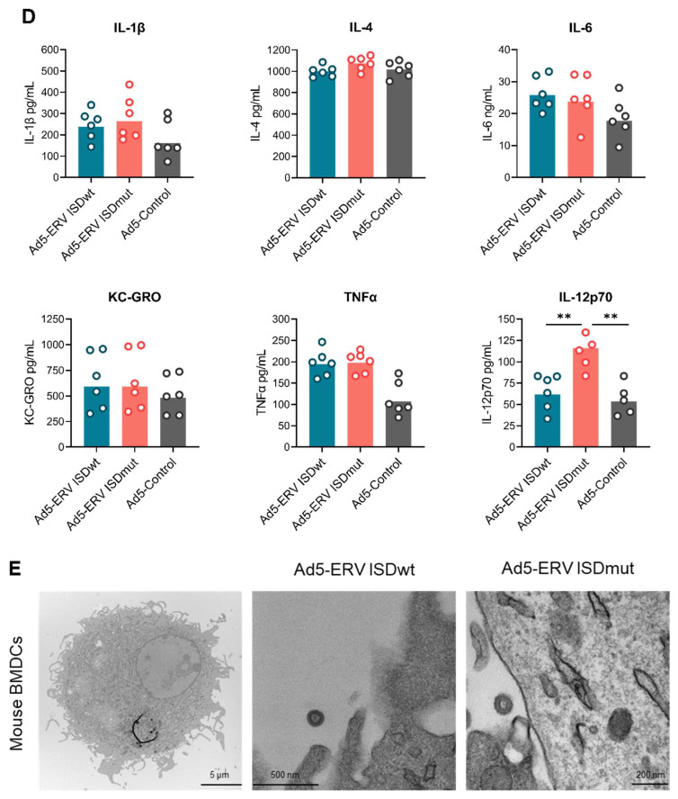
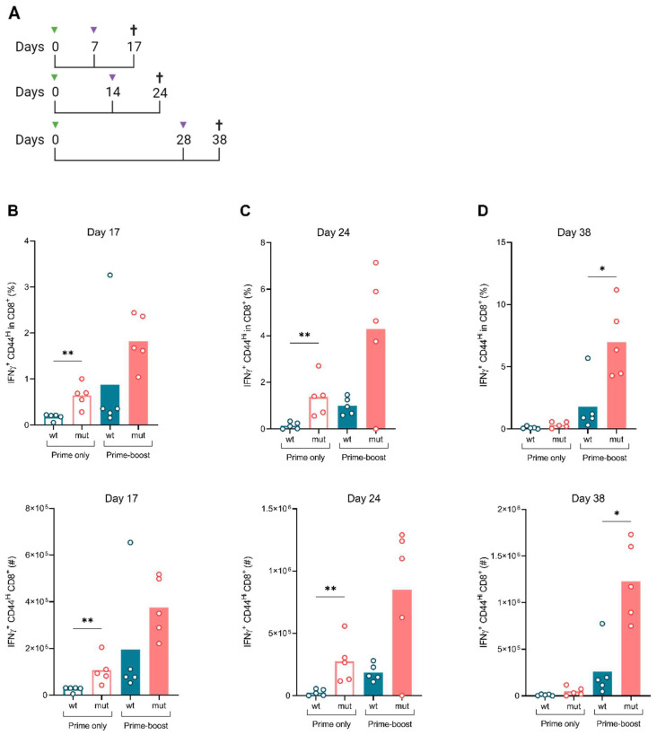
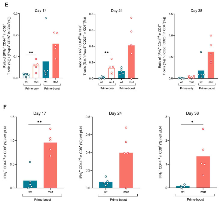
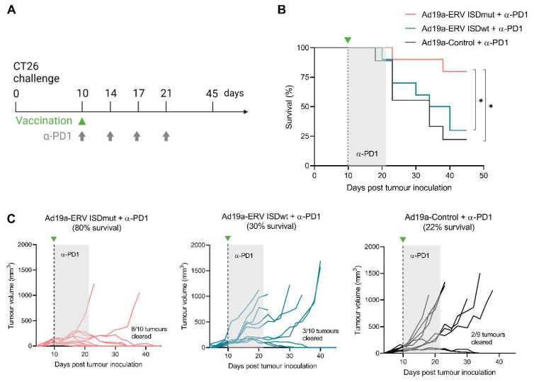
Endogenous retroviruses (ERVs) account for 8% of our genome, and, although they are usually silent in healthy tissues, they become reactivated and expressed in pathological conditions such as cancer. Several studies support a functional role of ERVs in tumour development and progression, specifically through their envelope (Env) protein, which contains a region described as an immunosuppressive domain (ISD). We have previously shown that targeting of the murine ERV (MelARV) Env using virus-like vaccine (VLV) technology, consisting of an adenoviral vector encoding virus-like particles (VLPs), induces protection against small tumours in mice. Here, we investigate the potency and efficacy of a novel MelARV VLV with a mutated ISD (ISDmut) that can modify the properties of the adenoviral vaccine-encoded Env protein. We show that the modification of the vaccine's ISD significantly enhanced T-cell immunogenicity in both prime and prime-boost vaccination regimens. The modified VLV in combination with an α-PD1 checkpoint inhibitor (CPI) exhibited excellent curative efficacy against large established colorectal CT26 tumours in mice. Furthermore, only ISDmut-vaccinated mice that survived CT26 challenge were additionally protected against rechallenge with a triple-negative breast cancer cell line (4T1), showing that our modified VLV provides cross-protection against different tumour types expressing ERV-derived antigens. We envision that translating these findings and technology into human ERVs (HERVs) could provide new treatment opportunities for cancer patients with unmet medical needs.

Keywords: adenoviral vectors; cancer; endogenous retroviruses; immunotherapy; murine melanoma-associated retrovirus; virus-like particles; virus-like-vaccines.


Conflict of interest statement

The project was sponsored by InProTher ApS, in collaboration with Sirion Biotech, that provided the used vaccines. L.N., P.J.H. and C.T. are co-inventors of the virus-like vaccine technology for immunotherapy against cancer. P.J.H. is the current CSO of InProTher ApS and has had the company as his primary employer since January 2019. P.J.H. and C.T. are major shareholders and board members of InProTher ApS. J.D., E.R., I.S., K.N.N., A.V.B., A.A. and L.N have been or are current employees and warrant holders of InProTher ApS. Despite this, the aforementioned facts had no influence on the design of the experimental experiments, nor the data and their representation.

Figures

Figure 1
Figure 1
Vaccine characterization. (A) The prototype vaccine (ISDwt) was modified by substituting E14 R14 and A20 F20 in the ISD of MelARV Env sequence (ISDmut), as described in Schlecht-Louf et al. [23]. (BD) Different mammalian cell lines were transduced with Ad19a/64 or Ad5-ERV ISDwt or ISDmut and were analysed for the expression of MelARV Env or Gag, as well as VLP formation and secretion. Adenoviral vaccines encoding an irrelevant transgene or no transgene (empty vector) were used as negative controls when transducing the cell lines. (B) Expression of the vaccine-encoded MelARV Env subunits on the surface of A549 cells 24 h post transduction. The primary antibodies 19F8 and MM2-9B6 were used to detect the respective Env subunits p15E/TM (ISD) and gp70/SU, by flow cytometry. (C) Expression and release of the vaccine-encoded MelARV Gag protein (detected by anti-P2A) as shown by Western blot after 48 h in cell lysates and VLPs purified form cell culture supernatants of transduced Vero cells. (D) Visualization of budding VLPs (circles of approximately 100 nm) in A549 cells at 48 h after transduction with Ad19a/64-ERV vaccines. Images were generated by TEM. Ad19a/64 is referred as Ad19 in the figure.
Figure 2
Figure 2
Impairment of the vaccine ISD increases the activation of BMDCs. (A) In vitro generation of mature DCs from murine BM cells. BM cells were isolated and differentiated into immature DCs by adding GM-CSF and IL-4 into the medium. DCs were matured by adding CpG and LPS in the culture and transduced at 1000 MOI or 250 MOI with the Ad19a/64- or Ad5-ERV ISDwt or ISDmut, respectively. Ad5- or Ad19a/64-empty vectors were used as negative controls. Twenty-four hours post transduction, the expression of MelARV Env and DC maturation/activation markers was assessed by means of flow cytometry. (B) Expression of MelARV Env gp70/SU subunit on the surface of transduced live DCs was assessed by means of flow cytometry using MM2-9B6 d. (C) Activation/maturation of live MelARV Env+ BMDCs was established by measuring MHCII and CD40 co-expression (fraction of double positive cells) using flow cytometry. (D) Secretion of the cytokines IL-1β, IL-4, IL-6, KC-GRO, TNF-α, and IL-12p70 was assessed in the supernatant of transduced mouse BMDCs using a V-PLEX assay. Data points represent samples from 6 independent mice. (E) VLPs (of approximately 100 nm size), secreted from mouse BMDCs, were visualized by TEM, 24 h after transduction. Ad19a/64 is referred to as Ad19 in the figure. **: p < 0.01—Mann–Whitney U test.
Figure 2
Figure 2
Impairment of the vaccine ISD increases the activation of BMDCs. (A) In vitro generation of mature DCs from murine BM cells. BM cells were isolated and differentiated into immature DCs by adding GM-CSF and IL-4 into the medium. DCs were matured by adding CpG and LPS in the culture and transduced at 1000 MOI or 250 MOI with the Ad19a/64- or Ad5-ERV ISDwt or ISDmut, respectively. Ad5- or Ad19a/64-empty vectors were used as negative controls. Twenty-four hours post transduction, the expression of MelARV Env and DC maturation/activation markers was assessed by means of flow cytometry. (B) Expression of MelARV Env gp70/SU subunit on the surface of transduced live DCs was assessed by means of flow cytometry using MM2-9B6 d. (C) Activation/maturation of live MelARV Env+ BMDCs was established by measuring MHCII and CD40 co-expression (fraction of double positive cells) using flow cytometry. (D) Secretion of the cytokines IL-1β, IL-4, IL-6, KC-GRO, TNF-α, and IL-12p70 was assessed in the supernatant of transduced mouse BMDCs using a V-PLEX assay. Data points represent samples from 6 independent mice. (E) VLPs (of approximately 100 nm size), secreted from mouse BMDCs, were visualized by TEM, 24 h after transduction. Ad19a/64 is referred to as Ad19 in the figure. **: p < 0.01—Mann–Whitney U test.
Figure 3
Figure 3
ISDmut vaccines elicit stronger CD8+ T cell responses both locally and systemically. (A) Schematic representation of the vaccine regimen. BALB/c mice were vaccinated s.c. (right paw) on day 0 with either the Ad19a/64-ERV ISDwt or the Ad19a/64-ERV ISDmut vaccine. Half of the primed mice were vaccinated s.c. (left paw) a second time (boost) with either the Ad5-ERV ISDwt or with the Ad5-ERV ISDmut on day 7, 14 or 28. Spleens and vaccine-draining pLN (at the site of booster injection) were collected 10 days after the boost to perform ICS against the MelARV Env gp70/SU H2-Ld-restricted T cell peptide AH1. (BD) Immune profiling of spleens in Ad19a/64 +/− Ad5-ERV ISDwt- or ISDmu-vaccinated mice. Frequency (upper graphs) and absolute number (bottom graphs) of IFNγ+ CD44Hi CD8+ T cells responding to AH1 MelARV peptide at day 17, 24 and 38 post prime. Day 17 was repeated in two independent experiments. (E) Ratio of the percentage of IFNγ+ CD44Hi in CD8+ T cells and the percentage of FoxP3+ CD25+ in CD4+ T cells (Tregs) in the spleen of prime-boosted mice. (F) Immune profiling of draining pLN in prime-boost (Ad19a/64- and Ad5-ERV ISDwt or ISDmut)-vaccinated mice showing the frequency of IFNγ+ CD44Hi CD8+ T cells responding to AH1 MelARV peptide at day 17, 24 and 38 after prime. N = 4–5, *: p < 0.05, **: p < 0.01—Mann–Whitney U test. The gating strategy can be found in Supplementary Figure S4A.
Figure 3
Figure 3
ISDmut vaccines elicit stronger CD8+ T cell responses both locally and systemically. (A) Schematic representation of the vaccine regimen. BALB/c mice were vaccinated s.c. (right paw) on day 0 with either the Ad19a/64-ERV ISDwt or the Ad19a/64-ERV ISDmut vaccine. Half of the primed mice were vaccinated s.c. (left paw) a second time (boost) with either the Ad5-ERV ISDwt or with the Ad5-ERV ISDmut on day 7, 14 or 28. Spleens and vaccine-draining pLN (at the site of booster injection) were collected 10 days after the boost to perform ICS against the MelARV Env gp70/SU H2-Ld-restricted T cell peptide AH1. (BD) Immune profiling of spleens in Ad19a/64 +/− Ad5-ERV ISDwt- or ISDmu-vaccinated mice. Frequency (upper graphs) and absolute number (bottom graphs) of IFNγ+ CD44Hi CD8+ T cells responding to AH1 MelARV peptide at day 17, 24 and 38 post prime. Day 17 was repeated in two independent experiments. (E) Ratio of the percentage of IFNγ+ CD44Hi in CD8+ T cells and the percentage of FoxP3+ CD25+ in CD4+ T cells (Tregs) in the spleen of prime-boosted mice. (F) Immune profiling of draining pLN in prime-boost (Ad19a/64- and Ad5-ERV ISDwt or ISDmut)-vaccinated mice showing the frequency of IFNγ+ CD44Hi CD8+ T cells responding to AH1 MelARV peptide at day 17, 24 and 38 after prime. N = 4–5, *: p < 0.05, **: p < 0.01—Mann–Whitney U test. The gating strategy can be found in Supplementary Figure S4A.
Figure 4
Figure 4
Ad19a/64-ERV ISDmut synergises with α-PD1 check point inhibitor and eradicates established colorectal tumours in mice. (A) Schematic representation of the tumour challenge and the therapeutic treatment. BALB/c mice were challenged s.c. (right flank) with 5 × 10⁵ CT26 cells and tumour size was evaluated every 2–3 days. Mice were randomized and vaccinated s.c. (left paw) on day 10 with either the Ad19a/64-ERV ISDwt (blue), the Ad19a/64-ERV ISDmut (coral) or the Ad19a/64-Irrelevant (black) vaccine. α-PD1 treatment was administered i.p. concomitant to the vaccination, and then repeated three times every 3–4 days. (B) Fraction of surviving mice over time after CT26 challenge and treatment. (C) Tumour volume (mm3) over time after challenge and treatment. N = 9–10, *: p < 0.05—Log-rank (Mantel–Cox) test. Ad19a/64 is referred to as Ad19a in the figure.
Figure 5
Figure 5
Ad19a/64-ERV ISDmut in combination with α-PD1 protects against rechallenge with a triple-negative breast cancer cell line. (A) Schematic representation of the tumour challenge and the therapeutic treatment. Ad19a/64-vaccinated BALB/c mice that survived CT26 challenge and naïve mice were (re)challenged with 1 × 10⁴ 4T1 tumour cells into the (left) thoracic mammary fat pad. Tumours were measured every 2–3 days. (B) Fraction of surviving mice over time after 4T1 rechallenge. (C) Measurements of tumour volume (mm3) over time after 4T1 rechallenge. N = 2–10, ***: p < 0.001—Log-rank (Mantel–Cox) test. Ad19a/64 is referred as Ad19 in the figure.

Similar articles

Cited by

References

    1. Sung H., Ferlay J., Siegel R.L., Laversanne M., Soerjomataram I., Jemal A., Bray F. Global Cancer Statistics 2020: GLOBOCAN Estimates of Incidence and Mortality Worldwide for 36 Cancers in 185 Countries. CA. Cancer J. Clin. 2021;71:209–249. doi: 10.3322/caac.21660. - DOI - PubMed
    1. Lander E.S., Linton L.M., Birren B., Nusbaum C., Zody M.C., Baldwin J., Devon K., Dewar K., Doyle M., FitzHugh W., et al. Initial sequencing and analysis of the human genome. Nature. 2001;409:860–921. doi: 10.1038/35057062. - DOI - PubMed
    1. Mouse Genome Sequencing Consortium. Waterston R.H., Lindblad-Toh K., Birney E., Rogers J., Abril J.F., Agarwal P., Agarwala R., Ainscough R., Alexandersson M., et al. Initial sequencing and comparative analysis of the mouse genome. Nature. 2002;420:520–562. doi: 10.1038/nature01262. - DOI - PubMed
    1. Stocking C., Kozak C.A. Murine endogenous retroviruses. Cell. Mol. Life Sci. 2008;65:3383–3398. doi: 10.1007/s00018-008-8497-0. - DOI - PMC - PubMed
    1. Nellåker C., Keane T.M., Yalcin B., Wong K., Agam A., Belgard T.G., Flint J., Adams D.J., Frankel W.N., Ponting C.P. The genomic landscape shaped by selection on transposable elements across 18 mouse strains. Genome Biol. 2012;13:R45. doi: 10.1186/gb-2012-13-6-r45. - DOI - PMC - PubMed

Publication types

Substances

Grants and funding

The study was funded by InProTher ApS and within kind contribution from Sirion Biotech GmbH. Innovation Fund Denmark supported the realization of this project with granting a scholarship (9065-00055B) to Joana Daradoumis.

LinkOut - more resources