

The Brain and Spinal Microvasculature in Normal Aging
- PMID: 37093786
- PMCID: PMC10395569
- DOI: 10.1093/gerona/glad107
The Brain and Spinal Microvasculature in Normal Aging
Abstract
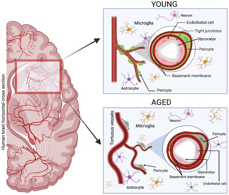
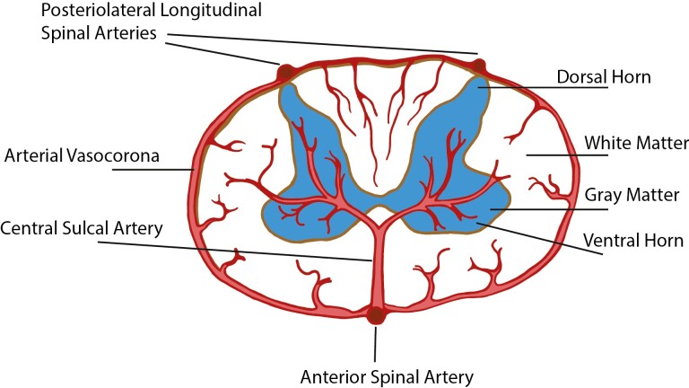
Changes in the brain and spinal cord microvasculature during normal aging contribute to the "sensitive" nature of aged central nervous system tissue to ischemic insults. In this review, we will examine alterations in the central nervous system microvasculature during normal aging, which we define as aging without a dominant pathology such as neurodegenerative processes, vascular injury or disease, or trauma. We will also discuss newer technologies to improve the study of central nervous system microvascular structure and function. Microvasculature within the brain and spinal cord will be discussed separately as anatomy and physiology differ between these compartments. Lastly, we will identify critical areas for future studies as well as key unanswered questions.
Keywords: Brain; Microvessels; Normal aging; Spinal cord.
© The Author(s) 2023. Published by Oxford University Press on behalf of The Gerontological Society of America. All rights reserved. For permissions, please e-mail: journals.permissions@oup.com.
Conflict of interest statement
None declared.
Figures

Similar articles
-
Three-dimensional imaging of microvasculature in the rat spinal cord following injury.Sci Rep. 2015 Jul 29;5:12643. doi: 10.1038/srep12643. Sci Rep. 2015. PMID: 26220842 Free PMC article.
-
Three-Dimensional Changes in Cervical Spinal Cord Microvasculature During the Chronic Phase of Hemicontusion Spinal Cord Injury in Rats.World Neurosurg. 2019 Jun;126:e385-e391. doi: 10.1016/j.wneu.2019.02.061. Epub 2019 Feb 26. World Neurosurg. 2019. PMID: 30822573
-
Spinal Arteriolosclerosis Is Common in Older Adults and Associated With Parkinsonism.Stroke. 2017 Oct;48(10):2792-2798. doi: 10.1161/STROKEAHA.117.017643. Epub 2017 Sep 20. Stroke. 2017. PMID: 28931619 Free PMC article.
-
Vascular disease of the spine.Neurologist. 2015 May;19(5):121-7. doi: 10.1097/NRL.0000000000000018. Neurologist. 2015. PMID: 25970833 Review.
-
Advancing Stem Cell Therapy for Repair of Damaged Lung Microvasculature in Amyotrophic Lateral Sclerosis.Cell Transplant. 2020 Jan-Dec;29:963689720913494. doi: 10.1177/0963689720913494. Cell Transplant. 2020. PMID: 32207340 Free PMC article. Review.
Cited by
-
Contrast-enhanced ultrasound imaging detects anatomical and functional changes in rat cervical spine microvasculature with normal aging.bioRxiv [Preprint]. 2024 Mar 14:2024.03.12.584672. doi: 10.1101/2024.03.12.584672. bioRxiv. 2024. Update in: J Gerontol A Biol Sci Med Sci. 2024 Dec 11;80(1):glae215. doi: 10.1093/gerona/glae215. PMID: 38559128 Free PMC article. Updated. Preprint.
