. 2023 Apr 12:2023:9067621.
doi: 10.1155/2023/9067621. eCollection 2023.

Human Infrapatellar Fat Pad Mesenchymal Stem Cell-Derived Extracellular Vesicles Inhibit Fibroblast Proliferation by Regulating MT2A to Reduce Knee Arthrofibrosis

Affiliations

Human Infrapatellar Fat Pad Mesenchymal Stem Cell-Derived Extracellular Vesicles Inhibit Fibroblast Proliferation by Regulating MT2A to Reduce Knee Arthrofibrosis

Dazhou Jia et al. Stem Cells Int. .

Abstract

Knee arthrofibrosis is one of the most serious complications of knee surgery; however, its pathogenesis is unclear, and current treatment methods have not achieved satisfactory results. Mesenchymal stem cells (MSCs) have good anti-inflammatory and antifibrotic properties, and studies have reported that human infrapatellar fat pad-derived MSCs (IPFSCs) have the advantages of strong proliferative and differentiating ability, ease of acquisition, and minimal harm to the donor. Increasing evidence has shown that MSCs function through their paracrine extracellular vesicles (EVs). Our study is aimed at exploring the effects of human IPFSC-derived EVs (IPFSC-EVs) on knee arthrofibrosis and the underlying mechanisms in vivo and in vitro. In the in vivo study, injecting IPFSC-EVs into the knee joint cavity effectively reduced surgery-induced knee arthrofibrosis in rats. In the in vitro study, IPFSC-EVs were found to inhibit the proliferation of fibroblasts in the inflammatory environment. Additionally, we screened a potential IPFSC-EV molecular target, metallothionein 2A (MT2A), using RNA sequencing. We found that silencing MT2A partially reversed the inhibitory effect of IPFSC-EVs on fibroblast proliferation in the inflammatory environment. In conclusion, IPFSC-EVs inhibit the progression of knee arthrofibrosis by regulating MT2A, which inhibits fibroblast proliferation in the inflammatory environment.


Conflict of interest statement

The authors declare that there is no conflict of interest regarding the publication of this paper.

Figures
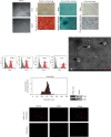
Figure 1
Figure 1
Identification of infrapatellar fat pad mesenchymal stem cells (IPFSCs) and IPFSC-EVs. (a) IPFSCs were isolated and cultured, showing a spindle-like shape under optical microscopy. (b) IPFSCs had adipogenic, osteogenic, and chondrogenic differentiation ability. (c) Characteristic antigens of IPFSCs detected using flow cytometry. (d) IPFSC-EVs showed a typical disc-shaped bilayer membrane structure under transmission electron microscopy. (e) NanoSight analysis showing the diameter range of the extracted EVs. (f) Western blot showing the expression levels of the characteristic EV proteins CD63 and CD81. (g) After PKH26 labeling of IPFSC-EVs, uptake of IPFSC-EVs by fibroblasts was observed under fluorescence microscopy.
Figure 2
Figure 2
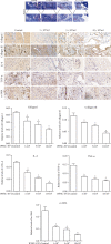
IPFSCs-EV treatment reduced the severity of knee arthrofibrosis in rats. (a) Hematoxylin and eosin staining showed that IPFSC-EVs reduced the degree of fibrosis and fibroblast number in the knee joint cavity. Masson staining showed that IPFSC-EVs reduced collagen content in the fibrotic tissues. (c and d) Immunohistochemical staining of collagens I and III revealed that IPFSC-EVs reduced collagen content in the fibrotic tissues in a concentration-dependent manner. (e and f) Immunohistochemical staining of IL-6 and TNF-α showed that IPFSC-EVs reduced the expression of proinflammatory factors in the fibrotic tissues in a concentration-dependent manner. (g) Immunohistochemical staining of α-SMA also revealed that IPFSC-EVs reduced the degree of fibrosis in the knee joint cavity in a concentration-dependent manner.
Figure 3
Figure 3
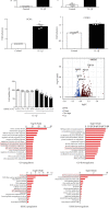
Construction of a fibroblast inflammatory model and screening of molecular targets for inhibiting knee arthrofibrosis. (a) Increased expression of the inflammatory factors IL-6 and TNF-α after IL-1β stimulation. (b) Increased expression of the proliferation-related genes PCNA and CCND1 after IL-1β stimulation. (c) IL-1β (10 ng/mL) was combined with various IPFSC-EV concentrations to react with fibroblasts for 24 h. CCK-8 assay revealed that fibroblast viability was suppressed in a concentration-dependent manner. (d) Volcano plot showing genes that were differentially expressed between IPFSC-EVs and controls. (e) GO analysis showing upregulated cell cycle phase transition and cell cycle G1/S phase transition and downregulated connective tissue development. (f) KEGG pathway analysis showing upregulated cell cycle and p53 signaling pathway and significantly downregulated extracellular matrix–receptor interactions. All data are presented as mean ± SD. P < 0.05 compared with the control group.
Figure 4
Figure 4
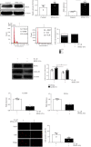
IPFSC-EVs inhibited fibroblast proliferation in the inflammatory environment. (a) Western blot and RT–PCR showing that IPFSC-EVs could promote MT2A expression. (b) Cell cycle analysis showing that IPFSC-EVs could arrest fibroblasts in the G1 phase in the inflammatory environment and inhibit cell cycle progression. (c and d) Western blot and RT–PCR results showing downregulated mRNA and protein expression levels of the proliferation-related genes CCND1 and PCNA after IPFSC-EV treatment of fibroblasts. (e) EdU cell proliferation assay showing a significantly lower proportion of EdU-positive cells in total cells after IPFSC-EV treatment of fibroblasts in the inflammatory environment for 24 h. All data are presented as mean ± SD. P < 0.05 compared with the control group.
Figure 5
Figure 5
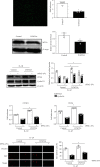
IPFSC-EVs inhibited fibroblast proliferation in the inflammatory environment by regulating MT2A. (a) Cell transfection efficiency under fluorescence microscopy. (b and c) Western blot and RT–PCR analyses showing decreased gene and protein expression of MT2A in the virus strain, indicating successful MT2A silencing. (d and e) Western blot and RT–PCR analysis showing the partially reversed tendency of IPFSC-EVs to inhibit CCDN1 and PCNA expression after silencing MT2A. (f) EdU cell proliferation assay showing the partially reversed ability of IPFSC-EVs to inhibit DNA synthesis in fibroblasts after silencing MT2A. All data are presented as mean ± SD. P < 0.05 compared with the control group.
Figure 6
Figure 6
A proposed underlying mechanism of IPFSC-EVs in attenuating the progression of knee arthrofibrosis.

Similar articles

References

    1. Bieger R., Kappe T., Fraitzl C. R., Reichel H. The aetiology of total knee arthroplasty failure influences the improvement in knee function. Archives of Orthopaedic and Trauma Surgery . 2013;133(2):237–241. doi: 10.1007/s00402-012-1647-8. - DOI - PubMed
    1. Faust I., Traut P., Nolting F., et al. Human xylosyltransferases - mediators of arthrofibrosis? New pathomechanistic insights into arthrofibrotic remodeling after knee replacement therapy. Scientific Reports . 2015;5(1, article 12537) doi: 10.1038/srep12537. - DOI - PMC - PubMed
    1. Li X., Chen S., Yan L., Wang J., Pei M. Prospective application of stem cells to prevent post-operative skeletal fibrosis. Journal of Orthopaedic Research . 2019;37(6):1236–1245. doi: 10.1002/jor.24266. - DOI - PMC - PubMed
    1. Blawat K., Mayr A., Hardt M., et al. Regulation of autophagic signaling by mechanical loading and inflammation in human PDL fibroblasts. International Journal of Molecular Sciences . 2020;21(24):p. 9446. doi: 10.3390/ijms21249446. - DOI - PMC - PubMed
    1. Wan Q., Chen H., Xiong G., et al. Artesunate protects against surgery-induced knee arthrofibrosis by activating Beclin-1-mediated autophagy via inhibition of mTOR signaling. European Journal of Pharmacology . 2019;854:149–158. doi: 10.1016/j.ejphar.2019.04.017. - DOI - PubMed

LinkOut - more resources