. 2023 Apr 6:14:1043581.
doi: 10.3389/fphys.2023.1043581. eCollection 2023.

βγ G-proteins, but not regulators of G-protein signaling 4, modulate opioid-induced respiratory rate depression

Affiliations

βγ G-proteins, but not regulators of G-protein signaling 4, modulate opioid-induced respiratory rate depression

Jamil Danaf et al. Front Physiol. .

Abstract

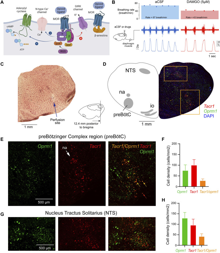
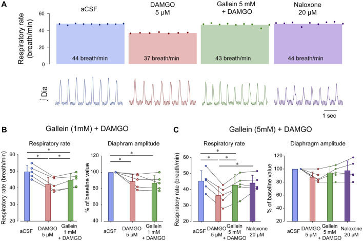
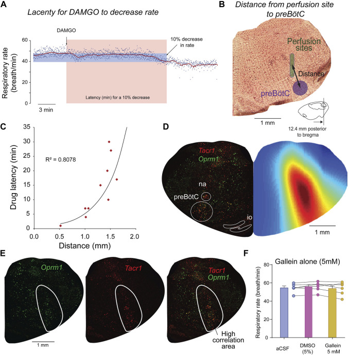
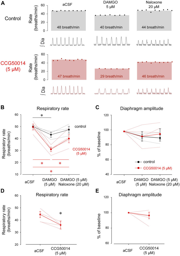
Opioid medications are the mainstay of pain management but present substantial side-effects such as respiratory depression which can be lethal with overdose. Most opioid drugs, such as fentanyl, act on opioid receptors such as the G-protein-coupled µ-opioid receptors (MOR). G-protein-coupled receptors activate pertussis toxin-sensitive G-proteins to inhibit neuronal activity. Binding of opioid ligands to MOR and subsequent activation G proteins βγ is modulated by regulator of G-protein signaling (RGS). The roles of G-proteins βγ and RGS in MOR-mediated inhibition of the respiratory network are not known. Using rodent models to pharmacologically modulate G-protein signaling, we aim to determine the roles of βγ G-proteins and RGS4. We showed that inhibition of βγ G-proteins using gallein perfused in the brainstem circuits regulating respiratory depression by opioid drugs results in complete reversal of respiratory depression. Blocking of RGS4 using CCG55014 did not change the respiratory depression induced by MOR activation despite co-expression of RGS4 and MORs in the brainstem. Our results suggest that neuronal inhibition by opioid drugs is mediated by G-proteins, but not by RGS4, which supports the concept that βγ G-proteins could be molecular targets to develop opioid overdose antidotes without the risks of re-narcotization often found with highly potent opioid drugs. On the other hand, RGS4 mediates opioid analgesia, but not respiratory depression, and RGS4 may be molecular targets to develop pain therapies without respiratory liability.

Keywords: regulators of G-protein-signaling; G-protein; breathing; medulla; opioid receptors; opioid-induced respiratory depression; preBotzinger complex.


Conflict of interest statement

The authors declare that the research was conducted in the absence of any commercial or financial relationships that could be construed as a potential conflict of interest.

Figures

FIGURE 1
FIGURE 1
Neural mechanisms of neuronal inhibition by MORs and expression of Oprm1 and Tacr1 (the gene encoding neurokinin-1 receptors) in the medulla. (A) Neuronal inhibition by MORs is regulated by G-protein-signaling including α, ß and γ proteins and regulators of G-protein signaling. (B) Microperfusion of the MOR agonist DAMGO directly into the preBötC depressed respiratory rate in anesthetized rats. (C) The location of the microperfusion site was identified with histological lesion made by the microdialysis probe shaft. Note that the probe extends beyond the guide by 0.5 mm. (D) The preBötC and the nucleus tractus solitarius (NTS) expressed Oprm1 (the gene coding for MORs) and Tacr1 (the gene coding for tachykinin-1 or neurokinin-1 receptors) mRNAs (E, F) The density of Oprm1 and Tacr1 were assessed in the preBötC and the NTS (G, H). MOR, µ-opioid receptor. GTP, guanine trisphosphate. GDP, guanine diphosphate. RGS, regulators of G-protein-signaling. GIRK, G-protein-activated inwardly rectifying potassium channels. Dia, diaphragm muscle. aCSF, artificial cerebrospinal fluid. Mean data are presented as means ± SEM. Panel a was created using Biorender.com.
FIGURE 2
FIGURE 2
Inhibition of βγ proteins reverses respiratory rate depression by the MOR agonist DAMGO. (A) DAMGO reduced respiratory rate by about 15.5% when compared to aCSF or vehicle, and this reduction was reversed by gallein at 5 mM, but only partially at 1 mM. Naloxone did not increase respiratory rate beyond the aCSF or gallein values. (B) DAMGO reduced respiratory rate compared to aCSF and gallein (1 mM) partially reversed respiratory rate. Although diaphragm amplitude was reduced by DAMGO, this reduction was not reversed by gallein. (C) DAMGO significantly depressed respiratory rate, an effect completely reversed by gallein at 5 mM. No effects were observed in diaphragm amplitude. aCSF, artificial cerebrospinal fluid. MOR, µ-opioid receptors. Bars show means ± SEM. Each circle represents an individual experiment. * indicate means significantly different with p < 0.05.
FIGURE 3
FIGURE 3
Site of action of DAMGO and gallein overlaps with Oprm1 and Tacr1 expressions. To determine the brainstem region where DAMGO is acting, we correlated the latency for DAMGO to decrease respiratory rate with the distances from the preBötC to the microperfusion sites. The latency in minutes for a 10% decrease from baseline respiratory rate was calculated for each experiment (A). For each corresponding experiment, the distance from the microperfusion site (indicated in green) to the center of the preBötC was calculated (B). The coordinates for the preBötC are 12.4 mm posterior, 6 mm ventral, and 2.0 lateral to Bregma. A significant exponential correlation was found between drug latencies and distances (R2 = 0.8078, p < 0.001, n = 10 experiments) (C). To determine whether other sites in the brainstem may mediate some of the respiratory depression induced by DAMGO, we generated a color map of the brainstem section where each pixel (every 50 µm) corresponds to a coefficient of correlation (blue equals low correlation, whereas red high correlation). For each set of coordinates in the section, the correlation coefficient was calculated using latencies and the distances from the microperfusion sites to the coordinates of the corresponding pixel. This correlation map shows that the area of the brainstem where the correlations are the highest corresponds to the region of the preBötC (D). The highly significant part of the correlation map (R2 > 0.5) overlapped with cells labelled with Oprm1 and Tacr1 (E). Control experiments were performed with microperfusion in the preBötC of aCSF, artificial spinal fluid, DMSO, dimethyl sulfoxide (5%) or gallein (5 mM) alone. See Methods for additional information on correlation maps as well as previous studies using this approach (Montandon et al., 2011; Montandon et al., 2016a).
FIGURE 4
FIGURE 4
Inhibition of RGS4 has limited impact on respiratory rate depression by DAMGO. (A) Inhibition of RGS4 potentiates MOR inhibition by the ligand DAMGO. (B) Pharmacological inhibition of RGS4 by CCG50014 significantly accentuated respiratory depression by DAMGO perfused in the preBötC compared to DAMGO alone. (C) The combination of DAMGO + CCG50014 (5 µM) induced a stronger respiratory rate depression than DAMGO alone (5 µM). Naloxone partially reversed respiratory rate depression by DAMGO and CCG50014. (D) Diaphragm amplitude was not significantly reduced by DAMGO with or without CCG50014. To determine whether RGS4 inhibition alone affects respiratory rate, we microperfused CCG50014 to the preBötC. CCG50014 (5 µM) decreased significantly respiratory rate (E) but not diaphragm amplitude (F). aCSF, artificial cerebrospinal fluid. MOR, µ-opioid receptors. RGS, regulators of G-protein-signaling. * indicate means significantly different. Circles indicate mean values ±SEM.
FIGURE 5
FIGURE 5
The site of action of DAMGO and CCG50014 overlaps with Oprm1 and Tacr1 expressing neurons. Using the same approach than in the gallein experiments, we determined the sites of drug perfusion and the latency for respiratory rate to decrease in response to DAMGO. Drugs were microperfused in the preBötC region as identified with expression of Tacr1 (gene for neurokinin-1 receptors) (A). A significant correlation was observed between distances from probe site to the centre of the preBötC and the latency for DAMGO to depress respiratory rate by 10% (B). Na, nucleus ambiguus.
FIGURE 6
FIGURE 6
Expression of Oprm1 and Rgs4 mRNAs in the preBötC and NTS. (A) The preBötC and the nucleus tractus solitarius (NTS) expressed Oprm1 (the gene coding for MORs, green) and Rgs4 (the gene coding for regulators of G-protein signaling 4, red) mRNAs. (B) Oprm1 and Rgs4 mRNAs were expressed in the preBötC and most cells expressing Oprm1 also co-expressed Rgs4 (orange). The density of Oprm1-positive cells was moderate compared to the high density of Oprm1 cells in the NTS (C). In the NTS, cells expressed Oprm1 as well as Rgs4 with substantial co-expression. Rgs4, mRNA of regulator of G-protein signaling 4. Oprm1, Gene encoding MORs. NTS, nucleus tractus solitarius. na, nucleus ambiguus. io, inferior olive.

Similar articles

References

    1. Abramow-Newerly M., Roy A. A., Nunn C., Chidiac P. (2006). RGS proteins have a signalling complex: Interactions between RGS proteins and GPCRs, effectors, and auxiliary proteins. Cell. Signal. 18 (5), 579–591. 10.1016/j.cellsig.2005.08.010 - DOI - PubMed
    1. Ballanyi K., Lalley P. M., Hoch B., Richter D. W. (1997). cAMP-dependent reversal of opioid- and prostaglandin-mediated depression of the isolated respiratory network in newborn rats. J. Physiol. 504, 127–134. 10.1111/j.1469-7793.1997.127bf.x - DOI - PMC - PubMed
    1. Blazer L. L., Roman D. L., Chung A., Larsen M. J., Greedy B. M., Husbands S. M., et al. (2010). Reversible, allosteric small-molecule inhibitors of regulator of G protein signaling proteins. Mol. Pharmacol. 78 (3), 524–533. 10.1124/mol.110.065128 - DOI - PMC - PubMed
    1. Bohn L. M., Lefkowitz R. J., Gainetdinov R. R., Peppel K., Caron M. G., Lin F. T. (1999). Enhanced morphine analgesia in mice lacking beta-arrestin 2. Science 286 (5449), 2495–2498. 10.1126/science.286.5449.2495 - DOI - PubMed
    1. Dahan A., Aarts L., Smith T. W. (2010). Incidence, reversal, and prevention of opioid-induced respiratory depression. Anesthesiology 112 (1), 226–238. 10.1097/ALN.0b013e3181c38c25 - DOI - PubMed

Grants and funding

This research was supported by a CIHR project grant, a Canadian Thoracic Society Grant-in-Aid, and a Pettit Block Term Grant from the Division of Respirology—University of Toronto.

LinkOut - more resources