

Bioluminogenic Probe for Rapid, Ultrasensitive Detection of β-Lactam-Resistant Bacteria
- PMID: 37083185
- PMCID: PMC10175212
- DOI: 10.1021/acs.analchem.3c00478
Bioluminogenic Probe for Rapid, Ultrasensitive Detection of β-Lactam-Resistant Bacteria
Abstract
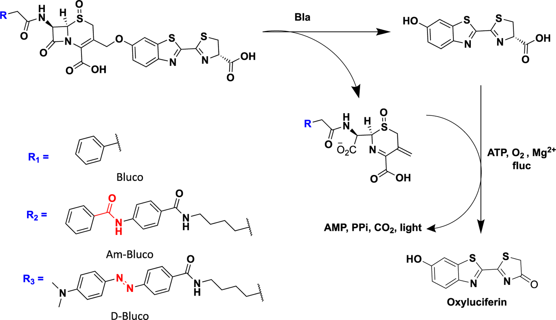
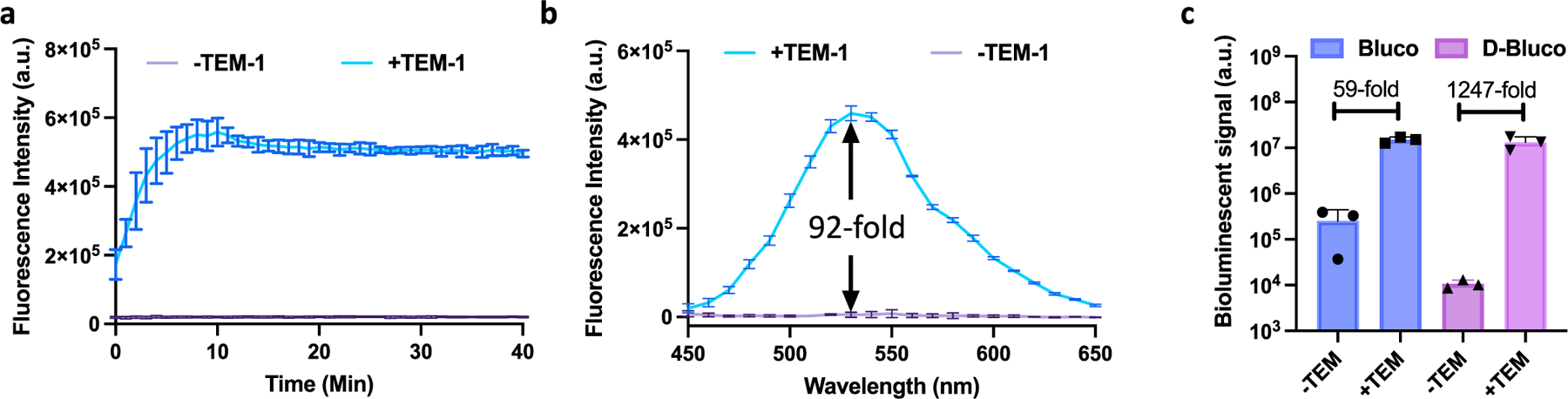
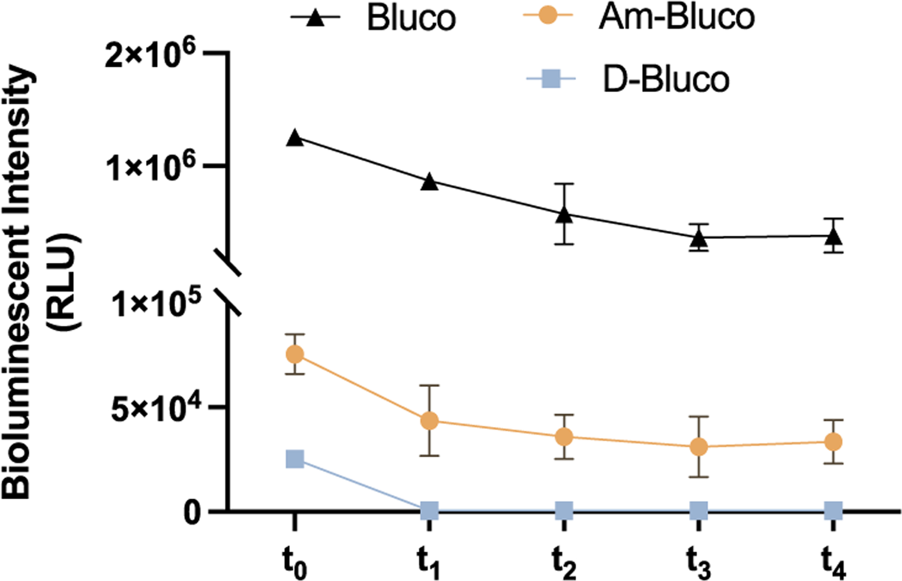
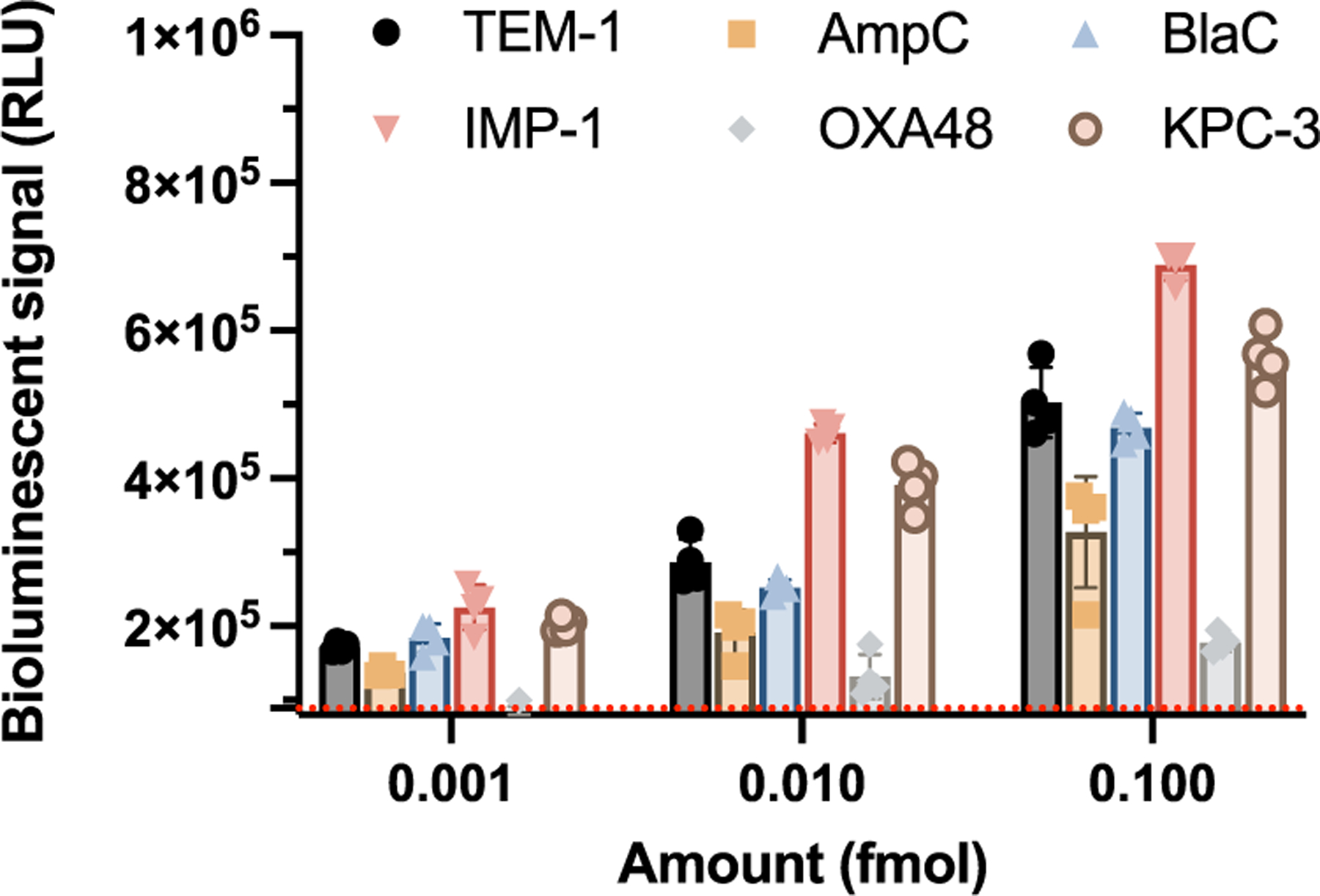
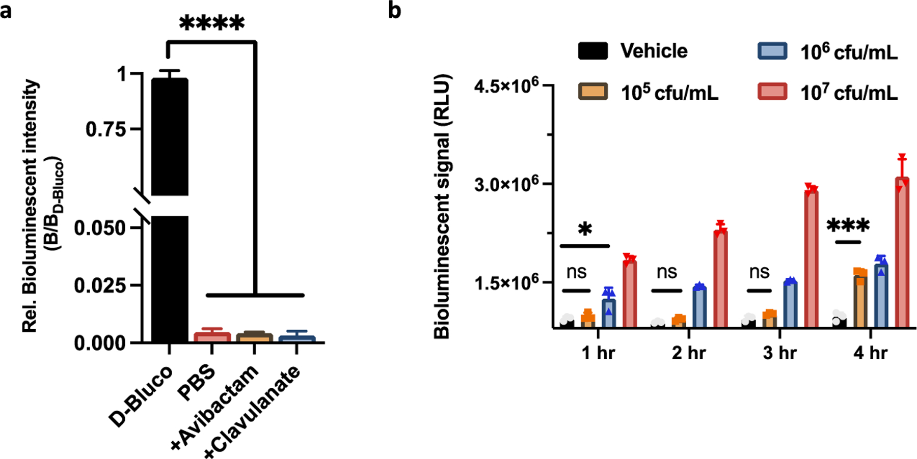
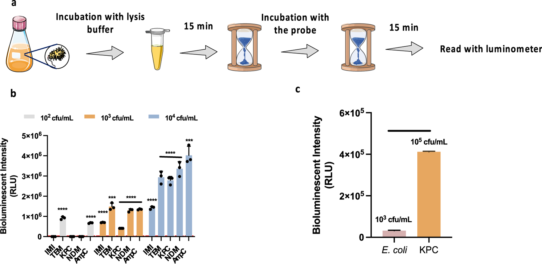
Increasingly difficult-to-treat infections by antibiotic-resistant bacteria have become a major public health challenge. Rapid detection of common resistance mechanisms before empiric antibiotic usage is essential for optimizing therapeutic outcomes and containing further spread of resistance to antibiotics among other bacteria. Herein, we present a bioluminogenic probe, D-Bluco, for rapid detection of β-lactamase activity in viable pathogenic bacteria. D-Bluco is a pro-luciferin caged by a β-lactamase-responsive cephalosporin structure and further conjugated with a dabcyl quencher. The caging and quenching significantly decreased the initial background emission and increased the signal-to-background ratio by more than 1200-fold. D-Bluco was shown to detect a broad range of β-lactamases at the femtomolar level. An ultrasensitive RAPID bioluminescence assay using D-Bluco can detect 102 to 103 colony forming unit per milliliter (cfu/mL) of β-lactamase-producing Enterobacterales in urine samples within 30 min. The high sensitivity and rapid detection make the assay attractive for the use of point-of-care diagnostics for lactam-resistant pathogens.
Conflict of interest statement
Notes
The authors declare the following competing financial interest(s): C.R.B. is a cofounder of Redwood Biosciences (a subsidiary of Catalent), Enable Biosciences, Palleon Pharmaceuticals, InterVenn Bio, Lycia Therapeutics, and OliLux Biosciences, and a member of the Board of Directors of Eli Lilly. All other authors declare no competing financial interest.
Figures
Similar articles
-
Rapid Detection of Bacterial Resistance to β-Lactam Antibiotics with a Relay-Response Chemiluminescence Assay.ACS Infect Dis. 2024 Jun 14;10(6):1970-1979. doi: 10.1021/acsinfecdis.3c00682. Epub 2024 May 31. ACS Infect Dis. 2024. PMID: 38819944
-
Deciphering the Evolution of Cephalosporin Resistance to Ceftolozane-Tazobactam in Pseudomonas aeruginosa.mBio. 2018 Dec 11;9(6):e02085-18. doi: 10.1128/mBio.02085-18. mBio. 2018. PMID: 30538183 Free PMC article.
-
Rapid optical determination of β-lactamase and antibiotic activity.BMC Microbiol. 2014 Apr 4;14:84. doi: 10.1186/1471-2180-14-84. BMC Microbiol. 2014. PMID: 24708478 Free PMC article.
-
Clinical data from studies involving novel antibiotics to treat multidrug-resistant Gram-negative bacterial infections.Int J Antimicrob Agents. 2022 Sep;60(3):106633. doi: 10.1016/j.ijantimicag.2022.106633. Epub 2022 Jul 1. Int J Antimicrob Agents. 2022. PMID: 35787918 Review.
-
Chemical sensors for the early diagnosis of bacterial resistance to β-lactam antibiotics.Bioorg Chem. 2024 Sep;150:107528. doi: 10.1016/j.bioorg.2024.107528. Epub 2024 Jun 4. Bioorg Chem. 2024. PMID: 38852309 Review.
Cited by
-
Sensing of Antibiotic-Bacteria Interactions.Antibiotics (Basel). 2023 Aug 19;12(8):1340. doi: 10.3390/antibiotics12081340. Antibiotics (Basel). 2023. PMID: 37627760 Free PMC article. Review.
References
-
- Fleming A Br. J. Exp. Pathol 1929, 10, 226–236.
Publication types
MeSH terms
Substances
Grants and funding
LinkOut - more resources
Full Text Sources
Medical
