Review
. 2023 Mar 30:17:910207.
doi: 10.3389/fncir.2023.910207. eCollection 2023.

Brainstem neural mechanisms controlling locomotion with special reference to basal vertebrates

Affiliations
Review

Brainstem neural mechanisms controlling locomotion with special reference to basal vertebrates

Philippe Lacroix-Ouellette et al. Front Neural Circuits. .

Abstract

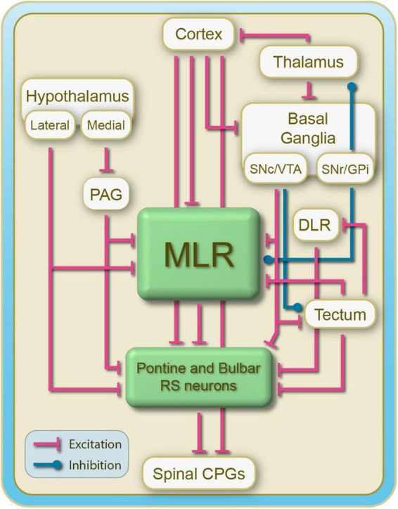
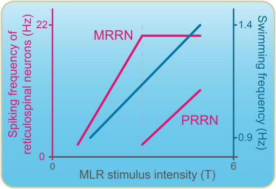
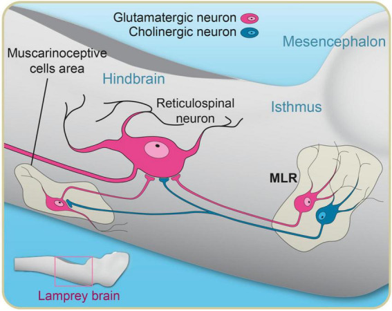
Over the last 60 years, the basic neural circuitry responsible for the supraspinal control of locomotion has progressively been uncovered. Initially, significant progress was made in identifying the different supraspinal structures controlling locomotion in mammals as well as some of the underlying mechanisms. It became clear, however, that the complexity of the mammalian central nervous system (CNS) prevented researchers from characterizing the detailed cellular mechanisms involved and that animal models with a simpler nervous system were needed. Basal vertebrate species such as lampreys, xenopus embryos, and zebrafish became models of choice. More recently, optogenetic approaches have considerably revived interest in mammalian models. The mesencephalic locomotor region (MLR) is an important brainstem region known to control locomotion in all vertebrate species examined to date. It controls locomotion through intermediary cells in the hindbrain, the reticulospinal neurons (RSNs). The MLR comprises populations of cholinergic and glutamatergic neurons and their specific contribution to the control of locomotion is not fully resolved yet. Moreover, the downward projections from the MLR to RSNs is still not fully understood. Reporting on discoveries made in different animal models, this review article focuses on the MLR, its projections to RSNs, and the contribution of these neural elements to the control of locomotion. Excellent and detailed reviews on the brainstem control of locomotion have been recently published with emphasis on mammalian species. The present review article focuses on findings made in basal vertebrates such as the lamprey, to help direct new research in mammals, including humans.

Keywords: acetylcholine; descending control; glutamate; locomotion; mesencephalic locomotor region (MLR); neuromodulation.


Conflict of interest statement

The authors declare that the research was conducted in the absence of any commercial or financial relationships that could be construed as a potential conflict of interest.

Figures

FIGURE 1
FIGURE 1
Schematics illustrating the general organization of supraspinal structures controlling locomotion in vertebrates. Reticulospinal cells constitute a large part of the locomotor-related descending inputs that activate the spinal locomotor CPGs. The MLR receives inputs from the basal ganglia and projects extensively to reticulospinal cells. The cortex projects down to the basal ganglia, the MLR and reticulospinal cells. The basal ganglia and the hypothalamus project down to the MLR as well as to reticulospinal neurons. There is another locomotor region, the DLR, that was identified in several species of vertebrates. Far less is known about this locomotor region that projects directly to reticulospinal neurons.
FIGURE 2
FIGURE 2
Mesopontine cholinergic cell populations have been described in the brainstem of mammals many decades ago. Here in the rat, much like in lampreys, the cells are arranged in a nearly continuous fashion but have been grouped into nuclei by neuroanatomists for simplicity. The exact names given to each of these nuclei somewhat vary from one species of mammal to the next. The CuN and PPN are traditionally associated with the MLR. (A) Schematic representation of a cross section of the isthmic region from the rat brainstem showing the mesopontine cholinergic cell populations (green) in reference to other landmarks in the region. (B) Overlay of brightfield and epifluorescence photomicrographs illustrating the cholinergic cells area labeled by immunofluorescence against the enzyme choline acetyltransferase. A black asterisk in the left bottom part of the illustration indicates immunofluorescence artifacts. The frame in panel (A) corresponds to the photographed area. CuN, cuneiform nucleus; IC, inferior colliculus; LDT, laterodorsal tegmental nucleus; Mes5, mesencephalic root of the trigeminal nerve; MLF, medial longitudinal fasciculus; nIV, trochlear nerve; SCP, superior cerebellar peduncle.
FIGURE 3
FIGURE 3
Dorsal view of the brain and rostral spinal cord of lampreys illustrating reticulospinal cells that were labeled by an injection of cobalt-lysine in the rostral spinal cord. (Left) Schematics illustrating the location of reticulospinal neurons (dark cells). (Right) Brain mounted on a microscope slide (wholemount preparation). Many of the reticulospinal cells are located just below the ventral surface of the fourth ventricle (PRRN, MRRN, and ARRN), making them easily accessible for intracellular/patch recordings/imaging as well as for local injections of drugs. Adapted from Bussières (1994).
FIGURE 4
FIGURE 4
A unilateral stimulation of the MLR in lampreys elicits coordinated and symmetrical swimming movements on both sides. Physiological experiments showed that the MLR on one side of the brainstem provides highly symmetrical glutamatergic and cholinergic inputs to paired, giant RS cells from opposite sides of the brainstem. Anatomical experiments confirm the presence of bilateral projections from the MLR to RS cells. (A1) Diagram of the hindbrain showing the bilateral connections between the MLR and paired, giant RS cells as well as the position of the electrodes for the electrophysiological experiments. (A2) Left side: the MLR on one side is stimulated at increasing intensities and the synaptic responses are recorded in paired, giant RS cells located in de pontine reticular formation (MRRN) on opposite sides of the brainstem. Note that the responses are remarkably similar on both sides. Right side: Same as on the left side, but for paired large RS cells in the bulbar reticular formation (PRRN). (B) Schematized illustration of the subthreshold synaptic responses elicited in reticulospinal neurons by stimulation of the MLR. Glutamatergic and cholinergic transmission were blocked or potentiated sequentially by adding different pharmacological agents. The EPSPs were increased under physostigmine, an inhibitor of the re-uptake of acetylcholine. The EPSPs were significantly decreased under D-tubocurarine, a nicotinic receptor antagonist, and were further decreased by the addition of a mixture of CNQX/AP5, glutamatergic receptors antagonists. A small response from unknown origin remained. Adapted from Brocard et al. (2010).
FIGURE 5
FIGURE 5
Recruitment pattern of reticulospinal neurons as the stimulation in the MLR is increased. The spiking frequency of reticulospinal neurons and swimming frequency are plotted against the intensity of MLR stimulation. Reticulospinal neurons in the pons (MRRN) are recruited at low MLR intensity and rapidly reach a discharge frequency plateau. Bulbar reticulospinal neurons (PRRN) are recruited at higher MLR stimulation intensities. They start discharging when the discharge frequency of pontine reticulospinal cells has reached a plateau. Adapted from Brocard and Dubuc (2003).
FIGURE 6
FIGURE 6
Schematic illustration of downward projections from the MLR to the pontine reticular formation. In addition to direct projections from glutamatergic and cholinergic MLR neurons to reticulospinal neurons, there is a muscarinic cholinergic projection from the MLR to a group of muscarinoceptive cells in the hindbrain that in turn provide extra excitation to reticulospinal neurons. This pathway is believed to boost the locomotor output at high swimming speeds, acting as a hyperdrive mechanism for locomotion. Adapted from Smetana et al. (2010).

Similar articles

Cited by

References

    1. Abols I. A., Basbaum A. I. (1981). Afferent connections of the rostral medulla of the cat: A neural substrate for midbrain-medullary interactions in the modulation of pain. J. Comp. Neurol. 201 285–297. 10.1002/cne.902010211 - DOI - PubMed
    1. Akay T. (2020). Sensory feedback control of locomotor pattern generation in cats and mice. Neuroscience 450 161–167. 10.1016/j.neuroscience.2020.05.008 - DOI - PubMed
    1. Akay T., Murray A. J. (2021). Relative contribution of proprioceptive and vestibular sensory systems to locomotion: opportunities for discovery in the age of molecular science. Int. J. Mol. Sci. 22:1467. 10.3390/ijms22031467 - DOI - PMC - PubMed
    1. Appell P. P., Behan M. (1990). Sources of subcortical GABAergic projections to the superior colliculus in the cat. J. Comp. Neurol. 302 143–158. 10.1002/cne.903020111 - DOI - PubMed
    1. Atwood H. L., Wiersma C. A. (1967). Command interneurons in the crayfish central nervous system. J. Exp. Biol. 46 249–261. 10.1242/jeb.46.2.249.PMID:6033993 - DOI - PubMed

Publication types

Grants and funding

LinkOut - more resources