. 2023 Jun 27;14(3):e0340822.
doi: 10.1128/mbio.03408-22. Epub 2023 Apr 13.

Porcine Epidemic Diarrhea Virus Antagonizes Host IFN-λ-Mediated Responses by Tilting Transcription Factor STAT1 toward Acetylation over Phosphorylation To Block Its Activation

Affiliations

Porcine Epidemic Diarrhea Virus Antagonizes Host IFN-λ-Mediated Responses by Tilting Transcription Factor STAT1 toward Acetylation over Phosphorylation To Block Its Activation

Jidong Xu et al. mBio. .

Abstract

Porcine epidemic diarrhea virus (PEDV) is the main etiologic agent causing acute swine epidemic diarrhea, leading to severe economic losses to the pig industry. PEDV has evolved to deploy complicated antagonistic strategies to escape from host antiviral innate immunity. Our previous study demonstrated that PEDV downregulates histone deacetylase 1 (HDAC1) expression by binding viral nucleocapsid (N) protein to the transcription factor Sp1, inducing enhanced protein acetylation. We hypothesized that PEDV inhibition of HDAC1 expression would enhance acetylation of the molecules critical in innate immune signaling. Signal transducer and activator of transcription 1 (STAT1) is a crucial transcription factor regulating expression of interferon (IFN)-stimulated genes (ISGs) and anti-PEDV immune responses, as shown by overexpression, chemical inhibition, and gene knockdown in IPEC-J2 cells. We further show that PEDV infection and its N protein overexpression, although they upregulated STAT1 transcription level, could significantly block poly(I·C) and IFN-λ3-induced STAT1 phosphorylation and nuclear localization. Western blotting revealed that PEDV and its N protein promote STAT1 acetylation via downregulation of HDAC1. Enhanced STAT1 acetylation due to HDAC1 inhibition by PEDV or MS-275 (an HDAC1 inhibitor) impaired STAT1 phosphorylation, indicating that STAT1 acetylation negatively regulated its activation. These results, together with our recent report on PEDV N-mediated inhibition of Sp1, clearly indicate that PEDV manipulates the Sp1-HDAC1-STAT1 signaling axis to inhibit transcription of OAS1 and ISG15 in favor of its replication. This novel immune evasion mechanism is realized by suppression of STAT1 activation through preferential modulation of STAT1 acetylation over phosphorylation as a result of HDAC1 expression inhibition. IMPORTANCE PEDV has developed sophisticated evasion mechanisms to escape host IFN signaling via its structural and nonstructural proteins. STAT1 is one of the key transcription factors in regulating expression of ISGs. We found that PEDV and its N protein inhibit STAT1 phosphorylation and nuclear localization via inducing STAT1 acetylation as a result of HDAC1 downregulation, which, in turn, dampens the host IFN signaling activation. Our study demonstrates a novel mechanism that PEDV evades host antiviral innate immunity through manipulating the reciprocal relationship of STAT1 acetylation and phosphorylation. This provides new insights into the pathogenetic mechanisms of PEDV and even other coronaviruses.

Keywords: acetylation; histone deacetylase 1; interferon-stimulated genes; porcine epidemic diarrhea virus; signal transducer and activator of transcription 1.


Conflict of interest statement

The authors declare no conflict of interest.

Figures

FIG 1
FIG 1
PEDV impairs poly(I·C)- and rIFN-λ3-induced transcription of OAS1 and ISG15 in IPEC-J2 cells. The IPEC-J2 cells were transfected with 0.1 μg/mL of poly(I·C) for 12 h and then infected with PEDV (MOI = 1) for 24 h. The cells were collected for extraction of total mRNA and were used to detect transcription of the ISGs IFITM1 (A), MX1 (B), OAS1 (C), and ISG15 (D). (E) Transcriptional changes of type I to III IFNs after PEDV infection for 24 h detected by qRT-PCR. (F and G) The IPEC-J2 cells were treated with recombinant IFN-α1, IFN-γ, and IFN-λ3 at the concentration of 50 ng/mL for 24 h, after which total mRNA was extracted for detecting OAS1 and ISG15 transcription level by qRT-PCR (F), and the whole-cell lysate was used for detecting ISG15 expression by Western blotting (G). (H) The cells were treated with recombinant IFN-α1, IFN-γ, and IFN-λ3 for 30 min as mentioned above, followed by PEDV infection; PEDV N expression level was detected by Western blotting after PEDV infection for 24 h. (I to L) IPEC-J2 cells were pretreated with rIFN-λ3 at the concentration of 50 ng/mL, followed by PEDV infection at the MOI of 1. Total RNA was extracted at 24 h post-PEDV infection. The transcription levels of IFITM1, MX1, OAS1, and ISG15 were detected by qRT-PCR. Transcription in the control cells without poly(I·C) or rIFN-λ3 treatment and PEDV infection was normalized to the housekeeping gene, GAPDH. The results were shown as means ± SD from three independent experiments. *, P < 0.05; **, P < 0.01.
FIG 2
FIG 2
Expression analysis of different STATs in PEDV-infected IPEC-J2 cells. (A to H) Transcription of different STAT genes (STAT1, STAT2, STAT3, STAT4, STAT5a, STAT5b, and STAT6) and IRF9 in IPEC-J2 cells infected with PEDV for 24 h detected by qRT-PCR. The uninfected cells with the same amount of trypsin treatment only were used as the mock control. The data were shown as means ± SD of three independent experiments. *, P < 0.05; **, P < 0.01. (I) Expression profiles of different STAT genes in uninfected IPEC-J2 cells by qRT-PCR, the STAT genes were normalized to GAPDH, and the data are shown as means ± SD of three independent experiments. (J) Expression of STAT1 at different time points after PEDV infection detected by Western blotting. PEDV replication was represented by its N protein expression, and β-actin was used as a loading control.
FIG 3
FIG 3
STAT1 overexpression inhibits PEDV replication and promotes ISG expression. The IPEC-J2 cells were transfected with pCMV-HA-STAT1 followed by PEDV infection for 24 h. Control vector-transfected cells or PEDV-infected cells were used as controls. Total RNA was extracted for detecting STAT1 (A), PEDV N (B), OAS1 (C), and ISG15 (D) transcription by qRT-PCR. (E) PEDV N, STAT1, and ISG15 expression detected by Western blotting. Density analysis shown as relative PEDV N (F) and ISG15 (G) expression to β-actin were obtained from 3 independent experiments of (E). Results of panels A, B, C, D, F, and G are shown as means ± SD. *, P < 0.05; **, P < 0.01.
FIG 4
FIG 4
STAT1 inhibition promotes PEDV replication via downregulating ISG expression. (A to C) The IPEC-J2 cells were pretreated with 1 μM FaraA followed by PEDV infection (MOI = 1) for 24 h. Total RNA was extracted for transcriptional analysis of PEDV N, OAS1, and ISG15 by qRT-PCR. Dimethyl sulfoxide (DMSO) treatment was used as a solvent control. (D) The same samples were also used for Western blotting to detect ISG15, PEDV N, total STAT1, and phosphorylated STAT1. (E to G) Densitometric analysis of p-STAT1/total STAT1, PEDV N/β-actin, and ISG15/β-actin of panel D. (H to L) The IPEC-J2 cells were transfected with STAT1 siRNA for 24 h followed by PEDV infection for qRT-PCR and Western blotting. (H) STAT1 knockdown efficiency detected by qPCR. (I) PEDV N transcription in STAT1 knockdown IPEC-J2 cells. (J) PEDV N, ISG15, and STAT1 expression in STAT1 knockdown and PEDV-infected cells detected by Western blotting. Density analysis shown as relative PEDV N (K) and ISG15 (L) expression to β-actin were obtained from 3 independent experiments of panel J. The results are shown as means ± SD from three independent experiments for panels A to C, E to G, H, and I. *, P < 0.05; **, P < 0.01.
FIG 5
FIG 5
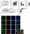
PEDV blocks STAT1 phosphorylation and translocation into the nuclei. (A) Changes of STAT1 phosphorylation in poly(I·C)-transfected and PEDV-infected IPEC-J2 cells. IPEC-J2 cells were inoculated with PEDV at the MOI of 1 for 4 h adsorption, after which poly(I·C) was transfected to the final concentration of 0.1 μg/mL for the next 20 h; poly(I·C) transfection or PEDV infection alone were used as controls, respectively. Total cell lysates were used to detect the expression of target proteins with β-actin as the loading control. (B to D) STAT1 expression, phosphorylation, and PEDV N expression as shown in panel A were analyzed and represented by STAT1/β-actin ratio (B), pSTAT1/STAT1 ratio (C), and PEDV N/β-actin ratio (D) based on 3 independent experiments, respectively. (E) STAT1 levels in the cytoplasmic and nuclear compartments as determined by Western blotting. (F) Ratios of STAT1 in the two compartments to total STAT1 in the whole-cell lysates based on panel E. Percentage of nuclear STAT1 is shown on top of the bars. The IPEC-J2 cells were treated as in panel A; β-actin and histone H3 were used as cytoplasmic and nuclear markers separately. (G) STAT1 subcellular localization in the cells treated as described above was also detected by immunocytochemistry with a confocal microscope; endogenous STAT1 and PEDV N were severally probed by rabbit anti-STAT1 and mouse anti-PEDV N antibodies. The nuclei were stained with DAPI.
FIG 6
FIG 6
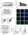
PEDV infection enhances significant STAT1 acetylation via inhibition of HDAC1 activity. (A) Western blotting detecting acetylated proteins in IPEC-J2 cells infected with PEDV or treated with MS-275. The IPEC-J2 cells were infected with PEDV at an MOI of 1 or treated with 1 μM MS-275 for 24 h. Total cell lysates were extracted for Western blotting using the acetyl-lysine antibody. Histone H3 and β-actin were used as loading controls. (B) STAT1 acetylation analyzed by co-IP using an acetyl-lysine antibody. Wild-type (WT) and HDAC1 knockdown (KD) IPEC-J2 cells were infected with PEDV for 24 h. Total cell lysates were immunoprecipitated with the acetyl-lysine antibody, followed by immunoblotting with the STAT1, β-actin, and histone H3 antibodies; STAT1, HDAC1, and β-actin detection of total IPEC-J2 cell lysates were used as the input (left). (C) STAT1 acetylation as shown in panel B was analyzed and shown by the ac-STAT1/STAT1 ratio from 3 independent experiments.
FIG 7
FIG 7
PEDV-enhanced STAT1 acetylation diminishes its phosphorylation. (A and B) STAT1 phosphorylation in IPEC-J2 cells affected by MS-275 treatment analyzed by Western blotting. The cells were pretreated with MS-275 at a concentration gradient, followed by poly(I·C) transfection for 24 h; MS-275 was maintained during the whole process. Total cell lysates were employed for SDS-PAGE followed by Western blotting using phosphorylated-STAT1 and total STAT1 antibodies; β-actin was used as the loading control. (C and D) STAT1 and HDAC1 subcellular localization measured and analyzed by cytoplasmic and nuclear fraction after poly(I·C) transfection and MS-275 treatment. The quality of cytoplasmic and nuclear fractionation was symbolized by β-actin and histone H3, respectively. (E) STAT1 cellular distribution was visualized by immunocytochemistry. The nuclei are shown by DAPI staining. (F) STAT1 phosphorylation in IPEC-J2 cells both treated with MS-275 and infected with PEDV. The cells were pretreated with 1 μM MS-275 for 1 h followed by PEDV infection. After 24 h postinfection, phosphorylated STAT1 together with total STAT1, PEDV N, and HDAC1 expression were measured by Western blotting. (G) The STAT1 phosphorylation and PEDV N expression were further obtained by densitometric analysis from three independent experiments. The results of panels G and H are shown as means ± SD. *, P < 0.05; **, P < 0.01.
FIG 8
FIG 8
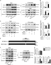
PEDV utilizes its N protein to induce STAT1 acetylation and suppress STAT1 phosphorylation. (A) STAT1 expression, acetylation, and phosphorylation were analyzed from the IPEC-J2 cells infected with PEDV followed by treatment of rIFN-λ3. The cells were first infected with PEDV at an MOI of 1; after 4 h absorption, the cells were treated with rIFN-λ3 to the concentration of 50 ng/mL for another 20 h, after which total protein was extracted. Acetylated STAT1 level was measured by acetylated lysine antibody enrichment followed by immunoblotting using total STAT1 antibody. STAT1 phosphorylation was detected using the phosphorylated STAT1 antibody at Y701. Expression of the other proteins was immunoblotted by specific antibodies mentioned in Materials and Methods. Histone H3 and β-actin were shown as loading controls. (B) STAT1 acetylation and phosphorylation as shown in panel A were analyzed and represented by the ac-STAT1/STAT1 ratio and p-STAT1/STAT1 ratio, separately. (C and D) STAT1 phosphorylation and acetylation were analyzed after PEDV N overexpression followed by rIFN-λ3 treatment, as the same procedures with panel A. (E) Schematic diagram showing NLS-deleted PEDV N mutant construction. (F and G) STAT1 expression, acetylation, and phosphorylation were analyzed from the IPEC-J2 cells exogenously expressed with WT and ΔNLS PEDV N followed by treatment of rIFN-λ3. The results of panels B, D, and G were obtained from 3 independent experiments of panels A, C, and F, respectively, and are shown as means ± SD. *, P < 0.05; **, P < 0.01.
FIG 9
FIG 9
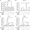
PEDV inhibits IFN-λ3-induced ISG expression in an HDAC1-dependent manner. (A to D) The wild-type IPEC-J2 cells and HDAC1 knockdown cells were infected with PEDV (MOI = 1) followed by rIFN-λ3 treatment (50 ng/mL) as shown in Fig. 8. The obtained total mRNA was used to detect transcription levels of HDAC1 (A), PEDV N (B), OAS1 (C), and ISG15 (D) by qRT-PCR; these genes were normalized to GAPDH. Data of panels A to D were shown as means ± SD from 3 independent experiments with the error bars representing SD. *, P < 0.05; **, P < 0.01; ***, P < 0.001.
FIG 10
FIG 10
PEDV antagonizes the expression of antiviral ISG15 and OAS1 by promoting STAT1 acetylation with subsequent reduction of its phosphorylation and nuclear translocation. (I) JAK-STAT1 signaling is directly activated by type III IFN stimulation (solid line), while it is indirectly activated by poly(I·C) (broken line). The phosphorylated STAT1 forms a heterodimer with activated STAT2, which further associates with IRF9 to form the ISGF3 complex. The complex is then translocated into the nucleus, where it binds to the target promoter and activates transcription of OAS1 and ISG15. (II) HDAC1 utilizes its deacetylase activity to facilitate STAT1 phosphorylation. (III) PEDV infection inhibits HDAC1 expression through the interaction of its N protein with Sp1, resulting in enhanced STAT1 acetylation. The acetylated STAT1 fails to be phosphorylated and is prevented from entering the nucleus. Consequently, ISG15 and OAS1 transcription is impaired. Therefore, PEDV evades the antiviral effect of ISGs by preventing STAT1 activation as a result of viral N protein-mediated suppression of the Sp1 transcriptional activity and decreased expression of HDAC1.

Similar articles

Cited by

References

    1. Song D, Park B. 2012. Porcine epidemic diarrhoea virus: a comprehensive review of molecular epidemiology, diagnosis, and vaccines. Virus Genes 44:167–175. doi:10.1007/s11262-012-0713-1. - DOI - PMC - PubMed
    1. Wang D, Fang L, Xiao S. 2016. Porcine epidemic diarrhea in China. Virus Res 226:7–13. doi:10.1016/j.virusres.2016.05.026. - DOI - PMC - PubMed
    1. Pensaert MB, de Bouck P. 1978. A new coronavirus-like particle associated with diarrhea in swine. Arch Virol 58:243–247. doi:10.1007/BF01317606. - DOI - PMC - PubMed
    1. Pensaert MB, Martelli P. 2016. Porcine epidemic diarrhea: a retrospect from Europe and matters of debate. Virus Res 226:1–6. doi:10.1016/j.virusres.2016.05.030. - DOI - PMC - PubMed
    1. Kocherhans R, Bridgen A, Ackermann M, Tobler K. 2001. Completion of the porcine epidemic diarrhoea coronavirus (PEDV) genome sequence. Virus Genes 23:137–144. doi:10.1023/A:1011831902219. - DOI - PMC - PubMed

Publication types