. 2023 Mar 30;28(7):3092.
doi: 10.3390/molecules28073092.

Intact Transition Epitope Mapping-Serological Inspection by Epitope EXtraction (ITEM-SIX)

Affiliations

Intact Transition Epitope Mapping-Serological Inspection by Epitope EXtraction (ITEM-SIX)

Agatino Zammataro et al. Molecules. .

Abstract

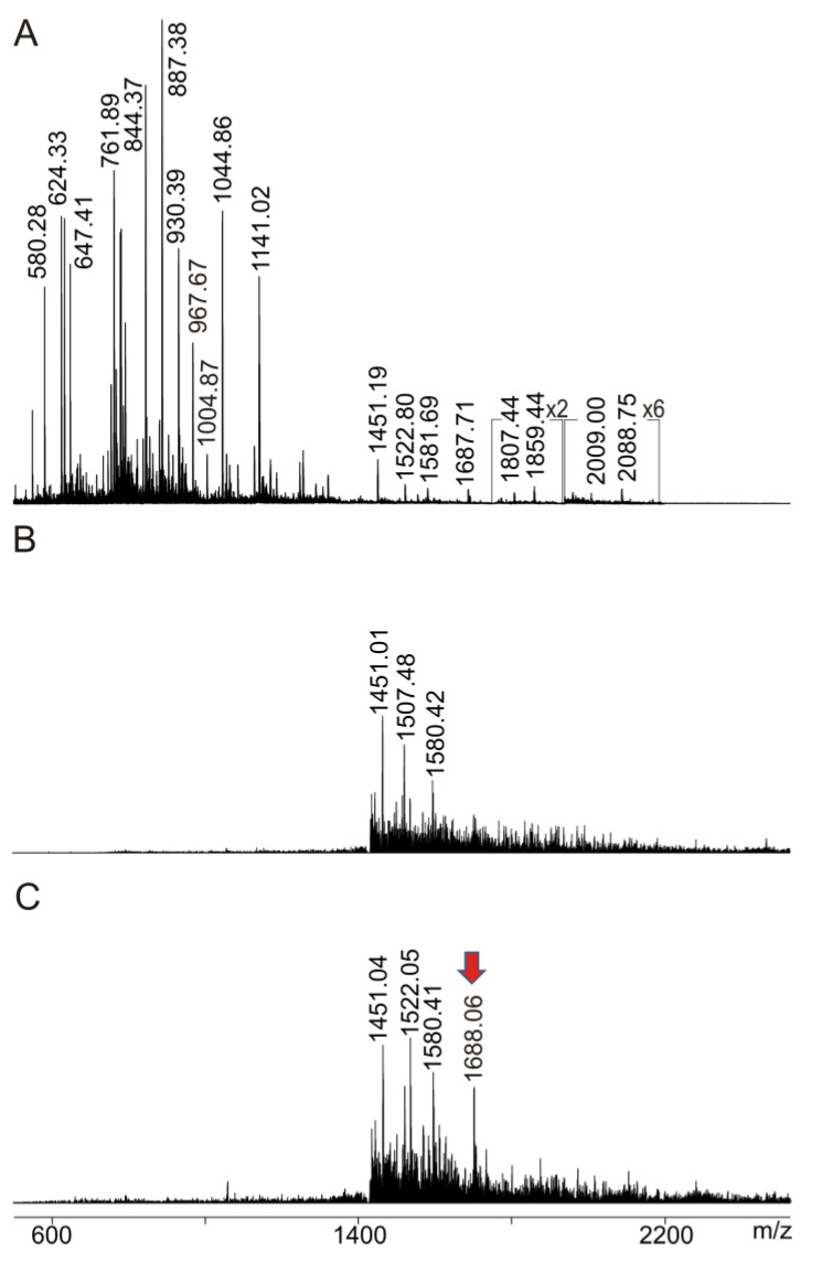
Precision medicine requests accurate serological inspections to precisely stratify patients for targeted treatment. Intact transition epitope mapping analysis proved surrogate seroconversion of a model organism's serum when spiked with a monoclonal murine anti-Ovalbumin antibody (mAb) with epitope resolution. Isolation of the IgG fraction from blood serum applied two consecutive protein precipitation steps followed by ultrafiltration and resulted in an ESI-MS analysis-ready IgG preparation. For epitope mapping by epitope extraction, the Ovalbumin antigen was digested with trypsin. After desalting, the peptide mixture was added to the ESI-MS-ready IgG preparation from mAb-spiked serum and the solution was incubated to form an immune complex between the Ovalbumin-derived epitope peptide and the anti-Ovalbumin mAb. Then, the entire mixture of proteins and peptides was directly electrosprayed. Sorting of ions in the mass spectrometer's gas phase, dissociation of the immune complex ions by collision-induced dissociation, and recording of the epitope peptide ion that had been released from the immune complex proved the presence of the anti-Ovalbumin mAb in serum. Mass determination of the complex-released epitope peptide ion with isotope resolution is highly accurate, guaranteeing high specificity of this novel analysis approach, which is termed Intact Transition Epitope Mapping-Serological Inspections by Epitope EXtraction (ITEM-SIX).

Keywords: blood serum; epitope mapping; immune complex analysis; nanoESI mass spectrometry; surrogate seroconversion.


Conflict of interest statement

The authors declare no conflict of interest.

Figures

Figure 1
Figure 1
Characterization of an IgG mix prepared from blood serum. (A) Nano-ESI mass spectrum of IgG mix (solution 0). Charge states and m/z values are provided for the multiply charged IgG ion signals. Quadrupole vacuum: 3.9 × 10⁻⁵ mbar. Insert: SDS-PAGE analysis of IgG mix prepared from serum under non-reducing conditions. Lane 1: molecular mass marker (PageRuler Prestained Protein Ladder). Apparent molecular masses are shown at the left. Lane 2: from blood-serum-isolated IgG mix (1 µg protein). Proteins were stained with colloidal Coomassie Brilliant Blue. The blue arrow points to the IgG band. The purple arrow head indicates residual serum albumin. (B) Nano-ESI mass spectrum of IgG mix (solution 0). Charge states and m/z values are provided for the multiply charged IgG ion signals. Quadrupole vacuum: 3.2 × 10⁻⁵ mbar. Insert: ion signals (m/z values and charge states) of residual monomeric serum albumin (#) and tetrameric transthyretin (*). Protein concentration is 0.34 µg/µL. 2.5 µL were loaded. Solvent: 200 mM ammonium acetate, pH 6.7. Recording time per spectrum is 1 min.
Figure 2
Figure 2
Characterization of anti-Ovalbumin antibody and IgG mix prepared from converted serum. (A) Nano-ESI mass spectrum of anti-Ovalbumin antibody (solution 1a). Charge states and m/z values are provided for the multiply charged IgG ion signals. Protein concentration is 0.28 µg/µL. 2.5 µL were loaded. (B) Nano-ESI mass spectrum of IgG mix (solution 1b) prepared from mAb-spiked serum. Charge states and m/z values are provided for the multiply charged IgG ion signals. Ion signals of residual tetrameric transthyretin (*) are marked. Protein concentration is 0.17 µg/µL. 2.5 µL were loaded. Solvent: 200 mM ammonium acetate, pH 6.7. Quadrupole vacuum: 3.2 × 10⁻⁵ mbar. Recording time per spectrum is 1 min.
Figure 3
Figure 3
Characterization of the Ovalbumin antigen. (A) NanoESI mass spectrum of ions from Ovalbumin upon tryptic digestion (solution 2′). Selected ion signals are labeled. Ion intensity multiplication factors are provided. Peptide concentration is 0.28 µg/µL. 2.5 µL were loaded. Solvent: 200 mM ammonium acetate, pH 6.7. Recording time is 1 min. (B) Amino acid sequence of chicken Ovalbumin (P01012; single letter code). Sequence parts that match with peptide ion signals are shaded in green (sequence coverage: 60%). Modified amino acid residues are highlighted in orange (cf. Table S2). The epitope peptide sequence is printed with red letters.
Figure 4
Figure 4
ITEM—ONE analysis. (A) NanoESI mass spectrum of ions from Ovalbumin upon tryptic digestion (solution 2′). Ion intensity multiplication factors are provided. (B,C) NanoESI mass spectrum of ions from mixture (solution 3a) of tryptically digested Ovalbumin (solution 2′) with anti-Ovalbumin mAb (solution 1a). Ion transmission below m/z 1450 is completely suppressed. (B) ∆CV: 3 V. (C): ∆CV: 20 V. Selected m/z values are provided. The red arrow points to the complex-released epitope peptide ion signal. The insert in (C) shows the isotope pattern of the complex-released peptide ion signal. Protein concentration is 0.22 µg/µL. 2.5 µL were loaded. Solvent: 200 mM ammonium acetate, pH 6.7. 1 min recording time per spectrum. 30% baseline subtraction.
Figure 5
Figure 5
ITEM—SIX analysis. (A) NanoESI mass spectrum of ions from Ovalbumin upon tryptic digestion (solution 2′). Ion intensity multiplication factors are provided. (B) NanoESI mass spectrum of ions from mixture (solution 3b) of tryptically digested Ovalbumin (solution 2′) with IgG mixture prepared from mAb-spiked blood serum (solution 1b). Ion transmission below m/z 1450 is completely suppressed. (B) ∆CV: 3 V. (C) ∆CV: 20 V. Selected m/z values are provided. The red arrow points to the complex-released epitope peptide. Protein concentration is 0.81 µg/µL. 2.5 µL were loaded. Solvent: 200 mM ammonium acetate, pH 6.7. Recording time per spectrum is 1 min. 50% baseline subtraction.

Similar articles

Cited by

References

    1. Wine Y., Horton A.P., Ippolito G.C., Georgiou G. Serology in the 21st century: The molecular-level analysis of the serum antibody repertoire. Curr. Opin. Immunol. 2015;35:89–97. doi: 10.1016/j.coi.2015.06.009. - DOI - PMC - PubMed
    1. Fox T., Geppert J., Dinnes J., Scandrett K., Bigio J., Sulis G., Hettiarachchi D., Mathangasinghe Y., Weeratunga P., Wickramasinghe D., et al. Antibody tests for identification of current and past infection with SARS-CoV-2. Cochrane Database Syst. Rev. 2022;11:1–1236. - PMC - PubMed
    1. Haselbeck A.H., Im J., Prifti K., Marks F., Holm M., Zellweger R.M. Serology as a Tool to Assess Infectious Disease Landscapes and Guide Public Health Policy. Pathogens. 2022;11:732. doi: 10.3390/pathogens11070732. - DOI - PMC - PubMed
    1. Prechl J. Why current quantitative serology is not quantitative and how systems immunology could provide solutions. Biol. Futur. 2021;72:37–44. doi: 10.1007/s42977-020-00061-1. - DOI - PMC - PubMed
    1. Sorohan B.M., Baston C., Tacu D., Bucșa C., Țincu C., Vizireanu P., Sinescu I., Constantinescu I. Non-HLA Antibodies in Kidney Transplantation: Immunity and Genetic Insights. Biomedicines. 2022;10:1506. doi: 10.3390/biomedicines10071506. - DOI - PMC - PubMed

LinkOut - more resources