. 2023 Apr 27;97(4):e0030223.
doi: 10.1128/jvi.00302-23. Epub 2023 Apr 11.

The SFTSV Nonstructural Proteins Induce Autophagy to Promote Viral Replication via Interaction with Vimentin

Affiliations

The SFTSV Nonstructural Proteins Induce Autophagy to Promote Viral Replication via Interaction with Vimentin

Sihua Liu et al. J Virol. .

Abstract

Severe fever with thrombocytopenia syndrome virus (SFTSV) is a newly identified phlebovirus associated with severe hemorrhagic fever in humans. Studies have shown that SFTSV nucleoprotein (N) induces BECN1-dependent autophagy to promote viral assembly and release. However, the function of other SFTSV proteins in regulating autophagy has not been reported. In this study, we identify SFTSV NSs, a nonstructural protein that forms viroplasm-like structures in the cytoplasm of infected cells as the virus component mediating SFTSV-induced autophagy. We found that SFTSV NSs-induced autophagy was inclusion body independent, and most phenuivirus NSs had autophagy-inducing effects. Unlike N protein-induced autophagy, SFTSV NSs was key in regulating autophagy by interacting with the host's vimentin in an inclusion body-independent manner. NSs interacted with vimentin and induced vimentin degradation through the K48-linked ubiquitin-proteasome pathway. This negatively regulating Beclin1-vimentin complex formed and promoted autophagy. Furthermore, we identified the NSs-binding domain of vimentin and found that overexpression of wild-type vimentin antagonized the induced effect of NSs on autophagy and inhibited viral replication, suggesting that vimentin is a potential antiviral target. The present study shows a novel mechanism through which SFTSV nonstructural protein activates autophagy, which provides new insights into the role of NSs in SFTSV infection and pathogenesis. IMPORTANCE Severe fever with thrombocytopenia syndrome virus (SFTSV) is a newly emerging tick-borne pathogen that causes multifunctional organ failure and even death in humans. As a housekeeping mechanism for cells to maintain steady state, autophagy plays a dual role in viral infection and the host's immune response. However, the relationship between SFTSV infection and autophagy has not been described in detail yet. Here, we demonstrated that SFTSV infection induced complete autophagic flux and facilitated viral proliferation. We also identified a key mechanism underlying NSs-induced autophagy, in which NSs interacted with vimentin to inhibit the formation of the Beclin1-vimentin complex and induced vimentin degradation through K48-linked ubiquitination modification. These findings may help us understand the new functions and mechanisms of NSs and may aid in the identification of new antiviral targets.

Keywords: Beclin1-vimentin complex; IBs; NSs; SFTSV; autophagy.


Conflict of interest statement

The authors declare no conflict of interest.

Figures
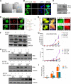
FIG 1
FIG 1
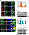
SFTSV induces complete autophagic flux to facilitate virus proliferation. (A and B) HeLa cells were infected with SFTSV (10 PFU/cell), collected, fixed, and stained at 6 h and examined under a TEM or laser confocal microscopy (the blue arrows indicate the autophagosomes). (C) After HeLa cells were infected with SFTSV for 6 h, the cells were collected to detect the expression of LC3 II by Western blotting. (D) HeLa cells were transfected with GFP-LC3B recombinant plasmid for 24 h and then infected with SFTSV (10 PFU/cell) for 12 h. Laser confocal microscopy was used to observe the colocalization of GFP-LC3B dots with Lyso-Tracker Red-stained lysosomes. The colocalization of autophagosomes and lysosomes in the white line region was analyzed with ImageJ software. (E) HeLa cells were transfected with GFP-mCherry-LC3B recombinant plasmids 24 h before SFTSV (10 PFU/cell) infection, and then the cells were fixed at 24 and 30 h. Laser confocal microscopy was used to observe the formation of the autophagosome (the white arrows indicate GFP+ mCherry+ dots, and the blue arrow indicates GFP- mCherry+ dots). The image in the dashed-line box on the right is a partial magnification of the left image. (F) Western blot detection of p62 and LC3 expression in HeLa cells infected or mock-infected with SFTSV (10 PFU/cell) and then cultured with or without bafA1 (20 nM). (G) HeLa cells were precultured with 10 mM 3-MA, 50 μM CQ, or 10 μM RAPA and then infected with SFTSV (0.1 PFU/cell) for 1 h; 10 mM 3-MA, 50 μM CQ, or 10 μM RAPA was added, and cells and supernatants were collected at 6, 12, 24, 36, and 48 h and analyzed by qRT-PCR. The control was the viral RNA expression level in the untreated group cells at 0 h. (H) Western blot detection of NSs expression in HeLa cells pretreated as in panel G but infected with 1 PFU/cell SFTSV. (I) The 293T cells were transfected with ATG5 siRNA for 24 h and then infected with SFTSV (0.1 PFU/cell). The cells were collected to detect viral RNA levels by qRT-PCR. The viral RNA expression level in the untreated group at 12 h was set as the control. Scale bar = 10 μm. (**, P < 0.01; ***, P < 0.001).
FIG 2
FIG 2
SFTSV NSs mediates autophagy. (A) HeLa cells were cotransfected with VR1012, NSs-HA, N-HA, Gc-HA, Gn-HA, L-HA, or GFP-LC3B recombinant plasmids for 24 h. Autophagosome formation was observed by laser confocal microscopy. (B) Statistical analysis of the number of autophagosomes. (C) HeLa cells were transfected with VR1012 or NSs-HA recombinant plasmids and collected at 12, 24, and 48 h. The expression of LC3 II was detected by Western blotting. (D) HeLa cells cotransfected with VR1012, SFTSV-NSs-HA, SFSV-NSs-HA, RVFV-NSs-HA, UUKV-NSs-HA, or GFP-LC3B recombinant plasmids for 24 h. Laser confocal microscopy was used to observe the autophagosomes. The images on the far right show the zoomed-in results in the white boxes. (E) Statistical analysis of the number of autophagosomes. (F) HeLa cells transfected with VR1012, SFTSV-NSs-HA, SFSV-NSs-HA, RVFV-NSs-HA, or UUKV-NSs-HA recombinant plasmids for 24 h. The expression of LC3 II was detected by Western blotting. LC3-II/GAPDH densitometric ratios were recorded. The blue arrows indicate autophagosomes. Scale bar = 10 μm. (***, P < 0.001).
FIG 3
FIG 3
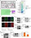
NSs interacts with vimentin and inhibits Beclin1-vimentin complex formation. (A) GO functional enrichment analysis of host proteins interacting with NSs. (B) KEGG pathway analysis of host proteins interacting with NSs. (C) Schematic representation of the AP-MS approach for identifying SFTSV-human PPIs. The green boxes indicate the interacting host proteins, the yellow circle indicates NSs, and the purple boxes indicate the autophagy-related regulatory proteins. (D) HeLa cells were transfected with VR1012 or NSs-HA recombinant plasmids. After 48 h, the cells were fixed, permeated, blocked, and incubated with antibodies. Laser confocal microscopy was used to observe the colocalization of vimentin and NSs and Beclin1 and NSs. The colocalization of NSs and vimentin or Beclin1 in the white line region was analyzed using ImageJ software. The blue arrows indicate the colocation peak formed by NSs IBs and vimentin. (E) The 293T cells were transfected with 6 μg VR1012 or 6 μg NSs-HA plasmids and collected at 48 h. The interactions of vimentin, Beclin1, AKT, and NSs were detected by immunoprecipitation. (F) The 293T cells were transfected with 6 μg VR1012 or 6 μg vimentin-plasmids before SFTSV (10 PFU/cell) infection and collected at 48 h. The interaction of vimentin and virally produced NSs was detected by immunoprecipitation. (G) The 293T cells were transfected with 3 μg VR1012 plus 3 μg NSs-HA, 3 μg VR1012 plus 3 μg vimentin-Flag, or 3 μg NSs-HA plus 3 μg vimentin-Flag recombinant plasmids. The cells were collected at 48 h, and the effect of NSs on the formation of Beclin1-vimentin complex was analyzed by immunoprecipitation. The quantified loss of Beclin1 binding to vimentin was analyzed by ImageJ and Prism software. P, Pearson correlation coefficient value of the whole picture. Scale bar = 10 μm. (**, P < 0.01).
FIG 4
FIG 4
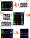
NSs-induced autophagy is not IB-dependent. (A) HeLa cells were cotransfected with GFP-LC3B and VR1012, NSs-WT-HA, NSs-V21/L23A-HA, or NSs-P66/69A-HA recombinant plasmids. After 24 h, the cells were fixed, permeated, blocked, and the antibodies were incubated. Laser confocal microscopy was used to observe the formation of the autophagosome. (B) Statistical analysis of the number of autophagosomes. (C) The expression of LC3 II was detected by Western blotting. LC3-II/GAPDH densitometric ratios were recorded. (D) The 293T cells were transfected with VR1012, NSs-WT-HA, NSs-V21/L23A-HA, or NSs-P66/69A-HA recombinant plasmids and collected at 48 h. The interaction of vimentin, NSs, NSs-V21/L23A-HA, and NSs-P66/69A-HA was detected by immunoprecipitation. (E) HeLa cells were cotransfected with GFP-LC3B and VR1012, NSs-WT-HA, NSs-ΔN35-HA, NSs-ΔN45-HA, NSs-ΔC50-HA, or NSs-ΔC80-HA recombinant plasmids. After 24 h, the cells were fixed, permeated, and blocked, and the antibodies were incubated. Laser confocal microscopy was used to observe the formation of the autophagosome. (F) Statistical analysis of the number of autophagosomes. (G) The expression of LC3 II was detected by Western blotting. LC3-II/GAPDH densitometric ratios were recorded. (H) HeLa cells were cotransfected with GFP-LC3B and VR1012, NSs-WT-HA, NSs-ΔN146-HA, or NSs-ΔC147-HA recombinant plasmids. After 24 h, the cells were fixed, permeated, and blocked, and the antibodies were incubated. Laser confocal microscopy was used to observe the formation of the autophagosome. (I) Statistical analysis of the number of autophagosomes (J) HeLa cells were transfected with VR1012 NSs-WT-HA, NSs-ΔN146-HA, or NSs-ΔC147-HA recombinant plasmids for 48 h. The cells were fixed, permeabilized, blocked, stained with vimentin and anti-HA antibodies, and incubated with anti-mouse minus and anti-rabbit plus PLA reagents, followed by a ligation and amplification mix. Images were captured with a laser confocal scanning microscope system (the blue arrows indicate autophagosomes, and the white arrow indicates that NSs IBs and LC3 are colocated. Scale bar = 10 μm; n.s., P > 0.05; ***, P < 0.001).
FIG 5
FIG 5
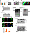
NSs degrades vimentin through the proteasome pathway and promotion of autophagy. (A) The 293T cells were transfected with VR1012 or NSs-HA recombinant plasmids for 24 h and 48 h. The cells were collected, and vimentin mRNA levels were detected by qRT-PCR. (B and C) The 293T cells were mock infected or infected with SFTSV (10 PFU/cell) or transfected with VR1012 or NSs-HA recombinant plasmids and collected at 48 h. The expression of vimentin was detected by Western blotting. (D) HeLa cells were transfected with GFP-LC3B recombinant plasmids 24 h before SFTSV (10 PFU/cell) infection, and then the cells were fixed at 24 h. Laser confocal microscopy was used to observe the formation of the autophagosome and colocalization with vimentin. (E) The 293T cells were transfected with VR1012 or NSs-HA recombinant plasmids for 24 h; MG123 (2 μM) and bafA1 (20 nM) inhibitors were added 12 h before cell collection. The expression of vimentin was detected by Western blotting. (F) The 293T cells were cotransfected with 4 μg VR1012 plus 2 μg Ub-K48-Myc, 2 μg VR1012 plus 2 μg vimentin-Flag plus 2 μg Ub-K48-Myc, 2 μg vimentin-Flag plus 2 μg Ub-K48-Myc plus 2 μg NSs-HA, or 2 μg vimentin-Flag plus 2 μg Ub-K48-Myc plus 2 μg N-HA recombinant plasmids, and the ubiquitin of vimentin was detected by immunoprecipitation. (G) The 293T cells were transfected with VR1012 or vimentin-Flag recombinant plasmids for 24 h and then infected with SFTSV (1 PFU/cell), and the expression of N proteins was detected by Western blotting. (H) HeLa cells were cotransfected with GFP-LC3B plus VR1012, GFP-LC3B plus NSs-HA, GFP-LC3B plus vimentin-Flag, or GFP-LC3B plus NSs-HA plus vimentin-Flag recombinant plasmids for 24 h. Autophagosome formations were observed by laser confocal microscopy. (I) Statistical analysis of the number of autophagosomes. (J) The expression of LC3 II was detected by Western blotting. (K) The 293T cells were transfected with VR1012 or vimentin-Flag for 24 h and then mock infected or infected with SFTSV (10 PFU/cell). The expression of LC3 II was detected by Western blotting. The blue arrows indicate autophagosomes. Scale bar = 10 μm. n.s., (P > 0.05; **, P < 0.01; ***, P < 0.001).
FIG 6
FIG 6
The key interacting domains of vimentin and NSs. (A) Schematic diagram of vimentin truncation. (B) The 293T cells were transfected with vimentin truncation recombinant plasmids for 48 h. The interaction of vimentin, vimentin truncation, and NSs was detected by immunoprecipitation. (C) HeLa cells were cotransfected with GFP-LC3B plus VR1012, GFP-LC3B plus NSs-HA, GFP-LC3B plus vimentin-WT-Flag, GFP-LC3B plus NSs-HA plus Vimentin-WT-Flag, GFP-LC3B plus vimentin-ΔM185~235-Flag, or GFP-LC3B plus vimentin-ΔM185~235-Flag plus NSs-HA recombinant plasmids for 24 h. Autophagosome formations were observed by laser confocal microscopy. (D) Statistical analysis of the number of autophagosomes (the blue arrows indicate autophagosomes). Scale bar = 10 μm; n.s., (P > 0.05; **, P < 0.01; ***, P < 0.001).
FIG 7
FIG 7
Proposed model of SFTSV NSs-induced autophagy. SFTSV infection induces complete autophagic flux, which is beneficial to virus proliferation. In addition, SFTSV NSs inhibits Beclin1-vimentin complex formation by sequestering and promoting vimentin ubiquitination and degradation, which frees Beclin1 to activate its downstream autophagy pathway.

Similar articles

Cited by

References

    1. Li DX. 2011. Fever with thrombocytopenia associated with a novel bunyavirus in China. Zhonghua Shi Yan He Lin Chuang Bing Du Xue Za Zhi 25:81–84. - PubMed
    1. Zhang YZ, Zhou DJ, Xiong Y, Chen XP, He YW, Sun Q, Yu B, Li J, Dai YA, Tian JH, Qin XC, Jin D, Cui Z, Luo XL, Li W, Lu S, Wang W, Peng JS, Guo WP, Li MH, Li ZJ, Zhang S, Chen C, Wang Y, de Jong MD, Xu J. 2011. Hemorrhagic fever caused by a novel tick-borne Bunyavirus in Huaiyangshan, China. Zhonghua Liu Xing Bing Xue Za Zhi 32:209–220. - PubMed
    1. Takahashi T, Maeda K, Suzuki T, Ishido A, Shigeoka T, Tominaga T, Kamei T, Honda M, Ninomiya D, Sakai T, Senba T, Kaneyuki S, Sakaguchi S, Satoh A, Hosokawa T, Kawabe Y, Kurihara S, Izumikawa K, Kohno S, Azuma T, Suemori K, Yasukawa M, Mizutani T, Omatsu T, Katayama Y, Miyahara M, Ijuin M, Doi K, Okuda M, Umeki K, Saito T, Fukushima K, Nakajima K, Yoshikawa T, Tani H, Fukushi S, Fukuma A, Ogata M, Shimojima M, Nakajima N, Nagata N, Katano H, Fukumoto H, Sato Y, Hasegawa H, Yamagishi T, Oishi K, Kurane I, Morikawa S, Saijo M. 2014. The first identification and retrospective study of severe fever with thrombocytopenia syndrome in Japan. J Infect Dis 209:816–827. doi:10.1093/infdis/jit603. - DOI - PMC - PubMed
    1. Li H, Zhang LK, Li SF, Zhang SF, Wan WW, Zhang YL, Xin QL, Dai K, Hu YY, Wang ZB, Zhu XT, Fang YJ, Cui N, Zhang PH, Yuan C, Lu QB, Bai JY, Deng F, Xiao GF, Liu W, Peng K. 2019. Calcium channel blockers reduce severe fever with thrombocytopenia syndrome virus (SFTSV) related fatality. Cell Res 29:739–753. doi:10.1038/s41422-019-0214-z. - DOI - PMC - PubMed
    1. Kim KH, Yi J, Kim G, Choi SJ, Jun KI, Kim NH, Choe PG, Kim NJ, Lee JK, Oh MD. 2013. Severe fever with thrombocytopenia syndrome, South Korea, 2012. Emerg Infect Dis 19:1892–1894. doi:10.3201/eid1911.130792. - DOI - PMC - PubMed

Publication types

MeSH terms