. 2023 Jun 15;11(3):e0450822.
doi: 10.1128/spectrum.04508-22. Epub 2023 Apr 10.

ISG15 Is Required for the Dissemination of Vaccinia Virus Extracellular Virions

Affiliations

ISG15 Is Required for the Dissemination of Vaccinia Virus Extracellular Virions

Martina Bécares et al. Microbiol Spectr. .

Abstract

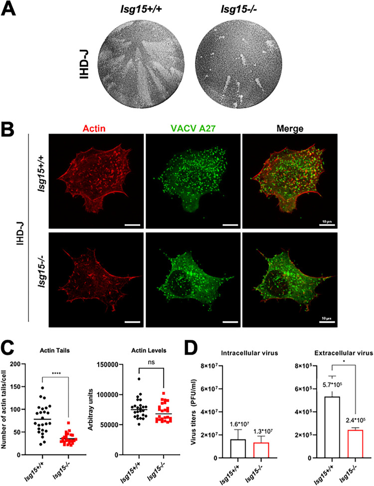
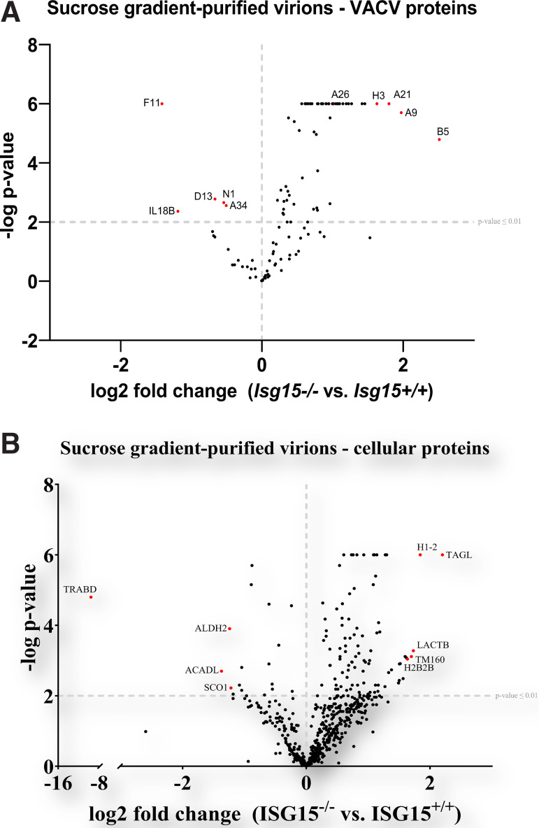
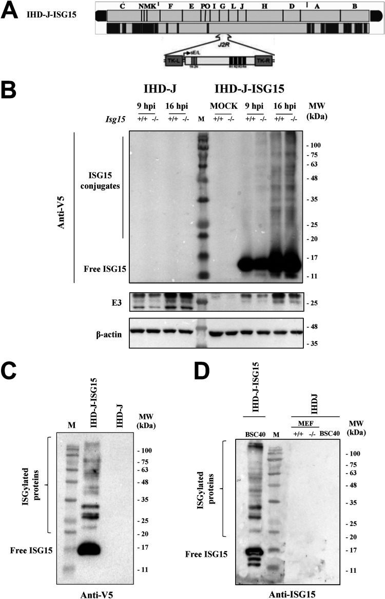
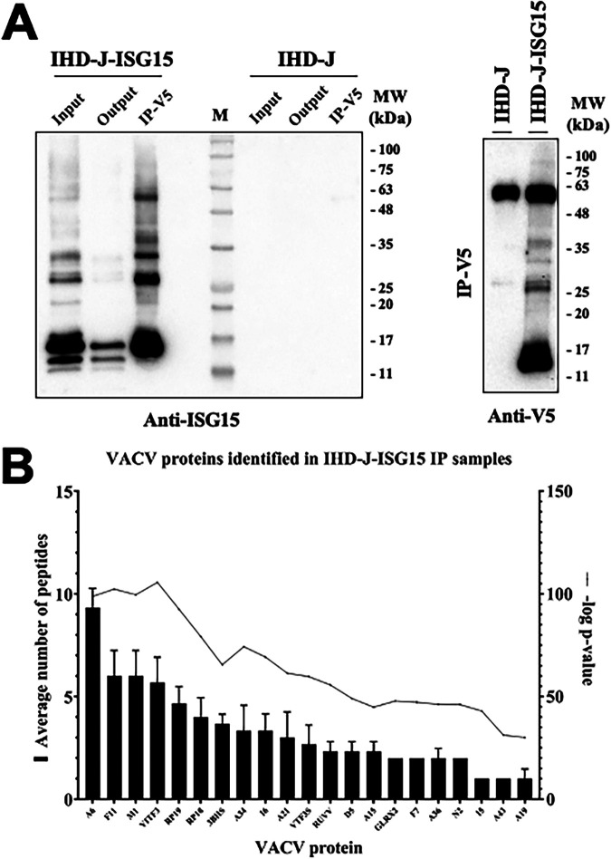
Viruses have developed many different strategies to counteract immune responses, and Vaccinia virus (VACV) is one of a kind in this aspect. To ensure an efficient infection, VACV undergoes a complex morphogenetic process resulting in the production of two types of infective virions: intracellular mature virus (MV) and extracellular enveloped virus (EV), whose spread depends on different dissemination mechanisms. MVs disseminate after cell lysis, whereas EVs are released or propelled in actin tails from living cells. Here, we show that ISG15 participates in the control of VACV dissemination. Infection of Isg15-/- mouse embryonic fibroblasts with VACV International Health Department-J (IHD-J) strain resulted in decreased EV production, concomitant with reduced induction of actin tails and the abolition of comet-shaped plaque formation, compared to Isg15+/+ cells. Transmission electron microscopy revealed the accumulation of intracellular virus particles and a decrease in extracellular virus particles in the absence of interferon-stimulated gene 15 (ISG15), a finding consistent with altered virus egress. Immunoblot and quantitative proteomic analysis of sucrose gradient-purified virions from both genotypes reported differences in protein levels and composition of viral proteins present on virions, suggesting an ISG15-mediated control of viral proteome. Lastly, the generation of a recombinant IHD-J expressing V5-tagged ISG15 (IHD-J-ISG15) allowed us to identify several viral proteins as potential ISG15 targets, highlighting the proteins A34 and A36, which are essential for EV formation. Altogether, our results indicate that ISG15 is an important host factor in the regulation of VACV dissemination. IMPORTANCE Viral infections are a constant battle between the virus and the host. While the host's only goal is victory, the main purpose of the virus is to spread and conquer new territories at the expense of the host's resources. Along millions of years of incessant encounters, poxviruses have developed a unique strategy consisting in the production two specialized "troops": intracellular mature virions (MVs) and extracellular virions (EVs). MVs mediate transmission between hosts, and EVs ensure advance on the battlefield mediating the long-range dissemination. The mechanism by which the virus "decides" to shed from the primary site of infection and its significant impact in viral transmission is not yet fully established. Here, we demonstrate that this process is finely regulated by ISG15/ISGylation, an interferon-induced ubiquitin-like protein with broad antiviral activity. Studying the mechanism that viruses use during infection could result in new ways of understanding our perpetual war against disease and how we might win the next great battle.

Keywords: ISG15; VACV; actin tails.


Conflict of interest statement

The authors declare no conflict of interest.

Figures

FIG 1
FIG 1
The absence of ISG15 impairs comet-shaped plaque, actin tail and EV production. (A) Comet-shaped plaque formation is dramatically reduced in ISG15−/− MEFs. Monolayers of immortalized ISG15+/+ and ISG15−/− MEFs were infected with IHD-J (0.0001 PFU/cell). At 48 hpi, cells were fixed and stained with 0.2% crystal violet in 10% formaldehyde for comet-shaped plaque visualization. (B and C) Actin tail formation is significantly reduced in ISG15−/− MEFs. Immortalized ISG15+/+ and ISG15−/− MEFs growing in coverslip were infected with IHD-J (2 PFU/cell). At 9 hpi, actin tails were visualized by phalloidin-staining (red), and viral particles were visualized using an anti-A27 antibody, followed by a fluorescent secondary antibody (green). Actin tails and actin levels were quantified using Leica AF or FIJI softwares (n = 25, each genotype in three different biologicals replicates), and actin tail numbers (means ± the SD) of individual cells are represented. The experiment has been carried out in triplicate and a representative image is shown. The specific values obtained are depicted in the graph. (D) EV release is significantly decreased in ISG15−/− MEFs. Immortalized ISG15+/+ and ISG15−/− MEFs were infected with IHD-J (2 PFU/cell). At 16 hpi, infectious viral particles from supernatants (extracellular virus) and cell extracts (intracellular virus) were titrated by plaque assay. Means ± the SD from three independent experiments are represented. *, P < 0.05; **, P < 0.01; ***, P < 0.005; ****, P < 0.0001. n.s, non significative.
FIG 2
FIG 2
IHD-J morphogenetic process is altered in ISG15−/− MEFs. (A) Viral protein synthesis is not affected by the absence of ISG15. Immortalized ISG15+/+ and ISG15−/− MEFs were infected with IHD-J (2 PFU/cell) and, at the indicated times postinfection, equal amounts of proteins from cell extracts were analyzed by Western blotting. Specific antibodies for VACV early protein E3 (E3L) and late proteins F13 (F13L) and A27 (A27L) were used. β-Actin and eiF2α were used as loading control. Molecular weights (MW) in kilodaltons (kDa) are indicated, based on protein standards. Protein levels of three independent experiments were analyzed with Fiji software. Means ± the SD are indicated. (B) Viral protein distribution does not change between ISG15+/+ and ISG15−/− MEFs. Confocal microscopy analysis of immortalized ISG15+/+ or ISG15−/− MEFs infected with IHD-J (2 PFU/cell) at 9 h postinfection. Viral proteins A27 (green) and F13 (red) were labeled by immunofluorescence; DAPI (blue) was used to stain the nuclear DNA. The experiment has been carried out in triplicate and a representative image is shown. (C) The absence of ISG15 causes accumulation of intracellular virions and a diminution in extracellular virus. Representative images of ISG15+/+ and ISG15−/− MEFs infected with IHD-J (2 PFU/cell; 9 h) are shown. Intracellular or extracellular viral particles per field were quantified with Fiji software (×2,500 magnification, n = 20, each genotype, in three different biologicals replicates). Means ± the SD are indicated. *, P < 0.05; **, P < 0.01; ***, P < 0.005; ****, P < 0.0001. The experiment was carried out in triplicate, and a representative image is shown. The specific values obtained are depicted in the graph.
FIG 3
FIG 3
The proteomes of purified virions change in the absence of ISG15. (A) The viral protein content of highly purified virions from ISG15+/+ and ISG15−/− MEFs differs between genotypes. Immortalized ISG15+/+ or ISG15−/− MEFs were infected with IHD-J (0.01 PFU/cell, 48 h), and intracellular virions were purified by ultracentrifugation through a 20% sucrose cushion, followed by ultracentrifugation through a sucrose gradient (20 to 45%). Purified virions were processed for LC-MS/MS analysis. Viral proteins are represented in the volcano plot. A P cutoff value of ≤0.01 is indicated on the graph. Proteins of interest significantly enriched in virions isolated from ISG15+/+ cells (upper left) or ISG15−/− cells (upper right) are labeled and highlighted in red. (B) The cellular protein content of highly purified virions from ISG15+/+ and ISG15−/− MEFs differs between genotypes. Immortalized ISG15+/+ or ISG15−/− MEFs were infected with IHD-J (0.01 PFU/cell, 48 h), and intracellular virions were purified by ultracentrifugation through a 20% sucrose cushion, followed by ultracentrifugation through a sucrose gradient (20 to 45%). Purified virions were processed for LC-MS/MS analysis. Cellular proteins are represented in the volcano plot. A P cutoff value of ≤0.01 is indicated on the graph. The most significantly enriched proteins in virions isolated from ISG15+/+ cells (upper left) or ISG15−/− cells (upper right) are labeled and highlighted in red.
FIG 4
FIG 4
Generation and in vitro characterization of IHD-J-ISG15. (A) Scheme of the IHD-J-ISG15 genome map. The full-length murine ISG15 with a V5 epitope fused to its N terminus was inserted within the VACV TK viral locus (J2R), and its expression was driven by the VACV E/L promoter (B) Expression of V5-ISG15 fusion protein. Immortalized ISG15+/+ or ISG15−/− MEFs were mock infected or infected at 2 PFU/cell with IHD-J or IHD-J-ISG15. At the indicated times postinfection, proteins were separated by SDS-PAGE, and the expression of viral early (E3) protein was analyzed by Western blotting with specific antibodies. Actin was used as a loading control. (C and D) Purified IHD-J-ISG15 virions contain ISGylated proteins and free ISG15. Purified IHD-J or IHD-J-ISG15 from BSC40 cells or immortalized ISG15+/+ and ISG15−/− MEFs were fractionated by SDS-PAGE, and the expression of V5-ISG15 (C) and ISG15 (D) was analyzed by Western blotting with specific antibodies. Molecular weights (MW) in kilodaltons (kDa) are indicated based on protein standards (M).
FIG 5
FIG 5
Several VACV proteins interact with ISG15. (A) Immunoprecipitation of ISG15 and ISGylated proteins from IHD-J-ISG15 virions. Sucrose gradient-purified IHD-J-ISG15 and IHD-J virions were processed, and V5-ISG15-conjugated viral proteins were subjected to immunoprecipitation with a V5 antibody. (Left panel) The total protein extract before (Input) and after (Output) the immunoprecipitation assay and the immunoprecipitated proteins (IP-V5) were fractionated by SDS-PAGE, and ISG15 and ISGylated proteins were analyzed by Western blotting with an ISG15-specific antibody. (Right panel) The immunoprecipitated proteins were analyzed by Western blotting with an anti-V5-specific antibody. Purified IHD-J virions were used as control. Molecular weights (MW) in kDa are indicated based on protein standards (M). (B) Identification of VACV proteins as potential targets of ISGylation. Immunoprecipitated protein extracts from sucrose gradient-purified IHD-J-ISG15 and IHD-J virions were analyzed by LC-ESI-MS/MS to identify VACV proteins that interact with ISG15. The graph collects 21 proteins that were found exclusively in IHD-J-ISG15 immunoprecipitated samples, identified as potential ISG15 interactors. The average numbers of peptides and –log P values from three biological replicates are represented for each protein.

Similar articles

Cited by

References

    1. Durfee LA, Huibregtse JM. 2012. The ISG15 conjugation system. Methods Mol Biol 832:141–149. doi:10.1007/978-1-61779-474-2_9. - DOI - PMC - PubMed
    1. Dos Santos PF, Mansur DS. 2017. Beyond ISGylation: functions of free intracellular and extracellular ISG15. J Interferon Cytokine Res 37:246–253. doi:10.1089/jir.2016.0103. - DOI - PubMed
    1. Nakashima H, Nguyen T, Goins WF, Chiocca EA. 2015. Interferon-stimulated gene 15 (ISG15) and ISG15-linked proteins can associate with members of the selective autophagic process, histone deacetylase 6 (HDAC6) and SQSTM1/p62. J Biol Chem 290:1485–1495. doi:10.1074/jbc.M114.593871. - DOI - PMC - PubMed
    1. Okumura A, Pitha PM, Harty RN. 2008. ISG15 inhibits Ebola VP40 VLP budding in an L-domain-dependent manner by blocking Nedd4 ligase activity. Proc Natl Acad Sci USA 105:3974–3979. doi:10.1073/pnas.0710629105. - DOI - PMC - PubMed
    1. Zhang X, Bogunovic D, Payelle-Brogard B, Francois-Newton V, Speer SD, Yuan C, Volpi S, Li Z, Sanal O, Mansouri D, Tezcan I, Rice GI, Chen C, Mansouri N, Mahdaviani SA, Itan Y, Boisson B, Okada S, Zeng L, Wang X, Jiang H, Liu W, Han T, Liu D, Ma T, Wang B, Liu M, Liu J-Y, Wang QK, Yalnizoglu D, Radoshevich L, Uzé G, Gros P, Rozenberg F, Zhang S-Y, Jouanguy E, Bustamante J, García-Sastre A, Abel L, Lebon P, Notarangelo LD, Crow YJ, Boisson-Dupuis S, Casanova J-L, Pellegrini S. 2015. Human intracellular ISG15 prevents interferon-alpha/beta over-amplification and auto-inflammation. Nature 517:89–93. doi:10.1038/nature13801. - DOI - PMC - PubMed

Publication types

LinkOut - more resources