Review
. 2023 May 11;48(1):99-112.
doi: 10.1247/csf.23016. Epub 2023 Apr 6.

Cell biology of protein-lipid conjugation

Affiliations
Review

Cell biology of protein-lipid conjugation

Jun-Ichi Sakamaki et al. Cell Struct Funct. .

Abstract

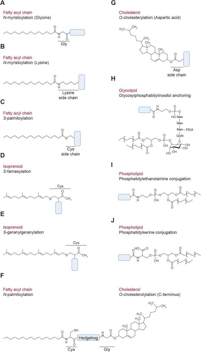
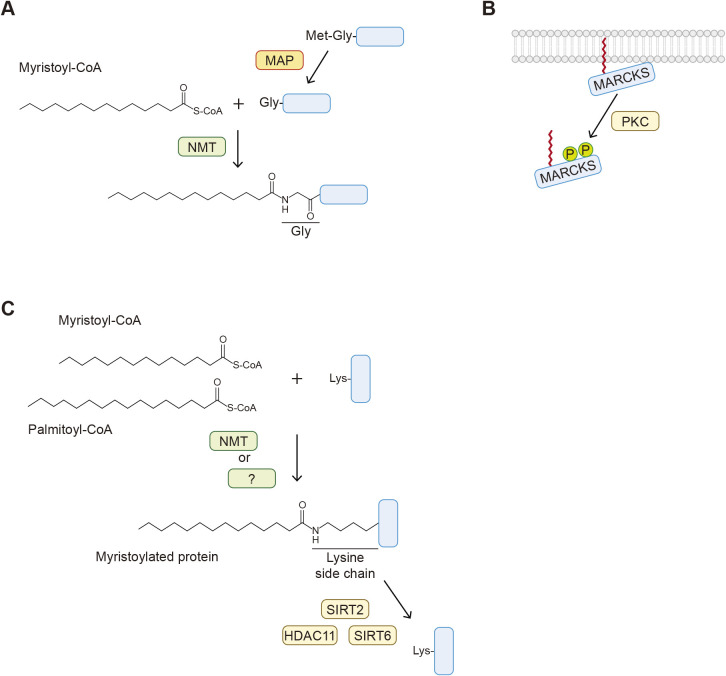
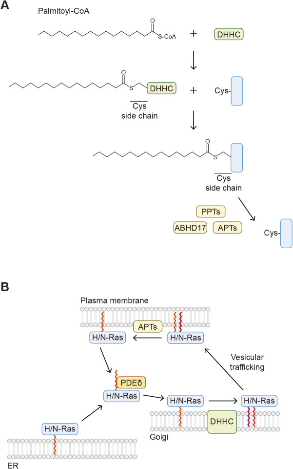
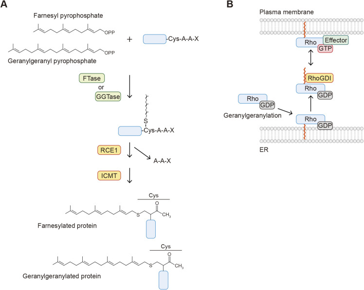
Protein-lipid conjugation is a widespread modification involved in many biological processes. Various lipids, including fatty acids, isoprenoids, sterols, glycosylphosphatidylinositol, sphingolipids, and phospholipids, are covalently linked with proteins. These modifications direct proteins to intracellular membranes through the hydrophobic nature of lipids. Some of these membrane-binding processes are reversible through delipidation or by reducing the affinity to membranes. Many signaling molecules undergo lipid modification, and their membrane binding is important for proper signal transduction. The conjugation of proteins to lipids also influences the dynamics and function of organellar membranes. Dysregulation of lipidation has been associated with diseases such as neurodegenerative diseases. In this review, we first provide an overview of diverse forms of protein-lipid conjugation and then summarize the catalytic mechanisms, regulation, and roles of these modifications.Key words: lipid, lipidation, membrane, organelle, protein modification.

Keywords: lipid; lipidation; membrane; organelle; protein modification.


Conflict of interest statement

The authors declare no competing interests.

Figures

Fig. 1
Fig. 1
Diverse types of protein–lipid conjugation A. Glycine N-myristoylation. Myristic acid forms an amide bond with glycine. B. Lysine N-myristoylation. Myristic acid is covalently linked to the side chain of lysine. C. Cysteine S-palmitoylation. Palmitic acid forms a thioester linkage with the side chain of cysteine. D and E. Cysteine S-prenylation. The farnesyl (D) or geranylgeranyl (E) group is covalently bound to the side chain of cysteine. F. Hedgehog is modified with palmitic acid and cholesterol at its N- and C- termini, respectively. G. The hydroxy group of cholesterol is covalently linked to the side chain of aspartic acid within Smoothened (SMO). H. The phosphoethanolamine moiety of glycosylphosphatidylinositol is covalently linked to the carboxyl group of the C-terminal amino acid via an amide bond. I and J. The carboxyl group of the C-terminal glycine residue of ubiquitin family proteins is covalently attached to the amino group of phosphatidylethanolamine (I) and phosphatidylserine (J).
Fig. 2
Fig. 2
Overview of N-myristoylation A. The mechanism of glycine N-myristoylation. The initiating methionine is cleaved by methionine amino peptidase (MAP), and myristic acid is covalently attached to the exposed glycine residue by N-myristoyl transferase (NMT) proteins. Myristoyl-CoA is the myristic acid donor. B. Myristoylated alanine-rich protein kinase C substrate (MARCKS) is associated with membranes through N-myristoylation and its positively charged basic amino acids. Phosphorylation within the basic amino acids by protein kinase C (PKC) causes the dissociation of MARCKS from membranes. C. The mechanism of lysine N-fatty acylation. Fatty acyl groups such as myristic and palmitic acids are covalently linked to the side chain of lysine residues by NMT (lysine N-myristoylation) and unknown enzymes. SIRT2, SIRT6, and HDAC11 remove fatty acyl groups from proteins.
Fig. 3
Fig. 3
Overview of S-palmitoylation A. The mechanism of cysteine S-palmitoylation. The palmitoyl group of palmitoyl-CoA forms an intermediate with the catalytic cysteine residue of the Asp-His-His-Cys (DHHC) protein and then is transferred to a cysteine residue in a substrate protein. B. Farnesylated H- and N-Ras proteins localized to the ER are dissociated from the membranes by PDEδ and transported to the Golgi. H-/N-Ras proteins are then modified with S-palmitoylation by DHHC and localized to the plasma membrane through vesicle trafficking. H-/N-Ras proteins are depalmitoylated by acyl-protein thioesterase (APT) and detached from the membranes.
Fig. 4
Fig. 4
Overview of S-prenylation A. The mechanism of S-prenylation. The farnesyl and geranylgeranyl groups of farnesyl pyrophosphate and geranylgeranyl pyrophosphate are covalently linked to the side chain of the cysteine residue within the CAAX motif. The AAX peptides are then removed by Ras-converting CAAX endopeptidase 1 (RCE1), and the C-terminal cysteine residue is methylated by isoprenylcysteine carboxylmethyltransferase (ICMT). B. Geranylgeranylated Rho proteins are dissociated from membranes by the RhoGDI protein. The GTP-bound Rho activates downstream effector proteins localized to the plasma membrane. RhoGDI sequesters Rho in the cytosol and locks it in the GDP-bound state.
Fig. 5
Fig. 5
Overview of O-cholesterylation A. Both N-palmitoylation and O-cholesterylation of hedgehog proteins. The N-terminal signal peptide is cleaved by a signal peptidase. The exposed cysteine residue is modified through N-palmitoylation by hedgehog acyltransferase (HHAT), while O-cholesterylation of hedgehog (Hh) is coupled with its autocleavage between glycine and cysteine residues. The C3 hydroxy group of cholesterol forms an ester bond with the carboxy group of the glycine residue. (The thioester bond formed is shown as “~” here.) B. Hh that is modified through N-palmitoylation and O-cholesterylation localizes to the outside surface of the plasma membrane. Hh is released from the membrane by the action of Dispatched 1 (DISP1) and signal peptide-CUB-epidermal growth factor-like domain-containing protein 2 (SCUBE2). The transfer of Hh to its receptor Patched 1 (PTCH1) is mediated by the coreceptors cell adhesion molecule-related/down-regulated by oncogenes (CDON)/brother of CDO (BOC) and growth arrest specific 1 (GAS1). This releases the inhibition of SMO by PTCH1 and activates the downstream signaling. Hh also promotes cholesterylation of Smoothened (SMO). This modification is important for its localization to cilia and function.
Fig. 6
Fig. 6
Conjugation of the ubiquitin family proteins to phospholipids A. ATG8 proteins are covalently linked to phosphatidylethanolamine (PE) in the autophagic membranes through the sequential reactions involving ATG7, ATG3, and the ATG12–ATG5–ATG16L1 complex. Cargo proteins are incorporated into the autophagic membranes, often via adaptor proteins. ATG8-PE is cleaved by ATG4 proteins and released from membranes. (The thioester bond formed is shown as “~” here.) B. Ubiquitin is covalently linked to PE in the endosomal and vacuolar membranes by the ubiquitin system enzymes, including Uba1, Ubc4/5, and Tul1. Ub-PE is turned over by either deubiquitination by Doa4 or degradation in the vacuole after being incorporated into intraluminal vesicles. Ub-PE recruits the ESCRT components to membranes in vitro and may be involved in intraluminal vesicle formation. (The thioester bond formed is shown as “~” here.)

Similar articles

Cited by

References

    1. Abrami, L., Audagnotto, M., Ho, S., Marcaida, M.J., Mesquita, F.S., Anwar, M.U., Sandoz, P.A., Fonti, G., Pojer, F., Dal Peraro, M., and van der Goot, F.G.. 2021. Palmitoylated acyl protein thioesterase APT2 deforms membranes to extract substrate acyl chains. Nat. Chem. Biol., 17: 438–447. - PMC - PubMed
    1. Antonny, B., Beraud-Dufour, S., Chardin, P., and Chabre, M.. 1997. N-terminal hydrophobic residues of the G-protein ADP-ribosylation factor-1 insert into membrane phospholipids upon GDP to GTP exchange. Biochemistry, 36: 4675–4684. - PubMed
    1. Bagchi, R.A., Robinson, E.L., Hu, T., Cao, J., Hong, J.Y., Tharp, C.A., Qasim, H., Gavin, K.M., Pires da Silva, J., Major, J.L., McConnell, B.K., Seto, E., Lin, H., and McKinsey, T.A.. 2022. Reversible lysine fatty acylation of an anchoring protein mediates adipocyte adrenergic signaling. Proc. Natl. Acad. Sci. U.S.A., 119: e2119678119. - PMC - PubMed
    1. Bas, L., Papinski, D., Licheva, M., Torggler, R., Rohringer, S., Schuschnig, M., and Kraft, C.. 2018. Reconstitution reveals Ykt6 as the autophagosomal SNARE in autophagosome-vacuole fusion. J. Cell Biol., 217: 3656–3669. - PMC - PubMed
    1. Briscoe, J. and Therond, P.P.. 2013. The mechanisms of Hedgehog signalling and its roles in development and disease. Nat. Rev. Mol. Cell Biol., 14: 416–429. - PubMed