. 2023 Apr 3;6(1):360.
doi: 10.1038/s42003-023-04639-y.

Targeting Enterococcus faecalis HMG-CoA reductase with a non-statin inhibitor

Affiliations

Targeting Enterococcus faecalis HMG-CoA reductase with a non-statin inhibitor

Sucharita Bose et al. Commun Biol. .

Abstract

HMG-CoA reductase (HMGR), a rate-limiting enzyme of the mevalonate pathway in Gram-positive pathogenic bacteria, is an attractive target for development of novel antibiotics. In this study, we report the crystal structures of HMGR from Enterococcus faecalis (efHMGR) in the apo and liganded forms, highlighting several unique features of this enzyme. Statins, which inhibit the human enzyme with nanomolar affinity, perform poorly against the bacterial HMGR homologs. We also report a potent competitive inhibitor (Chembridge2 ID 7828315 or compound 315) of the efHMGR enzyme identified by a high-throughput, in-vitro screening. The X-ray crystal structure of efHMGR in complex with 315 was determined to 1.27 Å resolution revealing that the inhibitor occupies the mevalonate-binding site and interacts with several key active site residues conserved among bacterial homologs. Importantly, 315 does not inhibit the human HMGR. Our identification of a selective, non-statin inhibitor of bacterial HMG-CoA reductases will be instrumental in lead optimization and development of novel antibacterial drug candidates.


Conflict of interest statement

The authors declare no competing interests.

Figures

Fig. 1
Fig. 1. efHMGR dimer architecture and domain swap.
a The apo efHMGR dimer structure: Two monomers interact in an intricate fashion to form the biologically active dimer, colored green and blue respectively. The N-terminal domain that is swapped is indicated by a red square. b In the liganded efHMGR dimer, the flap domain (orange) closes upon binding of HMG-CoA and NADP+ (represented as blue and yellow sticks). HMG-CoA primarily interacts with the large domain of one monomer (green) and NADP+ interacts with small domain (blue) of the other monomer. c The ENQISX3VP loop, unique in efHMGR, participates in various interactions at the crossover point. The interacting residues (Gln-39, Phe-40, Asn-42, Ala-44, Leu-45, Gln-57, Ile-58, Ser-59, Glu-60, Glu-62 and Lys-332) are represented in sticks. The H-bonds are shown as red dashed lines and the distances are shown in Å. d The ENVIG loop in pmHMGR (green) and DaHMGR (red) shown in cartoon representation suggest that presence of a proline residue preceding this loop along with other hydrophobic residues such as Met-44, Ala-47 and Val-54 prevent a complete domain swap. The dimer partner is shown in cyan. The “ENQIS loop” of efHMGR (grey) is shown as a reference. e Multiple sequence alignment of efHMGR with class II HMGR sequences from S. pneumoniae, S. aureus, P. mevalonii, Listeria monocytogenes, Delftia acidovorans, E. faecium, E. hirae show that the ENQIS loop (in red box) is unique to Enterococcus.
Fig. 2
Fig. 2. Structural determinants of NADP+ in efHMGR.
a Electrostatic surface potential map of efHMGR with NADP+ and HMG-CoA bound in the active site. Both HMG-CoA (green) and NADP+ (yellow) are represented as sticks. b Tyr-146, Ser-148 and Arg-152 residues represent the molecular determinants present in the small domain of efHMGR that interact with the oxygen atoms of the 2'-ribose-phosphate group of NADP+. Gln-410 originating from the flap also interacts with NADP+. The NADP+ molecule is encased in a 2Fo-Fc difference density at 1.0σ. Hydrogen bond interactions are represented as red dashes and the distances are shown in Å. c Class II HMGRs of S. pneumoniae, S. pyogenes, S. aureus and E. faecalis utilize NADPH as the cofactor. The residues (marked with an asterix) that determine the cofactor specificity are conserved in these bacterial enzymes including efHMGR. The P. mevalonii HMGR, which utilizes NADH has Asp-146, Leu-148 and Leu-152 as the molecular determinants.
Fig. 3
Fig. 3. Role of NADP+ in flap domain closure.
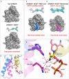
a The flap domain is disordered in the apo and NADP+ bound forms in efHMGR. With only NADP+ bound in the active site, the flap domain is disordered and the nicotinamide- ribose moiety in NADP+ does not have observable 2Fo-Fc density. b In the presence of HMG-CoA and NADP+, the flap domain is partially closed (blue). In this case the nicotinamide-ribose moiety of NADP+ is also disordered as seen by the 1.0σ 2Fo-Fc density. c The flap domain is fully closed (magenta) in the presence of mevalonate and a fully ordered NADP+ molecule as evident by the  2Fo-Fc density at 1.0σ. d Superposition of the HMG-CoA + NADP+ bound monomer on the mevalonate + NADP+ bound monomer suggests that the partially closed flap domain (blue) is rotated 15° as compared to the fully closed conformation (magenta). e The interactions that stabilize the fully closed flap (magenta) in the presence of a well-ordered NADP+ molecule are shown here. Several residues (His-376 and Gln-380) in the fully closed flap interact with the nicotinamide-ribose moiety of NADP+ via water-mediated interactions. Residue Asn-184 present in the small domain is seen to clamp down over the nicotinamide-ribose moiety and also participate in the water-mediated interactions. The catalytic His-376 is placed farther away in the partially closed flap (blue). Waters are shown as red spheres and hydrogen bond interactions are represented as red dashes and the distances are shown in Å.
Fig. 4
Fig. 4. 315 is a competitive inhibitor of efHMGR.
a Chemical structure of 315 (Chembridge2 ID 7828315): 5-{[(4-butylphenyl) amino] sulfonyl}-2-hydroxybenzoic acid. This compound is composed of a hydrophobic 4-butylphenyl group linked to the 2-hydroxy benzoic acid via an amino and a sulfonyl group. b Dose-response curves of 315 against E. faecalis HMG-CoA reductase. The IC50 value was estimated by Michaelis-Menten fitting of the inhibition % vs. inhibitor concentration data. Assays were conducted in triplicates c A Lineweaver plot shows that 315 is a competitive inhibitor of efHMGR. An analysis of this plot indicates that 315 inhibits the conversion of mevalonate to HMG-CoA with a Ki of 2 μM. Assays were conducted in duplicates and in the presence of 0, 5, 7.5 and 10 μM of 315 under otherwise standard conditions. Both datasets  were analyzed by Graphpad Prism 9.0 and presented as individual data points.
Fig. 5
Fig. 5. 315 binds in the mevalonate binding pocket in efHMGR.
a Electrostatic potential map showing 315 binding in the mevalonate binding pocket present at the dimer interface. The benzoic acid moiety is tucked inside a pocket lined with charged residues while the butylphenyl group of 315 is accommodated in a hydrophobic environment. b Ligplot diagram showing interactions between 315 and the mevalonate pocket residues in efHMGR. Hydrogen bond interactions are represented by green dashed lines, waters as cyan spheres and hydrophobic contacts as brown semicircles. c Hydrophilic interactions stabilize 315 in the mevalonate binding pocket of efHMGR. The large domain residues, Glu-86, Pro-87, Arg-257, Asn-267 and Gln-359, colored green, interact with the ligand via direct and water-mediated hydrogen bond interactions. The small domain residues from the adjacent monomer, Asn-187 and Asn-213, colored blue, also interact with the ligand via water molecules. Red spheres represent water molecules. Red dashes represent the hydrogen bond interactions with the numbers indicating the hydrogen bond distances in Å. d Hydrophobic interactions also stabilize 315 in the active site. Hydrophobic residues such as Ala-92, Ala-362, Ala-363, Ala-366, Ile-372 and His-376 pack against the hydrophobic butylphenyl group and stabilize the ligand. The residues are represented as sticks with Van der Waals radii as dotted spheres.

Similar articles

Cited by

References

    1. Hollenbeck BL, Rice LB. Intrinsic and acquired resistance mechanisms in enterococcus. Virulence. 2012;3:421–569. doi: 10.4161/viru.21282. - DOI - PMC - PubMed
    1. Paulsen IT, et al. Role of mobile DNA in the evolution of vancomycin-resistant Enterococcus faecalis. Science. 2003;299:2071–2074. doi: 10.1126/science.1080613. - DOI - PubMed
    1. Simor AE, et al. Staphylococcus aureus and vancomycin-resistant Enterococcus and of Clostridium difficile infection in Canadian hospitals. Infect. Hosp. Epidemiol. 2013;34:687–693. doi: 10.1086/670998. - DOI - PubMed
    1. Goldstein JL, Brown MS. Regulation of the mevalonate pathway. Nature. 1990;343:425–430. doi: 10.1038/343425a0. - DOI - PubMed
    1. Johnson EA, Schroeder WA. Microbial carotenoids. Adv. Biochem. Eng. Biotechnol. 1996;53:119–178. - PubMed

Publication types

MeSH terms

Substances