. 2023 Apr 11;120(15):e2206217120.
doi: 10.1073/pnas.2206217120. Epub 2023 Apr 3.

Neuronal activity regulates Matrin 3 abundance and function in a calcium-dependent manner through calpain-mediated cleavage and calmodulin binding

Affiliations

Neuronal activity regulates Matrin 3 abundance and function in a calcium-dependent manner through calpain-mediated cleavage and calmodulin binding

Ahmed M Malik et al. Proc Natl Acad Sci U S A. .

Abstract

RNA-binding protein (RBP) dysfunction is a fundamental hallmark of amyotrophic lateral sclerosis (ALS) and related neuromuscular disorders. Abnormal neuronal excitability is also a conserved feature in ALS patients and disease models, yet little is known about how activity-dependent processes regulate RBP levels and functions. Mutations in the gene encoding the RBP Matrin 3 (MATR3) cause familial disease, and MATR3 pathology has also been observed in sporadic ALS, suggesting a key role for MATR3 in disease pathogenesis. Here, we show that glutamatergic activity drives MATR3 degradation through an NMDA receptor-, Ca2+-, and calpain-dependent mechanism. The most common pathogenic MATR3 mutation renders it resistant to calpain degradation, suggesting a link between activity-dependent MATR3 regulation and disease. We also demonstrate that Ca2+ regulates MATR3 through a nondegradative process involving the binding of Ca2+/calmodulin to MATR3 and inhibition of its RNA-binding ability. These findings indicate that neuronal activity impacts both the abundance and function of MATR3, underscoring the effect of activity on RBPs and providing a foundation for further study of Ca2+-coupled regulation of RBPs implicated in ALS and related neurological diseases.

Keywords: ALS; FTD; NMDA; RNA binding protein.


Conflict of interest statement

The authors declare no competing interest.

Figures

Fig. 1.
Fig. 1.
Glutamatergic stimulation triggers a rapid MATR3 reduction in primary cortical neurons. (A and B) Application of 100 μM NMDA results in a rapid MATR3 reduction in DIV14-16 cortical neurons. n = 3; significance determined by sum of squares F test. (C and D) MATR3 reduction is recapitulated by immunostaining (vehicle, n = 547; NMDA, n = 419; glutamate, n = 337; ****P < 0.0001; one-way ANOVA with Tukey’s post hoc test). (Scale bars, 20 μm.) (EG) Both NMDA and glutamate elicit dose-dependent MATR3 clearance (n = 3 per dose; vehicle, y = 0.69−0.15x + 0.33; glutamate, y = 0.69−0.031x + 0.31; nonlinear one phase decay).
Fig. 2.
Fig. 2.
Neuronal activity dynamically regulates MATR3 across in vitro and ex vivo. (A and B) Neurons treated with NMDA show robust NMDA-mediated decrease in MATR3 only at DIV ≥ 10, coinciding with NMDAergic maturity (n = 3; ns, not significant; *P = 0.040, ****P < 0.0001, and ***P = 0.001; two-tailed t tests). (C) Baseline MATR3 levels in vehicle-treated cultures also decline with neuronal maturation (n = 3; ns, not significant; **P < 0.01; one-way ANOVA with Tukey’s post hoc test). (D and E) Neurons treated with NMDA and then subjected to washout fully recover MATR3 within 24 h, but recovery is impaired after stronger stimulus (n = 3; **P < 0.01, ***P < 0.001; ns, not significant; one-way ANOVA with Tukey’s post hoc test). (F and G) Calbindin-positive Purkinje neurons in cerebellar slice cultures displayed significant reductions in MATR3 staining intensity upon glutamate stimulation (vehicle, n = 73; glutamate, n = 76; ****P < 0.0001; two-tailed t test). (Scale bars [F], 20 μm.)
Fig. 3.
Fig. 3.
MATR3 reduction upon glutamatergic stimulation is NMDAR and Ca2+ dependent. (A) Schematic of ionotropic and metabotropic receptors activated by glutamate. (B and C) NMDAR activation is both necessary and sufficient for MATR3 clearance (n = 3; **P < 0.01; ns, not significant; one-way ANOVA with Tukey’s post hoc test). (D and E) The Ca2+ chelator BAPTA blocks NMDA-mediated MATR3 reduction (n = 3; ***P < 0.001; ns, not significant; one-way ANOVA with Tukey’s post hoc test). (F and G) Increasing intracellular Ca2+ with ionomycin (INMCN) is sufficient for MATR3 reduction in mature DIV14-16 and immature DIV4-5 neurons (n = 3; *P = 0.034, **P = 0.0080; two-tailed t test).
Fig. 4.
Fig. 4.
NMDA-triggered MATR3 reduction is accomplished posttranslationally via calpains. NMDA application does not significantly alter Matr3 transcript abundance (A; n = 4; P = 0.054; two-tailed t test), but exogenous FLAG-MATR3 (B) is reduced by NMDA application. (C and D) The proteasomal and cysteine protease inhibitor MG132, but not the autophagy inhibitor BAF, blocks MATR3 clearance in response to NMDA (n = 3; **P < 0.01; one-way ANOVA with Tukey’s post hoc test). (E and F) The selective proteasomal inhibitor BTZ fails to block NMDA-triggered MATR3 reduction (n = 3; ***P ≤ 0.001; one-way ANOVA with Tukey’s post hoc test). (G and H) The calpain inhibitor MDL28170 (MDL) effectively impairs MATR3 degradation upon NMDA treatment (n = 3; **P = 0.0040; one-way ANOVA with Tukey’s post hoc test).
Fig. 5.
Fig. 5.
MATR3 is a substrate for CAPN1, and the pathogenic S85C mutation renders it resistant to degradation. Transfection of HEK293T cells with calpain expressing constructs resulted in robust expression of CAPN1 (A) and CAPN2 (B) compared to controls. (C and D) CAPN1 but not CAPN2 overexpression resulted in MATR3 degradation (n = 3; *P = 0.010; one-way ANOVA with Tukey’s post hoc test). (E and F) While exogenous FLAG-MATR3(WT) is susceptible to cleavage by CAPN1 (n = 3; *P = 0.036; ns, not significant; one-way ANOVA with Tukey’s post hoc test), the pathogenic S85C mutation is resistant (n = 3; one-way ANOVA with Tukey’s post hoc test).
Fig. 6.
Fig. 6.
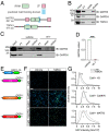
CaM binds to MATR3 in a Ca2+-dependent manner. (A) CaM is predicted to interact with the RRMs of both MATR3 and TDP43. (B) Ca2+-bound but not free apo-CaM binds to MATR3 and TDP43. (C and D) The conformational CaM inhibitor W-7 blocks the association between CaM and MATR3 (n = 3; ****P < 0.0001; two-tailed t test). (E) Schematic of split GFP constructs used for detecting in situ interaction of CaM and MATR3. (F and G) Application of NMDA increased the interaction between CaM-GFP1-10 and MATR3-GFP11, as determined by an increase in the reconstituted GFP signal. A similar although more subtle increase was detected in cells expressing CaM-GFP1-10 and CaMKIV-GFP11 but not those transduced with CaM-GFP1-10 and H3-GFP11. n = 200 neurons/condition, 2 reps/condition. (Scale bar, 50 µm.) ns, not significant; **P < 0.01 comparing vehicle to NMDA for each condition, one-sided Kolmogorov–Smirnov test with bootstrapping. ‡P < 0.0001 comparing CaM-GFP1-10 + H3-GFP11 to CaM-GFP1-10 + MATR3-GFP11, one-sided Kolmogorov–Smirnov test with bootstrapping.
Fig. 7.
Fig. 7.
Ca2+/CaM enriches in the nucleus of stimulated neurons and inhibits MATR3 RNA binding. (A and B) Ca2+/CaM is enriched within the nucleus of neurons treated with NMDA (vehicle, n = 67; NMDA, n = 48; ***P = 0.0001; two-tailed t test). (Scale bars [A], 20 μm.) (C) Schematic of ultraviolet (UV) cross-linking followed by IP (UV-CLIP). (DF) While NMDA treatment did not alter total levels of MATR3 target RNAs (n = 6 for each candidate), less RNA was cross-linked to MATR3 in stimulated conditions, indicating impaired RNA binding (n = 4 for each candidate; *P < 0.05; two-tailed t test). (GI) Pretreatment with CaM inhibitor W-7 prior to NMDA stimulation did not alter total levels of MATR3 target RNAs (n = 5 for each candidate). Although W-7 pretreatment increased Lsamp RNA cross-linked to MATR3 (*P = 0.045, two-tailed t test), it did not alter the amount of Grm7 or Ftx cross-linked to MATR3 (n = 5 per condition; ns, not significant; two-tailed t test).
Fig. 8.
Fig. 8.
A model depicting regulation of MATR3 abundance and RNA binding in a Ca2+/CaM-dependent manner. MATR3 possesses two ZF domains and two RRMs, with RRM2 being the dominant RNA-binding domain. Ca2+ influx through NMDARs activates calmodulin (CaM), which displaces bound RNA from MATR3 via overlapping interactions with RRM2. Simultaneously, Ca2+ activates calpain-1 (CAPN1), which is capable of degrading RNA-deficient MATR3. By virtue of its location within the predicted CAPN1 cleavage site, the pathogenic S85C mutation interferes with CAPN1-mediated MATR3 degradation.

Similar articles

Cited by

References

    1. Purice M. D., Taylor J. P., Linking hnRNP function to ALS and FTD pathology. Front. Neurosci.-switz 12, 326 (2018). - PMC - PubMed
    1. Arai T., et al. , TDP-43 is a component of ubiquitin-positive tau-negative inclusions in frontotemporal lobar degeneration and amyotrophic lateral sclerosis. Biochem. Biophys. Res. Commun. 351, 602–611 (2006). - PubMed
    1. Neumann M., et al. , Ubiquitinated TDP-43 in frontotemporal lobar degeneration and amyotrophic lateral sclerosis. Science 314, 130–133 (2006). - PubMed
    1. Kabashi E., et al. , TARDBP mutations in individuals with sporadic and familial amyotrophic lateral sclerosis. Nat. Genet. 40, 572–574 (2008). - PubMed
    1. Vance C., et al. , Mutations in FUS, an RNA processing protein, cause familial amyotrophic lateral sclerosis type 6. Science 323, 1208–1211 (2009). - PMC - PubMed

Publication types

MeSH terms