. 2023 Mar 17:14:1140703.
doi: 10.3389/fphar.2023.1140703. eCollection 2023.

Small-molecule BCL6 inhibitor protects chronic cardiac transplant rejection and inhibits T follicular helper cell expansion and humoral response

Affiliations

Small-molecule BCL6 inhibitor protects chronic cardiac transplant rejection and inhibits T follicular helper cell expansion and humoral response

Yuxuan Xia et al. Front Pharmacol. .

Abstract

Background: B cell lymphoma 6 (BCL6) is an important transcription factor of T follicular helper (Tfh) cells, which regulate the humoral response by supporting the maturation of germinal center B cells and plasma cells. The aim of this study is to investigate the expansion of T follicular helper cells and the effect of the BCL6 inhibitor FX1 in acute and chronic cardiac transplant rejection models. Methods: A mouse model of acute and chronic cardiac transplant rejection was established. Splenocytes were collected at different time points after transplantation for CXCR5+PD-1+ and CXCR5+BCL6+ Tfh cells detection by flow cytometry (FCM). Next, we treated the cardiac transplant with BCL6 inhibitor FX1 and the survival of grafts was recorded. The hematoxylin and eosin, Elastica van Gieson, and Masson staining of cardiac grafts was performed for the pathological analysis. Furthermore, the proportion and number of CD4+ T cells, effector CD4+ T cells (CD44+CD62L-), proliferating CD4+ T cells (Ki67+), and Tfh cells in the spleen were detected by FCM. The cells related to humoral response (plasma cells, germinal center B cells, IgG1+ B cells) and donor-specific antibody were also detected. Results: We found that the Tfh cells were significantly increased in the recipient mice on day 14 post transplantation. During the acute cardiac transplant rejection, even the BCL6 inhibitor FX1 did not prolong the survival or attenuate the immune response of cardiac graft, the expansion of Tfh cell expansion inhibit. During the chronic cardiac transplant rejection, FX1 prolonged survival of cardiac graft, and prevented occlusion and fibrosis of vascular in cardiac grafts. FX1 also decreased the proportion and number of splenic CD4+ T cells, effector CD4+ T cells, proliferating CD4+ T cells, and Tfh cells in mice with chronic rejection. Moreover, FX1 also inhibited the proportion and number of splenic plasma cells, germinal center B cells, IgG1+ B cells, and the donor-specific antibody in recipient mice. Conclusion: We found BCL6 inhibitor FX1 protects chronic cardiac transplant rejection and inhibits the expansion of Tfh cells and the humoral response, which suggest that BCL6 is a potential therapeutic target of the treatment for chronic cardiac transplant rejection.

Keywords: BCL6; FX1; T follicular helper cells; cardiac transplant rejection; humoral response.


Conflict of interest statement

The authors declare that the research was conducted in the absence of any commercial or financial relationships that could be construed as a potential conflict of interest.

Figures
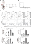
FIGURE 1
FIGURE 1
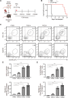
The expansion of T follicular helper cells during acute cardiac transplant rejection in mice (A). Illustration of the experimental design in (B–E). The murine intra-abdominal heterotopic cardiac transplantation model (BALB/c to B6 mice) was established, and the spleens of recipient mice were collected for flow detection at 0, 7, 14, and 21 days post-transplantation (B). The survival status of the grafts was observed every day through palpation. The survival time is illustrated in the survival curves (C). FCM analysis of the proportions of PD1+CXCR5+ Tfh and BCL6+CXCR5+ Tfh in the spleens of recipient mice at 0, 7, 14, and 21 days post-transplantation (D). The bar charts (n = 4) show the ratios and numbers of PD1+CXCR5+ Tfh cells in the spleens at different time points, as described in 1a (E). The bar charts (n = 4) show the ratios and numbers of BCL6+CXCR5+ Tfh cells in the spleens at different time points, as described in 1a.Error bars represent SD. ns, not significant, *p < 0.05, **p < 0.01, ***p < 0.001, ****p < 0.0001.
FIGURE 2
FIGURE 2
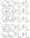
The expansion of T follicular helper cells during chronic cardiac transplant rejection in mice (A). Illustration of the experimental design in (B–E). The murine chronic cardiac transplant model was established. The mice were injected with 250 ug of CTLA-4-Ig or the equivalent amount of PBS intraperitoneally on the day after surgery, and their spleens were detected via FCM at 0, 7, 14, and 21 days post-transplantation. AuthorAnonymous, (B). The survival status of the grafts was observed every day through palpation. The survival time is illustrated in the survival curves (n = 6) (C). FCM analysis of the proportions of PD1+CXCR5+ Tfh and BCL6+CXCR5+ Tfh cells in the spleens of recipient mice at 0, 7, 14, and 21 days post-transplantation (D). The bar charts (n = 4) show the ratios and numbers of PD1+CXCR5+ Tfh cells in the spleens at different time points, as described in 2a (E). The bar charts (n = 4) show the ratios and numbers of BCL6+CXCR5+ Tfh cells in the spleens at different time points, as described in 2a.Error bars represent SD. ns, not significant, *p < 0.05, **p < 0.01, ***p < 0.001, ****p < 0.0001.
FIGURE 3
FIGURE 3
Small-molecule BCL inhibitor FX1 does not protect acute cardiac transplant rejection in mice, but it inhibits T follicular helper cell expansion (A). Illustration of the experimental design in (B–E). The murine acute cardiac transplant model was established, and the mice were administered CTLA-4-Ig or the equivalent amount of PBS for 3 consecutive days. The survival time of the cardiac grafts was observed, and the spleens were collected for FCM analysis on the 14th postoperative day (B). The survival status of the grafts was observed every day through palpation, and the survival time is illustrated in the survival curves (n = 6) (C). Graft specimens were obtained on the 6th postoperative day, and they were stained with H&E to visualize pathological changes in the grafts. International Society for Heart and Lung Transplantation grading was performed on tissue sections of each group (n = 6) (D). The bar charts (n = 4) show the ratios and numbers of PD1+CXCR5+ Tfh and BCL6+CXCR5+ Tfh cells in the spleens on the 6th day post-transplantation. Error bars represent SD. ns, not significant, *p < 0.05, **p < 0.01, ***p < 0.001, ****p < 0.0001.
FIGURE 4
FIGURE 4
FX1 protects chronic cardiac transplant rejection in mice (A). Illustration of the experimental design in (B–E). The murine chronic cardiac transplant model was established, and the mice were treated with FX1 or the equivalent amount of PBS three times. The survival time of the cardiac grafts was observed, and the spleens were collected for FCM analysis on the 28th postoperative day (B). The survival status of the grafts was observed every day through palpation, and the survival time is illustrated in the survival curves (n = 6) (C). Graft specimens were obtained on the 28th postoperative day, and vascular occlusion and fibrosis in the cardiac grafts was assessed by H&E, elastica van Gieson, and Masson staining (D). The bar charts shows the vascular occlusion and fibrosis of the cardiac grafts (n = 6). Error bars represent SD. ns, not significant, *p < 0.05, **p < 0.01, ***p < 0.001, ****p < 0.0001.
FIGURE 5
FIGURE 5
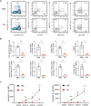
FX1 inhibits T follicular helper cell expansion during chronic cardiac transplant rejection in mice (A). The spleens were obtained from the recipient mice on postoperative day 28 and were ground into single-cell suspensions for FCM detection. The contour plots demonstrate the ratios of characteristic CD3+CD4+ T cells, CD44+CD62L-T cells, and CD44+Ki67 + T cells in both groups, while the bar charts (n = 4) illustrate the ratios between the two groups (B). The contour plots demonstrate the ratios of characteristic PD1+CXCR5+ Tfh and BCL6+CXCR5+ Tfh in both groups, while the bar charts (n = 4) illustrate the ratios of Tfh cells between the two groups. Error bars represent SD. ns, not significant, *p < 0.05, **p < 0.01, ***p < 0.001, ****p < 0.0001.
FIGURE 6
FIGURE 6
FX1 inhibits humoral response during chronic cardiac transplant rejection in mice (A). Spleens were obtained from the recipient mice on postoperative day 28, ground into single-cell suspensions for FCM detection, and stained for the ratios of CD19CD138+ plasma cells, GL7+CD95+B cells, PNA+CD95+B cells, and IgG1+CD19+B cells (B). The bar charts (n = 4) illustrate the ratios and number of CD19CD138+ plasma cells, GL7+CD95+ GC B cells, PNA+CD95+ GC B cells, and IgG1+CD19+B cells in both groups (C). The sera of the mice in both groups were collected 0, 14, 21, and 28 days postoperatively to measure the concentrations of anti-MHC-I and anti-MHC-II antibodies.

Similar articles

Cited by

References

    1. Borges T. J., Murakami N., Lape I. T., Gassen R. B., Liu K., Cai S., et al. (2021). Overexpression of PD-1 on T cells promotes tolerance in cardiac transplantation via ICOS-dependent mechanisms. JCI insight 6 (24), e142909. 10.1172/jci.insight.142909 - DOI - PMC - PubMed
    1. Cai Y., Watkins M. A., Xue F., Ai Y., Cheng H., Midkiff C. C., et al. (2020). BCL6 BTB-specific inhibition via FX1 treatment reduces Tfh cells and reverses lymphoid follicle hyperplasia in Indian rhesus macaque (Macaca mulatta). J. Med. Primatol. 49 (1), 26–33. 10.1111/jmp.12438 - DOI - PMC - PubMed
    1. Carbone J. (2018). Complement genomics and antibody-mediated rejection in heart recipients. J. Heart Lung Transpl. 37 (4), 439–440. 10.1016/j.healun.2017.08.008 - DOI - PubMed
    1. Cardenas M. G., Yu W., Beguelin W., Teater M. R., Geng H., Goldstein R. L., et al. (2016). Rationally designed BCL6 inhibitors target activated B cell diffuse large B cell lymphoma. J. Clin. Invest. 126 (9), 3351–3362. 10.1172/JCI85795 - DOI - PMC - PubMed
    1. Chhabra M., Alsughayyir J., Qureshi M. S., Mallik M., Ali J. M., Gamper I., et al. (2018). Germinal center alloantibody responses mediate progression of chronic allograft injury. Front. Immunol. 9, 3038. 10.3389/fimmu.2018.03038 - DOI - PMC - PubMed

Grants and funding

This study was supported by the grants from the National Natural Science Foundation of China [Grants 31871154, 91849120].