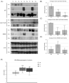
Murine herpesvirus-68-related growth factors treatment correlates with decrease of p53 and HIF-1α protein levels
- PMID: 36997335
- PMCID: PMC10093997
- DOI: 10.1093/femspd/ftad004
Murine herpesvirus-68-related growth factors treatment correlates with decrease of p53 and HIF-1α protein levels
Abstract
Murine herpesvirus 68 (MHV-68) belongs to the subfamily Gammaherpesvirinae of the family Herpesviridae. This exceptional murine herpesvirus is an excellent model for the study of human gammaherpesvirus infections. Cells infected with MHV-68 under nonpermissive conditions for viral replication produce substances designated as MHV-68 growth factors (MHGF-68), that can cause transformation of the cells, or on the other side, turn transformed cells into normal. It was already proposed, that the MHGF-68 fractions cause transformation, disruption of the cytoskeleton and slower growth of the tumors in nude mice. Here, we examined newly extracted fractions of MHGF-68 designated as F5 and F8. Both fractions proved to inhibit the growth of the spheroids and also tumours induced in nude mice. What more, the fractions caused the decrease of the protein levels of wt p53 and HIF-1α. Decreased levels of p53 and HIF-1α activity leads to decreased vascularization, slower tumour growth, and lower adaptation to hypoxic conditions. This would propose MHGF-68 fractions, or their human herpesvirus equivalents, as a potential anticancer drugs in combined chemotherapy.
Keywords: HIF-1α; MHGF-68; MHV-68; growth factors; mdm2; p53.
© The Author(s) 2023. Published by Oxford University Press on behalf of FEMS.
Conflict of interest statement
The authors declare no conflict of interest. The funders had no role in the design of the study; in the collection, analyses, or interpretation of data; in the writing of the manuscript, or in the decision to publish the results’.
Figures


Similar articles
-
Cells transformed by murine herpesvirus 68 (MHV-68) release compounds with transforming and transformed phenotype suppressing activity resembling growth factors.Acta Virol. 2015 Dec;59(4):418-22. doi: 10.4149/av_2015_04_418. Acta Virol. 2015. PMID: 26666191
-
Murid herpesvirus 4 (MuHV-4, prototype strain MHV-68) as an important model in global research of human oncogenic gammaherpesviruses.Acta Virol. 2020;64(2):167-176. doi: 10.4149/av_2020_206. Acta Virol. 2020. PMID: 32551785 Review.
-
Pathogenetic characterization of a mouse herpesvirus isolate Sumava.Acta Virol. 2002;46(1):41-6. Acta Virol. 2002. PMID: 12197633
-
Analysis of a novel strain of murine gammaherpesvirus reveals a genomic locus important for acute pathogenesis.J Virol. 2001 Jun;75(11):5315-27. doi: 10.1128/JVI.75.11.5315-5327.2001. J Virol. 2001. PMID: 11333912 Free PMC article.
-
Murine gammaherpesvirus 68: a model for the study of gammaherpesvirus pathogenesis.Trends Microbiol. 1998 Jul;6(7):276-82. doi: 10.1016/s0966-842x(98)01306-7. Trends Microbiol. 1998. PMID: 9717216 Review.
References
-
- Abràmoff MD, Magalhães PJ, Ram SJ. Image processing with imageJ. Biophotonics Int. 2004;11:36–42.
-
- Ala-Aho R, Grénman R, Seth Pet al. . Adenoviral delivery of p53 gene suppresses expression of collagenase-3 (MMP-13) in squamous carcinoma cells. Oncogene. 2002;21:1187–95. - PubMed
-
- An WG, Kanekal M, Simon MCet al. . Stabilization of wild-type p53 by hypoxia-inducible factor 1α. Nature. 1998;392:405–8. - PubMed
-
- Bergers G, Javaherian K, Lo KMet al. . Effects of angiogenesis inhibitors on multistage carcinogenesis in mice. Science. 1999;284:808–12. - PubMed
-
- Boehm T, Folkman J, Browder Tet al. . Antiangiogenic therapy of experimental cancer does not induce acquired drug resistance. Nature. 1997;390:404–7. - PubMed
Publication types
MeSH terms
Substances
LinkOut - more resources
Full Text Sources
Research Materials
Miscellaneous

