

Viral Conjunctivitis
- PMID: 36992385
- PMCID: PMC10057170
- DOI: 10.3390/v15030676
Viral Conjunctivitis
Abstract
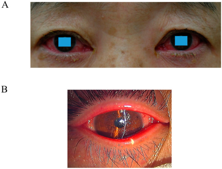
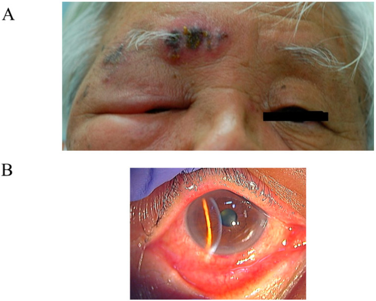
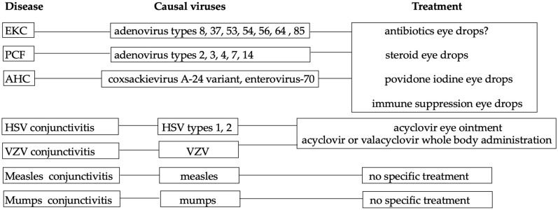
Viruses account for 80% of all cases of acute conjunctivitis and adenovirus; enterovirus and herpes virus are the common causative agents. In general, viral conjunctivitis spreads easily. Therefore, to control the spread, it is crucial to quickly diagnose illnesses, strictly implement hand washing laws, and sanitize surfaces. Swelling of the lid margin and ciliary injection are subjective symptoms, and eye discharge is frequently serofibrinous. Preauricular lymph node swelling can occasionally occur. Approximately 80% of cases of viral conjunctivitis are caused by adenoviruses. Adenoviral conjunctivitis may become a big global concern and may cause a pandemic. Diagnosis of herpes simplex viral conjunctivitis is crucial for using corticosteroid eye solution as a treatment for adenovirus conjunctivitis. Although specific treatments are not always accessible, early diagnosis of viral conjunctivitis may help to alleviate short-term symptoms and avoid long-term consequences.
Keywords: adenovirus; conjunctivitis; enterovirus; herpes virus.
Conflict of interest statement
The authors declare no conflict of interest.
Figures
Similar articles
-
Concurrent adenoviral and herpetic ocular infections.Ann Ophthalmol. 1989 Nov;21(11):432-8. Ann Ophthalmol. 1989. PMID: 2559639
-
Prevalence of acute conjunctivitis caused by chlamydia, adenovirus, and herpes simplex virus in an ophthalmic casualty department.Br J Ophthalmol. 1984 Sep;68(9):653-5. doi: 10.1136/bjo.68.9.653. Br J Ophthalmol. 1984. PMID: 6087876 Free PMC article.
-
Viral conjunctivitis: a retrospective study in an Australian hospital.Cont Lens Anterior Eye. 2019 Dec;42(6):679-684. doi: 10.1016/j.clae.2019.07.001. Epub 2019 Jul 9. Cont Lens Anterior Eye. 2019. PMID: 31300283
-
Adenoviral keratoconjunctivitis.Surv Ophthalmol. 2015 Sep-Oct;60(5):435-43. doi: 10.1016/j.survophthal.2015.04.001. Epub 2015 May 5. Surv Ophthalmol. 2015. PMID: 26077630 Review.
-
Acute red eye. Differentiating viral conjunctivitis from other, less common causes.Postgrad Med. 1997 May;101(5):185-6, 189-92, 195-6. doi: 10.3810/pgm.1997.05.246. Postgrad Med. 1997. PMID: 9158614 Review.
Cited by
-
Identification of coxsackievirus A-24 GIV C5 strain as the cause of acute hemorrhagic conjunctivitis outbreak in Hyderabad, India in 2022.Heliyon. 2024 Jun 1;10(11):e32254. doi: 10.1016/j.heliyon.2024.e32254. eCollection 2024 Jun 15. Heliyon. 2024. PMID: 38947457 Free PMC article.
-
Teleost Eye Is the Portal of IHNV Entry and Contributes to a Robust Mucosal Immune Response.Int J Mol Sci. 2023 Dec 21;25(1):160. doi: 10.3390/ijms25010160. Int J Mol Sci. 2023. PMID: 38203332 Free PMC article.
-
Unraveling the conjunctivitis crisis: understanding the spiking incidence in Karachi and Lahore-Pakistan.Ann Med Surg (Lond). 2023 Dec 13;86(2):920-922. doi: 10.1097/MS9.0000000000001623. eCollection 2024 Feb. Ann Med Surg (Lond). 2023. PMID: 38333251 Free PMC article. Review.
-
Vitamin D and tear fluid cytokines in predicting outcomes in viral conjunctivitis - A new outlook.Indian J Ophthalmol. 2024 Jul 1;72(Suppl 4):S702-S708. doi: 10.4103/IJO.IJO_2345_23. Epub 2024 Apr 16. Indian J Ophthalmol. 2024. PMID: 38622859 Free PMC article.
-
Acute Conjunctivitis among Patients Visiting the Outpatient Department of Ophthalmology in a Tertiary Care Centre.JNMA J Nepal Med Assoc. 2024 Jan 2;62(269):24-26. doi: 10.31729/jnma.8391. JNMA J Nepal Med Assoc. 2024. PMID: 38410017 Free PMC article.
References
Publication types
MeSH terms
Grants and funding
LinkOut - more resources
Full Text Sources
Medical
