Review
. 2023 Feb 28;15(3):653.
doi: 10.3390/v15030653.

CryoEM of Viral Ribonucleoproteins and Nucleocapsids of Single-Stranded RNA Viruses

Affiliations
Review

CryoEM of Viral Ribonucleoproteins and Nucleocapsids of Single-Stranded RNA Viruses

Andrea Modrego et al. Viruses. .

Abstract

Single-stranded RNA viruses (ssRNAv) are characterized by their biological diversity and great adaptability to different hosts; traits which make them a major threat to human health due to their potential to cause zoonotic outbreaks. A detailed understanding of the mechanisms involved in viral proliferation is essential to address the challenges posed by these pathogens. Key to these processes are ribonucleoproteins (RNPs), the genome-containing RNA-protein complexes whose function is to carry out viral transcription and replication. Structural determination of RNPs can provide crucial information on the molecular mechanisms of these processes, paving the way for the development of new, more effective strategies to control and prevent the spread of ssRNAv diseases. In this scenario, cryogenic electron microscopy (cryoEM), relying on the technical and methodological revolution it has undergone in recent years, can provide invaluable help in elucidating how these macromolecular complexes are organized, packaged within the virion, or the functional implications of these structures. In this review, we summarize some of the most prominent achievements by cryoEM in the study of RNP and nucleocapsid structures in lipid-enveloped ssRNAv.

Keywords: cryogenic electron microscopy (cryoEM); genome packaging; nucleoprotein (NP); ribonucleoprotein (RNP); single-stranded RNA virus (ssRNAv); virus assembly.


Conflict of interest statement

The authors declare no conflict of interest.

Figures

Figure 1
Figure 1
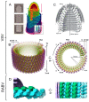
CryoEM structure of ebolavirus (EBOV) and Marburg virus (MARV) nucleocapsids from intact virions. (A) Overview of the EBOV nucleocapsid (NC). For ease of visualization, adjacent rungs are shown in dark and light gray, and a single subunit is highlighted in pink. The structure has 11.9 subunits per turn. (B) Segmentation of an EBOV NC subunit; two adjacent nucleoproteins (NPs) are shown in blue and cyan, two VP24 are shown in orange and purple, and the putative density corresponding to the RNA is shown in yellow. (C) Docking of the atomic structure of NP (blue and cyan) and VP24 (orange and purple) in the cryoEM map [18]. (D) Top view of another helical structure formed by overexpression of EBOV NP [20]. The structure has 42.4 NP subunits per turn. (E) Overview of the MARV NC; colors follow the same criteria as in A. The structure has 14.8 subunits per turn. (F) Segmentation of a subunit of the MARV NC; colors follow the same criteria as in B, and the green region is an extra disordered density present in the MARV NC. (G) Docking of the NP and VP24 atomic structures; colors follow the same criteria as in C [18]. (H) Top view of the helical structure of the NP-RNA complex of MARV; an NP monomer is highlighted in yellow and the RNA chain is highlighted in red [22]. The structure has 30.50 NP subunits per turn.
Figure 2
Figure 2
CryoEM structure of the vesicular stomatitis virus (VSV) and rabies virus (RABV) nucleocapsids. (A) 2D averages of the conical tip, trunk, and base of VSV and a full 3D model of the virion. The nucleoprotein (N) is depicted in red-yellow-green according to radial distance to the center, the matrix protein (M) in light-dark blue (according also to the radial distance), and the inner (2) and outer (1) membrane leaflets in purple and pink, respectively [27]. (B) Helical nucleocapsid structure with 38.5 N monomers per turn in lateral and top views. The N protein is colored green, the RNA in red, and the two layers of matrix protein (M1 and M2) are colored orange and purple, respectively. (C) Reconstruction of the VSV nucleocapsid tip without the imposition of any symmetry. To show the interior, the tip is cut in half [28]. (D) Two orthogonal views of the RABV nucleocapsid reconstruction. The densities corresponding to N, M, and membrane proteins are colored in cyan, green, and purple respectively. The atomic structure of three N proteins (PDB: 2GTT, cyan) are docked in the electron density map [33].
Figure 3
Figure 3
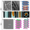
CryoEM structure of measles morbillivirus (MeV) and mumps virus (MuV). (A) Representative electron micrographs of MeV-infected (left) or nucleoprotein (N)- and matrix (M)-cotransfected (right) cell lysates prepared by immunosorbent electron microscopy. (B) Isosurface representations of the MeV nucleocapsid structure viewed laterally (first three images), and a slice taken across the axis (final image). The outer and inner parts (blue and orange, putatively M and N) show the helical twist, and the stars represent the five-star helical arrangement [38]. (C) Nucleocapsid-like helical particle structure obtained from the interaction of overexpressed MeV N protein in the presence of RNA. A side view (above) and a cutaway view (below) are shown [41]. (D) Representative cryoEM micrograph of helical structures obtained from overexpressed MuV N protein interacting with RNA; three different arrangements termed dense, hyperdense, and ring-stacked filaments are highlighted in blue, green, and red boxes, respectively. (E) 3D reconstruction of the dense helical filament and the respective atomic model [43].
Figure 4
Figure 4
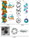
CryoEM structure of different RNPs of viruses of the order Bunyavirales. (A) Structure of an RNP-like particle obtained from overexpression of Hantaan orthohantavirus (HTNV) nucleoprotein (NP) interacting with host cell RNA. The upper part of the cryoEM map is colored by NP domain; cyan represents the N-terminal lobe, pink the N-terminal hinge, green the N-terminal arm, orange the C-terminal lobe, red the C-terminal arm, navy blue the C-terminal hinge and yellow the RNA, while the lower part is colored by NP protomer. The NP surrounded by dotted lines corresponds to Figure 4B orientation. (B) Two views of the NP domains of a monomer extracted from the helical assembly (color code as in A) [55]. (C) CryoEM structure of the Bunyamwera orthobunyavirus (BUNV) RNP at 13 Å resolution. (D) Pseudoatomic model of the BUNV RNP obtained after flexible fitting of an NP model; distance between rungs of the helix is indicated [57]. (E) Negative staining EM image of native Crimean-Congo hemorrhagic fever orthonairovirus (CCHFV) RNPs extracted from virions, showing the high flexibility of these complexes. (F) SPA cryoEM 3D reconstruction of a CCHFV NP oligomer obtained from overexpression, with the crystal structure (PDB 3U3I) docked in the map [60].
Figure 5
Figure 5
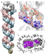
3D structure of the native RNP of influenza A virus (IAV) and its arrangement in the virion. (A) Model of the segment 8 RNP [67]. The RdRp is shown in gray, the antiparallel nucleoprotein (NP) strands are shown in red and blue, and the closing loop is in yellow. The positions of the 3′ and 5′ ends are defined as described in [72]. (B) Docking of the NP monomer (PDB:2IQH) in the helical region of the RNP. In the lower strand NP, monomers are represented as electrostatic potential surfaces, and in the upper strand as ribbons; the modeled RNA template is depicted as a red thread. (C) Segmented tomogram of an IAV virion in which subtomogram-averaged RNP segments (in purple) are placed back on the position, with orientation computed in the alignment [67].
Figure 6
Figure 6
3D reconstructions of different conformations of the helical part of Influenza A virus (IAV) RNPs and the transcription process mechanism. (A) The docking of the nucleoprotein atomic structure (PDB:2IQH) is shown on the opposite strands in red and blue. Scale bar, 50 Å. (B) The nucleoprotein (NP) head and body domains are outlined in yellow and green, respectively, showing the variation of the relative position of two NP monomers from the opposite strands. Red arrows indicate the direction of displacement. (C) Scheme of the RNP during the different transcription steps: (i) initiation; (ii) elongation; (iii) polyadenylation. Green arrows indicate the relative movement between the strands [10].
Figure 7
Figure 7
The native assembly and tentative model of SARS-CoV-2 RNPs [82]. (A) Ultrastructure of the RNP hexon and tetrahedron assemblies. At the top, seven RNPs are packed against the viral envelope (gray), forming an “eggs-in-a-nest” hexagonal assembly. At the bottom, four RNPs are packed as a membrane-free tetrahedral assembly. (B) Representative projection of RNP hexons assembling into a spherical virus (top) and tetrahedrons into an ellipsoidal virus (bottom). (C) 13.1 Å-resolution RNP showing a reverse G-shaped architecture, measuring 15 nm in diameter and 16 nm in height. N-terminal (6WKP) and C-terminal (6WJI) domains of N are fitted into the map in each head-to-tail reverse L-shaped density.

Similar articles

Cited by

References

    1. Kühlbrandt W. The Resolution Revolution. Science. 2014;343:1443–1444. doi: 10.1126/science.1251652. - DOI - PubMed
    1. Liu C., Zhou D., Nutalai R., Duyvesteyn H.M.E., Tuekprakhon A., Ginn H.M., Dejnirattisai W., Supasa P., Mentzer A.J., Wang B., et al. The antibody response to SARS-CoV-2 Beta underscores the antigenic distance to other variants. Cell Host Microbe. 2022;30:53–68.e12. doi: 10.1016/j.chom.2021.11.013. - DOI - PMC - PubMed
    1. Wandzik J.M., Kouba T., Karuppasamy M., Pflug A., Drncova P., Provaznik J., Azevedo N., Cusack S. A Structure-Based Model for the Complete Transcription Cycle of Influenza Polymerase. Cell. 2020;181:877–893.e21. doi: 10.1016/j.cell.2020.03.061. - DOI - PubMed
    1. Wandzik J.M., Kouba T., Cusack S. Structure and Function of Influenza Polymerase. Cold Spring Harb. Perspect. Med. 2021;11:a038372. doi: 10.1101/cshperspect.a038372. - DOI - PMC - PubMed
    1. Carrasco-Hernandez R., Jácome R., López Vidal Y., Ponce de León S. Are RNA Viruses Candidate Agents for the Next Global Pandemic? A Review. ILAR J. 2017;58:343–358. doi: 10.1093/ilar/ilx026. - DOI - PMC - PubMed

Publication types

LinkOut - more resources