Integration of multiple prognostic predictors in a porcine spinal cord injury model: A further step closer to reality
- PMID: 36970513
- PMCID: PMC10030512
- DOI: 10.3389/fneur.2023.1136267
Integration of multiple prognostic predictors in a porcine spinal cord injury model: A further step closer to reality
Abstract
Introduction: Spinal cord injury (SCI) is a devastating neurological disorder with an enormous impact on individual's life and society. A reliable and reproducible animal model of SCI is crucial to have a deeper understanding of SCI. We have developed a large-animal model of spinal cord compression injury (SCI) with integration of multiple prognostic factors that would have applications in humans.
Methods: Fourteen human-like sized pigs underwent compression at T8 by implantation of an inflatable balloon catheter. In addition to basic neurophysiological recording of somatosensory and motor evoked potentials, we introduced spine-to-spine evoked spinal cord potentials (SP-EPs) by direct stimulation and measured them just above and below the affected segment. A novel intraspinal pressure monitoring technique was utilized to measure the actual pressure on the cord. The gait and spinal MRI findings were assessed in each animal postoperatively to quantify the severity of injury.
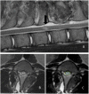
Results: We found a strong negative correlation between the intensity of pressure applied to the spinal cord and the functional outcome (P < 0.0001). SP-EPs showed high sensitivity for real time monitoring of intraoperative cord damage. On MRI, the ratio of the high-intensity area to the cross-sectional of the cord was a good predictor of recovery (P < 0.0001).
Conclusion: Our balloon compression SCI model is reliable, predictable, and easy to implement. By integrating SP-EPs, cord pressure, and findings on MRI, we can build a real-time warning and prediction system for early detection of impending or iatrogenic SCI and improve outcomes.
Keywords: balloon compression; intraspinal pressure; magnetic resonance imaging; motor behavior; porcine model; spinal cord injury; spine-to-spine evoked potentials.
Copyright © 2023 Hu, Chen, Wang, Sun and Wu.
Conflict of interest statement
The authors declare that the research was conducted in the absence of any commercial or financial relationships that could be construed as a potential conflict of interest.
Figures


Similar articles
-
Neurophysiological detection of impending spinal cord injury during scoliosis surgery.J Bone Joint Surg Am. 2007 Nov;89(11):2440-9. doi: 10.2106/JBJS.F.01476. J Bone Joint Surg Am. 2007. PMID: 17974887
-
[Changes of somatosensory and transcranial magnetic stimulation motor evoked potentials in experimental spinal cord injury].Zhonghua Yi Xue Za Zhi. 2008 Mar 18;88(11):773-7. Zhonghua Yi Xue Za Zhi. 2008. PMID: 18683688 Chinese.
-
Development of a motor and somatosensory evoked potentials-guided spinal cord Injury model in non-human primates.J Neurosci Methods. 2019 Jan 1;311:200-214. doi: 10.1016/j.jneumeth.2018.10.030. Epub 2018 Oct 25. J Neurosci Methods. 2019. PMID: 30393204
-
Direct Wave Intraoperative Neuromonitoring for Spinal Tumor Resection: A Focused Review.World Neurosurg X. 2022 Sep 15;17:100139. doi: 10.1016/j.wnsx.2022.100139. eCollection 2023 Jan. World Neurosurg X. 2022. PMID: 36217537 Free PMC article. Review.
-
Intraoperative neurophysiological monitoring of the spinal cord during spinal cord and spine surgery: a review focus on the corticospinal tracts.Clin Neurophysiol. 2008 Feb;119(2):248-64. doi: 10.1016/j.clinph.2007.09.135. Epub 2007 Nov 28. Clin Neurophysiol. 2008. PMID: 18053764 Review.
Cited by
-
A novel reconstruction model for thoracic spinal cord injury in swine.PLoS One. 2024 Sep 26;19(9):e0308637. doi: 10.1371/journal.pone.0308637. eCollection 2024. PLoS One. 2024. PMID: 39325721 Free PMC article.
-
Porcine Models of Spinal Cord Injury.Biomedicines. 2023 Aug 4;11(8):2202. doi: 10.3390/biomedicines11082202. Biomedicines. 2023. PMID: 37626699 Free PMC article. Review.
References
-
- Allen A. Surgery of experimental lesion of spinal cord equivalent to crush injury of fracture dislocation of spinal column: a preliminary report. J Am Med Assoc. (1911) LVII:878–80. 10.1001/jama.1911.04260090100008 - DOI
LinkOut - more resources
Full Text Sources

