Effect of prenatal stress and extremely low-frequency electromagnetic fields on anxiety-like behavior in female rats: With an emphasis on prefrontal cortex and hippocampus
- PMID: 36942730
- PMCID: PMC10097060
- DOI: 10.1002/brb3.2949
Effect of prenatal stress and extremely low-frequency electromagnetic fields on anxiety-like behavior in female rats: With an emphasis on prefrontal cortex and hippocampus
Abstract
Objective: Prenatal stress (PS) is a problematic situation resulting in psychological implications such as social anxiety. Ubiquitous extremely low-frequency electromagnetic fields (ELF-EMF) have been confirmed as a potential physiological stressor; however, useful neuroregenerative effect of these types of electromagnetic fields has also frequently been reported. The aim of the present study was to survey the interaction of PS and ELF-EMF on anxiety-like behavior.
Method: A total of 24 female rats 40 days of age were distributed into four groups of 6 rats each: control, stress (their mothers were exposed to stress), EMF (their mothers underwent to ELF-EMF), and EMF/stress (their mothers concurrently underwent to stress and ELF-EMF). The rats were assayed using elevated plus-maze and open field tests.
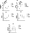
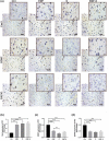
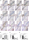
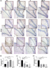
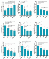
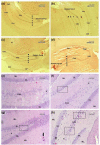
Results: Expressions of the hippocampus GAP-43, BDNF, and caspase-3 (cas-3) were detected by immunohistochemistry in Cornu Ammonis 1 (CA1) and dentate gyrus (DG) of the hippocampus and prefrontal cortex (PFC). Anxiety-like behavior increased in all treatment groups. Rats in the EMF/stress group presented more serious anxiety-like behavior. In all treatment groups, upregulated expression of cas-3 was seen in PFC, DG, and CA1 and downregulated expression of BDNF and GAP-43 was seen in PFC and DG and the CA1. Histomorphological study showed vast neurodegenerative changes in the hippocampus and PFC.
Conclusion: The results showed ,female rats that underwent PS or/and EMF exhibited critical anxiety-like behavior and this process may be attributed to neurodegeneration in PFC and DG of the hippocampus and possibly decreased synaptic plasticity so-called areas.
Keywords: BDNF; GAP-43; anxiety-like behavior; cas-3; electromagnetic fields; female rat; hippocampus; prefrontal cortex; prenatal stress.
© 2023 The Authors. Brain and Behavior published by Wiley Periodicals LLC.
Conflict of interest statement
The authors declare that they have not any conflict of interest.
Figures




Similar articles
-
Maternal stress induced anxiety-like behavior exacerbated by electromagnetic fields radiation in female rats offspring.PLoS One. 2022 Aug 23;17(8):e0273206. doi: 10.1371/journal.pone.0273206. eCollection 2022. PLoS One. 2022. PMID: 35998127 Free PMC article.
-
Impact of ketamine administration on chronic unpredictable stress-induced rat model of depression during extremely low-frequency electromagnetic field exposure: Behavioral, histological and molecular study.Brain Behav. 2023 May;13(5):e2986. doi: 10.1002/brb3.2986. Epub 2023 Apr 9. Brain Behav. 2023. PMID: 37032465 Free PMC article.
-
Anxiety-like behavioural effects of extremely low-frequency electromagnetic field in rats.Environ Sci Pollut Res Int. 2017 Sep;24(27):21693-21699. doi: 10.1007/s11356-017-9710-1. Epub 2017 Jul 29. Environ Sci Pollut Res Int. 2017. PMID: 28756602
-
Cellular stress response to extremely low-frequency electromagnetic fields (ELF-EMF): An explanation for controversial effects of ELF-EMF on apoptosis.Cell Prolif. 2021 Dec;54(12):e13154. doi: 10.1111/cpr.13154. Epub 2021 Nov 6. Cell Prolif. 2021. PMID: 34741480 Free PMC article. Review.
-
Cellular and molecular effects of non-ionizing electromagnetic fields.Rev Environ Health. 2023 Apr 7;39(3):519-529. doi: 10.1515/reveh-2023-0023. Print 2024 Sep 25. Rev Environ Health. 2023. PMID: 37021652 Review.
Cited by
-
Effect of prenatal exposure to stress and extremely low-frequency electromagnetic field on hippocampal and serum BDNF levels in male adult rat offspring.Iran J Basic Med Sci. 2024;27(9):1115-1123. doi: 10.22038/IJBMS.2024.75459.16357. Iran J Basic Med Sci. 2024. PMID: 39055879 Free PMC article.
References
-
- Aarts, L. , Schotman, P. , Verhaagen, J. , Schrama, L. , & Gispen, W. H. (1998). The role of the neural growth associated protein B‐50/GAP‐43 in morphogenesis. Molecular and Cellular Mechanisms of Neuronal Plasticity, 446, 85–106. - PubMed
-
- Bagheri Hosseinabadi, M. , Khanjani, N. , Ebrahimi, M. H. , Haji, B. , & Abdolahfard, M. (2019). The effect of chronic exposure to extremely low‐frequency electromagnetic fields on sleep quality, stress, depression and anxiety. Electromagnetic Biology and Medicine, 38(1), 96–101. - PubMed
MeSH terms
Substances
LinkOut - more resources
Full Text Sources
Medical
Research Materials
Miscellaneous

