. 2023 Feb 16:28:321-333.
doi: 10.1016/j.omto.2023.02.005. eCollection 2023 Mar 16.

Seneca Valley virus replicons are packaged in trans and have the capacity to overcome the limitations of viral transgene expression

Affiliations

Seneca Valley virus replicons are packaged in trans and have the capacity to overcome the limitations of viral transgene expression

Jeffrey D Bryant et al. Mol Ther Oncolytics. .

Abstract

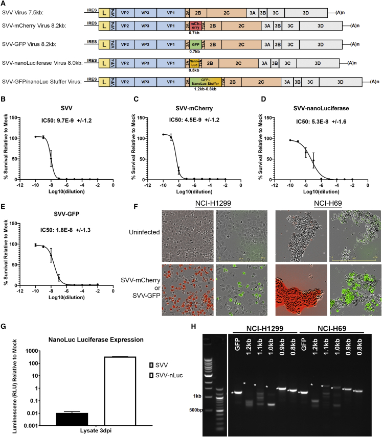
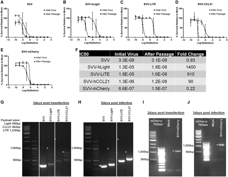
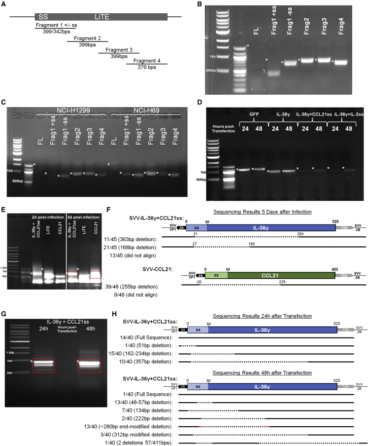
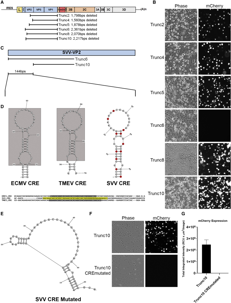
Oncolytic viruses (OVs) promote the anti-tumor immune response as their replication, and the subsequent lysis of tumor cells, triggers the activation of immune-sensing pathways. Arming OVs by expressing transgenes with the potential to promote immune cell recruitment and activation is an attractive strategy to enhance OVs' therapeutic benefit. For picornaviruses, a family of OVs with clinical experience, the expression of a transgene is limited by multiple factors: genome physical packaging limits, high rates of recombination, and viral-mediated inhibition of transgene secretion. Here, we evaluated strategies for arming Seneca Valley virus (SVV) with relevant immunomodulatory transgenes. Specificially in the contex of arming SVV, we evaluated transgene maximum size and stabiltity, transgene secretion, and the impact of transgene inclusion on viral fitness. We find that SVV is not capable of expressing secreted payloads and has a transgene packaging capacity of ∼10% of viral genome size. To enable transgene expression, we developed SVV replicons with greater transgene size capacity and secretion capabilities. SVV replicons can be packaged in trans by virus in co-infected cells to express immunomodulatory transgenes in surrounding cells, thus providing a means to enhance the potential of this therapeutic to augment the anti-tumor immune response.

Keywords: CRE; RNA therapeutics; SVV; Seneca Valley virus; cancer; oncolytic viral therapy; replicon; signal sequence; stable transgene expression; transgene.


Conflict of interest statement

J.D.B., J.S.L. (at the time the study was conducted), A.D.A., J.J., C.-H.C. (at the time the study was conducted), W.F., C.Q. (at the time the study was conducted), L.L. (at the time the study was conducted), and E.M.K. are all employees of Oncorus, Inc.

Figures

None
Graphical abstract
Figure 1
Figure 1
SVV is capable of expression of reporter transgenes that are within the packaging limit (A) Schematic representation of transgene-containing SVV constructs. Transgenes are inserted after the SVV 2A self-cleaving protein and 3′ flanked by a T2A peptide. Inserted are the reporter genes mCherry (red), GFP (green), nLuc (orange), and a fusion of GFP to nLuc (green to orange), which is inserted full sized (1.2 kb) or as 100 bp truncated forms down to 0.8 kb. Viral genes are in shaded boxes, the leader protein is yellow, the capsid proteins are light blue, and the non-structural proteins are divided into P2 (peach) and P3 (gray). IRES, internal ribosome entry site; p(A)n, polyadenylation tail; GFP, green fluorescent protein. (B–E) Dose-response infection curves of SVV and SVV encoding reporter transgenes were done on NCI-H446 cells with cell survival determined by quantification of ATP by CellTiter-Glo (Promega) assay. Calculated IC50 ± standard deviation mean values are shown with error bars displaying SEM from four replicates. (F) mCherry and GFP reporter expression was observed by 10× microscopy images in NCI-H1299 and NCI-H69 cells 24 h post 10 MOI infection with SVV reporter transgene viruses. Representative images were chosen from two replicate experiments. (G) Luciferase expression was detected 3 days post-infection with 5 μL transfection supernatant containing recovered SVV-nLuc virus relative to media treatment (mock) in infected NCI-H446 cells. Mean values are shown with error bars displaying SEM from two replicates. (H) Payload composed of GFP alone (0.7 kb) or nLuc-GFP fusion of different lengths (0.8–1.2 kb) was inserted into SVV and analyzed by RT-PCR to determine status of the transgene at the sequence level in recovered virus-containing supernatants collected 48 hr. The RNA in the supernatant was recovered and reverse transcribed to DNA, specific primers PCR amplified the transgene insertion region, and the resulting PCR product was run on a 0.7% TAE gel, with the expected band sizes for the transgenes marked with an asterisk. Representative gel images were chosen from three replicate experiments.
Figure 2
Figure 2
Immunomodulatory transgenes that are within the packaging limit are not stably retained as SVV paloads (A–F) TCID50 curves for SVV and SVV-mCherry or immunomodulatory transgenes in NCI-H446 cells were determined. The initial viruses recovered following RNA transfection (circles) and after passage infection with 5 μL transfection supernatant (squares) were plotted for comparison. IC50 values were calculated as well as the fold change between the virus before and after passage. Mean values are shown with error bars displaying SEM from four replicates. (G–J) Analysis for SVV transgenes retention by RT-PCR in NCI-H1299 cells after transfection and after infection in recovered supernatants. Transgene sizes are listed with expected sizes of bands marked with an asterisk on the gel images. Representative gel images were chosen from two replicate experiments.
Figure 3
Figure 3
Transgene signal sequences are detrimental to viral fitness and selected against (A) Diagram of the five DLL3 LiTE diagnostic transgene fragments including fragment 1 with and without the signal sequence (ss) to be tested as payloads in SVV. (B and C) Full-length (FL) and the LiTE gene fragments were tested for transgene retention by transfection in NCI-H1299 cells and infection in NCI-H1299 and NCI-H69 cells with RT-PCR analysis of supernatants. Representative gel images were chosen from two replicate experiments. (D) RT-PCR for detection of the retention of GFP or IL-36γ transgenes with/without the addition of the signal sequence from CCL21 or IL-2 in supernatants following transfection of NCI-H1299 cells. Representative gel images were chosen from two replicate experiments. (E and F) Analysis of transgene deletions after infection with SVV IL-36γ+CCL21-ss (blue) and CCL21 (green) transgene by sequencing individual PCR products from RT-PCR gel isolated bands (red boxes in E). DNA from indicated bands was isolated and cloned by Taq polymerase TA cloning to get individual colonies containing DNA inserts from single PCR products. 40 colonies were picked and sequenced. Discovered deletions are shown as dotted segments in diagrams, with numbers corresponding to bp of the beginning and end of detected deletion. (G and H) Analysis of transgene deletions after transfection with SVV IL-36γ+CCL21-ss (blue) transgene by sequencing individual PCR products from RT-PCR gel isolated bands (red boxes). Discovered deletions are shown as dotted segments in diagrams. The 48 h results (13/40–280-end-modified deletion) had a few bp at both ends of the deletion that did not align with the parental sequence, which is marked with a short red line. Expected sizes for all gels are marked with an asterisk.
Figure 4
Figure 4
Identification of the SVV CRE locus. (A) Diagram displaying the SVV-mCherry deletion constructs removing capsid proteins VP1 and VP3 and varying amounts of the VP2 protein referred to as Trunc 2, 4, 5, 6, 8, and 10. (B)2 μg of the listed Trunc SVVmCherry replicon candidate RNAs were transfected into NCI-H1299 cells and 24 h later assessed by 10× microscopy, with images showing both phase contrast and black-and-white mCherry images. Representative images were chosen from two replicate experiments. (C and D) The 114 bp sequence gap in SVV VP2 between Trunc6 and Trunc10 constructs was structurally aligned with the VP2 CRE regions of two closely related cardioviruses, TMEV and EMCV. The published EMCV and TMEV CRE sequences and structures are shaded in gray with the potential CRE sequence of SVV highlighted in yellow, with the predicted RNA stem-loop structure shown. Predicted structures and structural alignment performed by TurboFold. (E)TurboFold structural prediction of SVV with the CRE mutated. The base pairs annotated in red in (D)’s SVV CRE structure were changed to obliterate the CRE stem loop without changing the amino acid sequence encoded. (F and G) 1 μg of the listed Trunc SVVmCherry replicon candidate RNAs were transfected into NCI-H1299 cells and 24 h later assessed by 10× microscopy, with images showing both phase contrast and black-and-white mCherry images. mCherry expression was quantified by IncuCyte image analysis software. Mean values are shown with error bars displaying SEM, and the representative images are from two replicate experiments.
Figure 5
Figure 5
SVV replicons are trans-encapsidated with expression, and secretion of transgenes upon transduction (A) Schematic representation of SVV and the SVV-mCherry replicon. The Trunc10 deletion of VP2-VP1 is combined with the reporter mCherry transgene inserted after the SVV 2A self-cleaving protein and followed 3′ by a T2A peptide. (B) Procedural diagram detailing trans-encapsidation experiments. Cells are transfected with a mix of SVV , virus or control, and/or SVV-replicon RNA. After transfection, the cell supernatant is collected, 0.45 μm filtered, and used to infect a new set of cells. Red cells after infection indicate that the replicon genome has been trans-encapsidated. Created with Biorender.com (C) mCherry expression detected by microscopy following 24. h infection with 100 μL supernatants from mixed transfection (collected 48 h post-transfection) with SVV and/or SVV-mCherry replicon RNA as indicated. Representative images were chosen from two replicate experiments. (D) Schematic representation of SVVmCherry virus and the SVV murine IL-12 replicons with and without the signal sequence deleted. The locations of primers used for PCR detection of payloads are indicated by F and R. (E) PCR products from primers F and R in (D) from cDNA made from RNA collected from lysates of cells 48 h after infection with 100 μL supernatants from transfection or 48 h after transfection with 1 μg SVV-mCherry or SVV-mIL-12-replicon alone or in the mixed transfection of 0.5 μg SVV-mCherry with 0.5 μg SVV-mIL-12 replicon. Transfections were performed in NSCLC NCI-H1299 cells, while trans-encapsidation infections were done in NCI-H1299 and SCLC lines NCI-69 and NCI-H1963. Expected sizes for SVV-mCherry viral payload and SVV-mIL-12 replicon are indicated on images with an asterisk (∗) or a plus symbol (+), respectively. Representative gel images were chosen from four replicate experiments. (F–H) Detection by ELISA of mIL-12 in trans-encapsidation infection supernatants collected before cell lysis, 8 h for NCI-H1299 and 48 h for NCI-H69 and NCI-H1963. Transfection and infection experiments were performed as previously described, done here with both the mIL-12 replicon (SS) and the signal sequence deleted (ΔSS) version. Furthermore, the ratios of replicon to SVVmCherry viral RNA were varied with increased replicon and decreased viral RNA, i.e., 0.8 μg replicon/ 0.2 μg SVVmCh viral RNA compared with the previously used 0.5/0.5 μg RNA ratios. Total transfected RNA remained at 1 μg for all variations. Mock treatments were transfected with 1 μg SVV-NEG RNA. ∗p > 0.05 in unpaired two-tailed t tests. Mean values are shown with error bars displaying SEM from four replicates.

Similar articles

Cited by

References

    1. Ribas A., Dummer R., Puzanov I., VanderWalde A., Andtbacka R.H.I., Michielin O., Olszanski A.J., Malvehy J., Cebon J., Fernandez E., et al. Oncolytic virotherapy promotes intratumoral T cell infiltration and improves anti-PD-1 immunotherapy. Cell. 2018;174:1031–1032. doi: 10.1016/j.cell.2018.07.035. - DOI - PubMed
    1. Andtbacka R.H.I., Kaufman H.L., Collichio F., Amatruda T., Senzer N., Chesney J., Delman K.A., Spitler L.E., Puzanov I., Agarwala S.S., et al. Talimogene laherparepvec improves durable response rate in patients with advanced melanoma. J. Clin. Oncol. 2015;33:2780–2788. doi: 10.1200/JCO.2014.58.3377. - DOI - PubMed
    1. Peters C., Rabkin S.D. Designing herpes viruses as oncolytics. Mol. Ther. Oncolytics. 2015;2:15010. doi: 10.1038/mto.2015.10. - DOI - PMC - PubMed
    1. Fu X., Rivera A., Tao L., De Geest B., Zhang X. Construction of an oncolytic herpes simplex virus that precisely targets hepatocellular carcinoma cells. Mol. Ther. 2012;20:339–346. doi: 10.1038/mt.2011.265. - DOI - PMC - PubMed
    1. Lee C.Y.F., Rennie P.S., Jia W.W.G. MicroRNA regulation of oncolytic herpes simplex virus-1 for selective killing of prostate cancer cells. Clin. Cancer Res. 2009;15:5126–5135. doi: 10.1158/1078-0432.CCR-09-0051. - DOI - PubMed

LinkOut - more resources