

doi: 10.4103/1673-5374.363827.
Tissue optical clearing for neural regeneration research
Affiliations
- PMID: 36926711
- PMCID: PMC10233757
- DOI: 10.4103/1673-5374.363827
Item in Clipboard
Tissue optical clearing for neural regeneration research
Neural Regen Res.
2023 Sep.
No abstract available
Conflict of interest statement
None
Figures
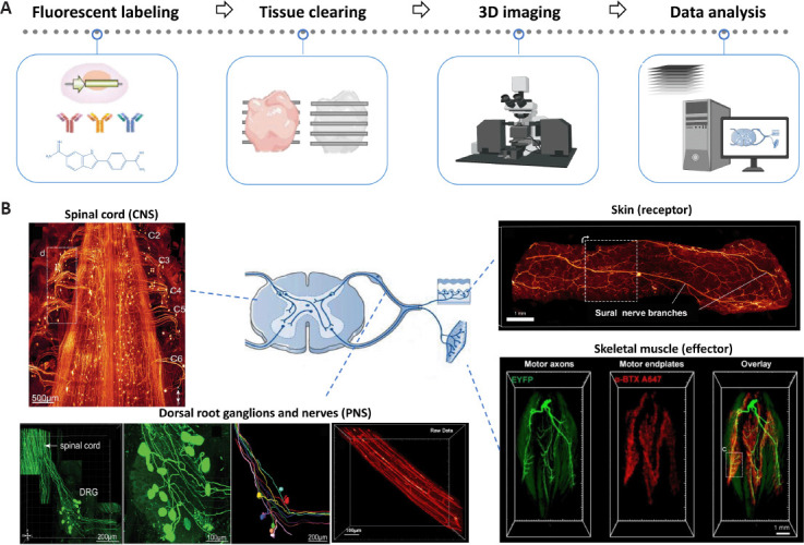
Overview of tissue optical clearing scheme for 3D imaging and its applications in neural tissues and target organs. (A) Overview of tissue optical clearing scheme for three-dimensional (3D) imaging. The main steps include whole-mount fluorescent labeling, tissue clearing, 3D imaging with optical sectioning microscopy, and data analysis of image stacks. (B) Tissue optical clearing provides novel tools for 3D morphological analyses of neural tissues and target organs for outcome assessments in neural regeneration research, such as the 3D visualization of the spinal cord (central nervous system [CNS]), dorsal root ganglions and nerves (peripheral nervous system [PNS]), as well as the skin and skeletal muscle (target organs). Images are adapted with permissions from Jing et al. (2018), Qi et al. (2019), and Daeschler et al. (2022).
Similar articles
-
Optical Tissue Clearing Enables Three-Dimensional Morphometry in Experimental Nerve Regeneration Research.Methods Mol Biol. 2023;2593:163-169. doi: 10.1007/978-1-0716-2811-9_10. Methods Mol Biol. 2023. PMID: 36513930
-
Recent progress in optical clearing of eye tissues.Exp Eye Res. 2021 Nov;212:108796. doi: 10.1016/j.exer.2021.108796. Epub 2021 Oct 15. Exp Eye Res. 2021. PMID: 34662543 Review.
-
Tissue Optical Clearing for Biomedical Imaging: From In Vitro to In Vivo.Adv Exp Med Biol. 2021;3233:217-255. doi: 10.1007/978-981-15-7627-0_11. Adv Exp Med Biol. 2021. PMID: 34053030
-
Salamander-Eci: An optical clearing protocol for the three-dimensional exploration of regeneration.Dev Dyn. 2021 Jun;250(6):902-915. doi: 10.1002/dvdy.264. Epub 2020 Oct 31. Dev Dyn. 2021. PMID: 33084146
-
Tissue optical clearing for 3D visualization of vascular networks: A review.Vascul Pharmacol. 2021 Dec;141:106905. doi: 10.1016/j.vph.2021.106905. Epub 2021 Sep 20. Vascul Pharmacol. 2021. PMID: 34506969 Review.
References
-
- Ertürk A, Mauch CP, Hellal F, Förstner F, Keck T, Becker K, Jährling N, Steffens H, Richter M, Hübener M, Kramer E, Kirchhoff F, Dodt HU, Bradke F. Three-dimensional imaging of the unsectioned adult spinal cord to assess axon regeneration and glial responses after injury. Nat Med. 2012;18:166–171. - PubMed
-
- Hilton BJ, Blanquie O, Tedeschi A, Bradke F. High-resolution 3D imaging and analysis of axon regeneration in unsectioned spinal cord with or without tissue clearing. Nat Protoc. 2019;14:1235–1260. - PubMed
LinkOut - more resources
Full Text Sources
