. 2023 Mar;615(7953):720-727.
doi: 10.1038/s41586-023-05824-z. Epub 2023 Mar 15.

A swapped genetic code prevents viral infections and gene transfer

Affiliations

A swapped genetic code prevents viral infections and gene transfer

Akos Nyerges et al. Nature. 2023 Mar.

Abstract

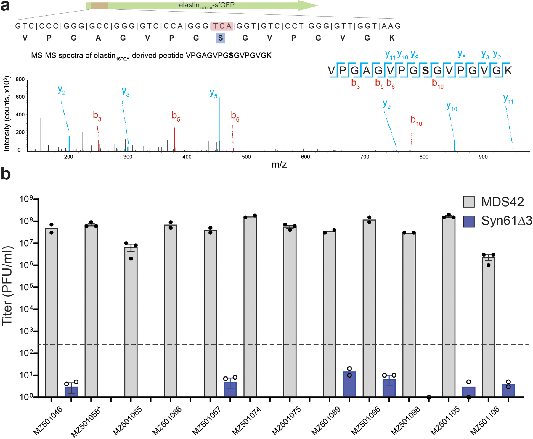
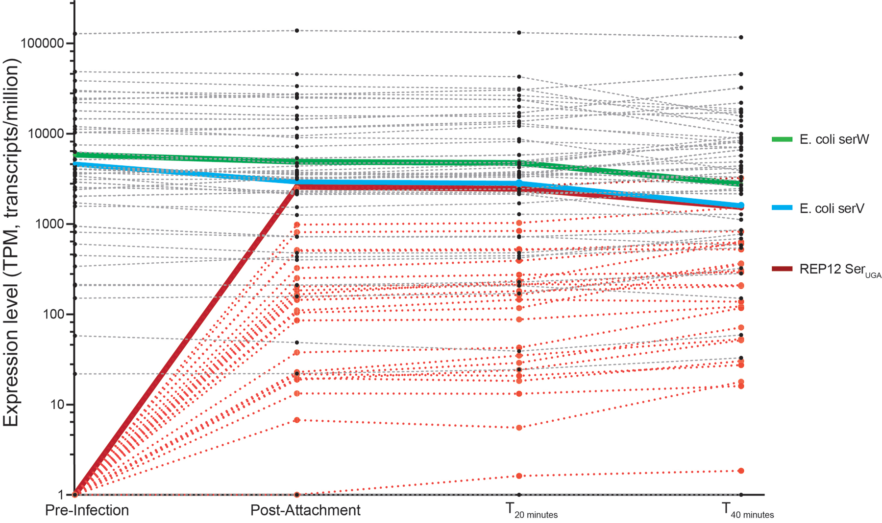
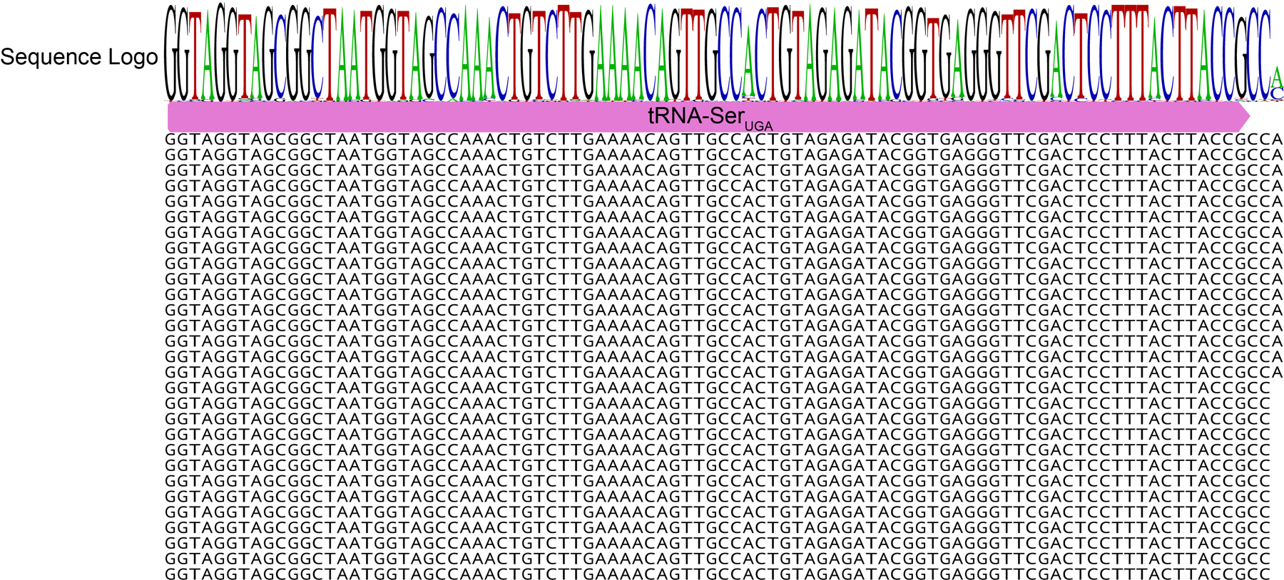
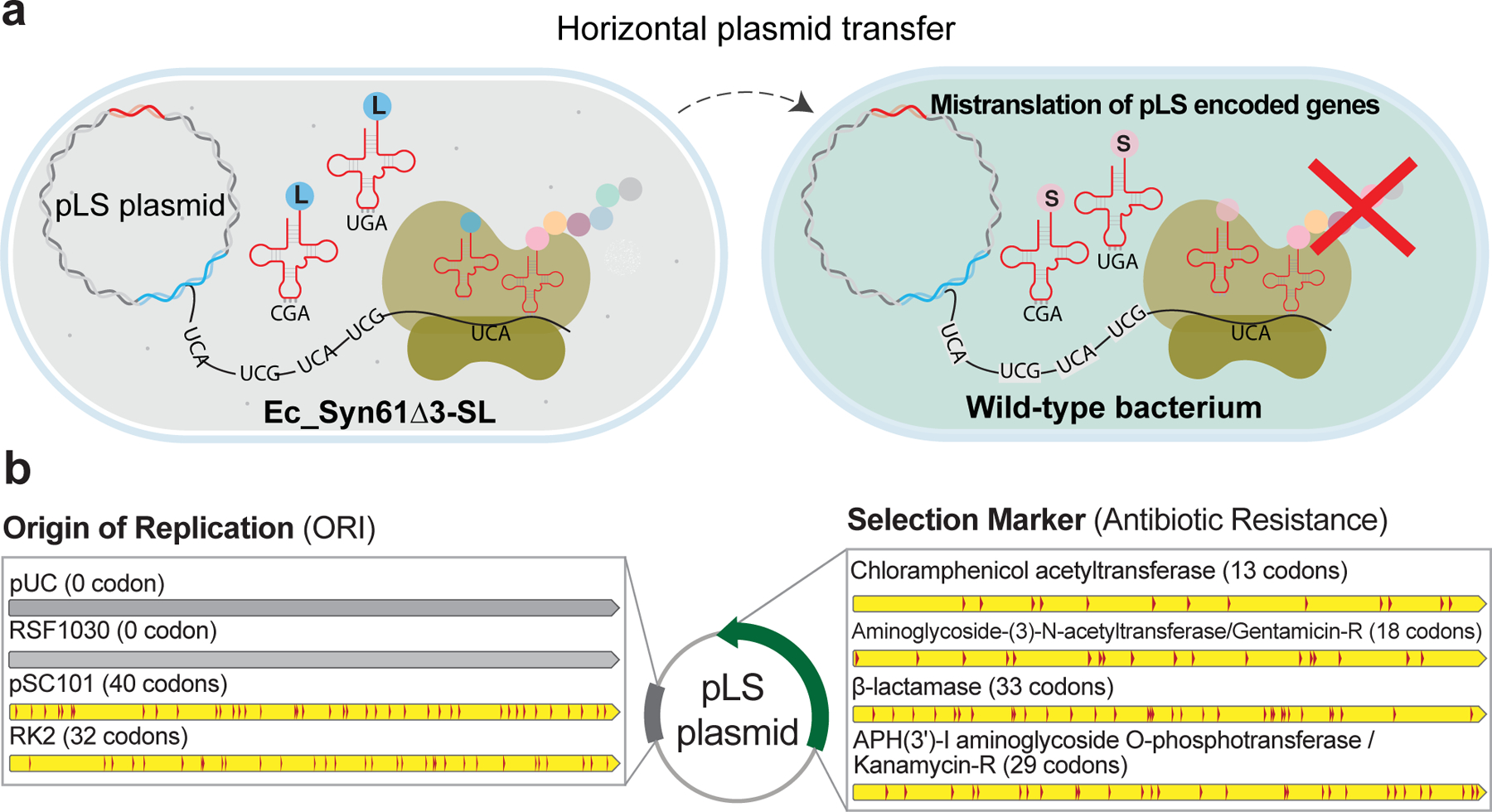
Engineering the genetic code of an organism has been proposed to provide a firewall from natural ecosystems by preventing viral infections and gene transfer1-6. However, numerous viruses and mobile genetic elements encode parts of the translational apparatus7-9, potentially rendering a genetic-code-based firewall ineffective. Here we show that such mobile transfer RNAs (tRNAs) enable gene transfer and allow viral replication in Escherichia coli despite the genome-wide removal of 3 of the 64 codons and the previously essential cognate tRNA and release factor genes. We then establish a genetic firewall by discovering viral tRNAs that provide exceptionally efficient codon reassignment allowing us to develop cells bearing an amino acid-swapped genetic code that reassigns two of the six serine codons to leucine during translation. This amino acid-swapped genetic code renders cells resistant to viral infections by mistranslating viral proteomes and prevents the escape of synthetic genetic information by engineered reliance on serine codons to produce leucine-requiring proteins. As these cells may have a selective advantage over wild organisms due to virus resistance, we also repurpose a third codon to biocontain this virus-resistant host through dependence on an amino acid not found in nature10. Our results may provide the basis for a general strategy to make any organism safely resistant to all natural viruses and prevent genetic information flow into and out of genetically modified organisms.


Conflict of interest statement

Competing Interests statement

The authors declare competing financial interests. Harvard Medical School has filed a provisional patent application related to this work on which A.N., S.V., and G.M.C. are listed as inventors. M.L., K.C., and F.H. are employed by GenScript USA Inc., but the company had no role in designing or executing experiments. G.M.C. is a founder of the following companies in which he has related financial interests: GRO Biosciences, EnEvolv (Ginkgo Bioworks), and 64x Bio. Other potentially relevant financial interests of G.M.C. are listed at http://arep.med.harvard.edu/gmc/tech.html.

Figures

Extended Data Figure 1.
Extended Data Figure 1.. Viral serine tRNAs decode TCA codons as serine, but Syn61Δ3 obstructs the replication of viruses containing genomic tRNA-SerUGA.
a) Viral TCR suppressor tRNAs decode TCA codons as serine. The amino acid identity of the translated TCR codon within elastin16 TCA-sfGFP-His6 was confirmed by tandem mass spectrometry from Syn61Δ3 expressing the tRNA-SerUGA of Escherichia phage IrisVonRoten (GenBank ID MZ501075). The figure shows the amino acid sequence and MS/MS spectrum of the analyzed elastin16 TCA peptide. MS/MS data was collected once. b) Syn61Δ3 obstructs the replication of viruses containing genomic tRNA-SerUGA. Figure shows the titer of twelve tRNA gene-containing coliphages, after 24 hours of growth on MDS42 and Syn61Δ3. All analyzed bacteriophages, except MZ501058, contain a genomic tRNA-SerUGA tRNA that provides TCR suppressor activity based on our screen (Figure 1b, Supplementary Data 1). Early exponential phase cultures of MDS42 and Syn61Δ3 were infected at an MOI of 0.001 with the corresponding phages, and free phage titers were determined after 24 hours of incubation. Measurements were performed in n=3 independent experiments (i.e., MDS42 + MZ501058, MZ501065, MZ501075, MZ501105, MZ501106; Syn61Δ3 + MZ501046, MZ501067, MZ501066, MZ501096, MZ501074, MZ501098) or in n=2 independent experiments (i.e., MDS42 + MZ501046, MZ501066, MZ501067, MZ501074, MZ501089, MZ501096, MZ501098; Syn61Δ3 + MZ501058, MZ501065, MZ501075, MZ501089, MZ501105, MZ501106); dashed line represents input phage titer without bacterial cells (i.e., a titer of 420 PFU/ml); dots represent data from independent experiments; bar graphs represent the mean; error bars represent the SEM based on n=3 independent experiments.
Extended Data Figure 2.
Extended Data Figure 2.. Viruses overcome genetic-code-based resistance by rapidly complementing the cellular tRNA pool with virus-encoded tRNAs.
The time-course kinetics of host and viral tRNA expression in Syn61Δ3 cells following REP12 phage infection was quantified using tRNAseq (Methods). The endogenous serV and serW tRNAs of the host Syn61Δ3 are highlighted in green and blue, respectively, while the tRNA-SerUGA of the REP12 virus is highlighted in red. REP12 viral tRNAs are shown in light red; endogenous tRNAs of Syn61Δ3 are shown in gray. Data represent mean TPM (transcript/million). Source data is available within this paper.
Extended Data Figure 3.
Extended Data Figure 3.. tRNAseq-based detection of CCA tRNA tail addition to REP12 tRNA-SerUGA.
The tRNA tail is modified into CCA even if the phage-encoded tRNA-SerUGA does not encode a CCA end. The sequence of genomic phage-encoded tRNA-SerUGA is highlighted in magenta. Black letters indicate example tRNAseq sequencing reads from REP12 infected Syn61Δ3 cells directly after phage attachment based on a single experiment.
Extended Data Figure 4.
Extended Data Figure 4.. Transcriptomic changes in Syn61Δ3 following phage infection.
Volcano plot shows the differential expression between uninfected and REP12-infected Syn61Δ3 cells, 40 minutes post-infection, based on n=3 independent experiments (Methods). QueG encodes epoxyqueuosine reductase that catalyzes the final step in the de novo synthesis of queuosine in tRNAs. TrmJ encodes tRNA Cm32/Um32 methyltransferase that introduces methyl groups at the 2’-O position of U32 of several tRNAs, including tRNA-SerUGA. Differential expression and −Log10 adjusted p-values were calculated using the DESeq2 algorithm. Source data is available within this paper.
Extended Data Figure 5.
Extended Data Figure 5.. Multiple sequence alignment of leuUYGA and phage-derived tRNA-LeuYGA variants.
a) Multiple sequence alignment of leuUYGA variants selected in aminoglycoside O-phosphotransferase expression screen, compared to E. coli leuU and the YGA anticodon-swapped E. coli leuU tRNA variant. Grey shading indicates the anticodon region, and the host’s LeuS leucine-tRNA-ligase identity elements are shown in blue. Sequence information of the leuUYGA variants is available in Supplementary Data 3. b) Multiple sequence alignment of phage-derived tRNA-LeuYGA variants selected in the aph3Ia29×Leu→TCR aminoglycoside O-phosphotransferase expression screen, compared to endogenous E. coli leucine tRNAs. Grey shading indicates the anticodon region, while the host’s LeuS leucine-tRNA-ligase identity elements are shown in blue. Sequence information of the phage-derived tRNA-LeuYGA variants is available in Supplementary Data 3.
Extended Data Figure 6.
Extended Data Figure 6.. tRNAseq-based quantification of viral Leu-tRNAUGA and Leu-tRNACGA in the tRNAome of Ec_Syn61Δ3-SL.
tRNA levels of Ec_Syn61Δ3-SL were quantified using tRNAseq (Methods). Viral Leu-tRNAUGA and Leu-tRNACGA are highlighted in red, and the host’s endogenous E. coli serV tRNA is highlighted in orange. tRNAseq data was collected once. Data represent TPM (transcript/million). Source data is available within this paper.
Extended Data Figure 7.
Extended Data Figure 7.. Serine-to-leucine mistranslation of TCT codons in Ec_Syn61Δ3-SL cells.
a) MS/MS spectrum of the serine-to-leucine mistranslated TufA peptide. b) MS/MS spectrum of the wild-type TufA peptide. The figure shows the amino acid sequence and MS/MS spectrum of the Ec_Syn61Δ3-SL-expressed TufA protein fragment, together with its genomic sequence, in which the serine-coding TCT codon (as shown in panel b) is partially mistranslated as leucine (as shown in panel a). The experiment was performed by analyzing the total proteome of Ec_Syn61Δ3-SL cells, expressing Leu9-tRNAYGA from Escherichia phage OSYSP (GenBank ID MF402939.1) by tandem mass spectrometry (Methods). MS/MS data was collected once.
Extended Data Figure 8.
Extended Data Figure 8.. Doubling time and growth curves of Syn61Δ3(ev5), Syn61Δ3(ev5)ΔrecA, Syn61Δ3(ev5)ΔrecA(ev1), and Ec_Syn61Δ3-SL.
a) Doubling times of Syn61Δ3(ev5), Syn61Δ3(ev5)ΔrecA, Syn61Δ3(ev5)ΔrecA(ev1), and Ec_Syn61Δ3-SL, calculated based on growth curves (shown in panels b, c, d) in rich bacterial media under standard laboratory conditions. b) Growth curves of Syn61Δ3(ev5), Syn61Δ3(ev5)ΔrecA, and Syn61Δ3(ev5)ΔrecA(ev1) in LBL broth. c) Growth curves of Syn61Δ3(ev5) and Syn61Δ3(ev5)ΔrecA(ev1) in 2×YT broth. d) Growth curves of Ec_Syn61Δ3-SL in 2×YT broth containing 50 μg/ml kanamycin. Three independent cultures were grown aerobically in vented shake flasks at 37 °C, and OD600 measurements were taken during exponential growth (Methods). Data curves and bars represent the mean. Error bars show standard deviation based on n=3 independent experiments.
Extended Data Figure 9.
Extended Data Figure 9.. Ec_Syn61Δ3-SL resists viruses in environmental samples.
Phage enrichment experiment using Ec_Syn61Δ3-SL, expressing KP869110.1 tRNA24 LeuYGA and MF402939.1 tRNA9 LeuYGA, as host. (b) Phage enrichment experiment using Ec_Syn61Δ3-SL, expressing KP869110.1 tRNA24 LeuYGA and MF402939.1 tRNA21 LeuYGA, as host. Phage enrichment experiments were performed by mixing early exponential cultures of Ec_Syn61Δ3-SL with 10 ml environmental sample mix containing the mixture of Sample 2–13 from our study (Extended Data Table 1a). After two enrichment cycles (Methods, Supplementary Note), filter-sterilized culture supernatants were mixed with phage-susceptible E. coli MDS42 cells in top agar and plated on LBL agar plates to determine viral titer. Enrichment experiments were performed in n=2 independent replicates with the same result. (c) Lytic E. coli MDS42 phage plaques after 103-fold dilution of the environmental sample mix. d) Lytic phage titer of the environmental sample mix, before and after enrichment on Ec_Syn61Δ3-SL. Dots represent the viral titer of the unenriched sample based on three independent experiments, measured on E. coli MDS42 cells. ND represents no plaques detected. Bar represents the mean. Error bar shows standard deviation based on n=3 independent experiments.
Figure 1.
Figure 1.. Discovery of mobile TCR codon translating tRNAs in E. coli Syn61Δ3.
(a). We screened the mobile tRNAome for tRNAs that can simultaneously translate TCA and TCG (together TCR) codons by computationally identifying tRNA genes in mobile genetic elements (1.) and then synthesizing select candidates as an oligonucleotide library and cloning these variants into a plasmid vector carrying a nptII40TCA,68TCG,104TCA,251TCG marker (conferring kanamycin resistance) (2.). Following the transformation of this library into Syn61Δ3 (3.), in which the deletion of serU (encoding tRNA-SerCGA) and serT (encoding tRNA-SerUGA) makes TCG and TCA codons unreadable, only variants carrying functional TCR suppressor tRNAs survive kanamycin selection (4.). Finally, high-throughput sequencing of the tRNA inserts from kanamycin-resistant clones identified suppressor tRNAs (5.). (b). Multiple sequence alignment of mobile TCR codon translating tRNAs. Grey shading indicates the anticodon region, while the host’s serine-tRNA-ligase identity elements are shown in blue. (c). Viral serine tRNAUGA translates the TCA codon. Syn61Δ3 expressing elastin16 GCA(alanine)-sfGFP-His6 served as wild-type expression control, and the elastin16 TCR-sfGFP-His6 expression was compared with and without the coexpression of the tRNA-SerUGA of Escherichia phage IrisVonRoten. xxx marks the analyzed codon, TCA or GCA. Bar graph represents the mean; error bars represent SD based on n=3 independent experiments; a.u. denotes arbitrary fluorescence units. (d). The expression of viral TCR suppressor tRNAs and serU (tRNA-SerCGA) restores the replication of T6 bacteriophage in Syn61Δ3. Dots represent data from n=3 independent experiments, error bars represent SD, and the bar graph represents the mean. ND represents below the detection limit (i.e., <103 PFU/ml); ns indicates lack of significance (p = 0.116) based on unpaired two-sided Student’s t-test.
Figure 2.
Figure 2.. Lytic phages of Syn61Δ3.
(a). Titer of Syn61Δ3 phage isolates after replication on Syn61Δ3. Dots represent data from n=2 independent experiments; bar graphs represent the mean. (b). Single-step growth curve of REP12 lytic Syn61Δ3 phage. Single-step growth was performed in n=3 independent experiments, red line represents the mean, and dots represent the total viral titer. (c). Genomic maps of tRNA operons in lytic Syn61Δ3 phages. Magenta arrows represent predicted tRNA genes; * denotes tRNA genes identified in our earlier TCR codon suppressor screen (Figure 1b); green arrow represents homing endonuclease gene, while orange arrows represent protein-coding genes. Phage operon numbers correspond to the following REP phages: 1.=REP1; 2.=REP2; 3.=REP4; 4.=REP6; 5.=REP12. (d). Viral tRNA operon-expressed tRNAs translate TCR codons. Syn61Δ3 expressing elastin16GCA(alanine)-sfGFP-His6 served as wild-type expression control, and the elastin16 TCR-sfGFP-His6 expression was compared with and without the coexpression of the REP12 viral tRNA operon. xxx marks the analyzed codon, TCA, TCG, or GCA. A.u. denotes arbitrary fluorescence units; bar graphs represent the mean. Error bars represent SD based on n=3 independent experiments. (e). Viral tRNA operon-expressed tRNAs decode TCR codons as serine. The amino acid identity of the translated TCA codon within elastin16 TCA-sfGFP-His6 was confirmed by tandem mass spectrometry from Syn61Δ3 cells containing the REP12 tRNA operon and its cognate promoter. The figure shows the amino acid sequence and MS/MS spectrum of the analyzed elastin16 TCR peptide. MS/MS data was collected once.
Figure 3.
Figure 3.. An amino-acid-swapped genetic code provides multi-virus resistance.
(a). The creation of an E. coli GRO, Ec_Syn61Δ3-SL, in which both TCA and TCG—naturally serine-meaning—codons are translated as leucine. The introduction of bacteriophage-derived Leu-tRNAUGA and Leu-tRNACGA to Syn61Δ3 reassigns TCA and TCG codons to leucine, while the reassignment of the TAG stop codon to encode L-4,4’-biphenylalanine (bipA) in an essential gene of the host ensures the biocontainment of Ec_Syn61Δ3-SL. (b). Schematic of viral infection in Ec_Syn61Δ3-SL. The reassignment of sense codons TCA and TCG to leucine in Ec_Syn61Δ3-SL provides multivirus resistance by mistranslating the viral proteome. (c). Bacteriophage-derived Leu-tRNAUGA and Leu-tRNACGA expression in Syn61Δ3 provides multivirus resistance. The figure shows the titer of lytic Syn61Δ3 phages following the infection of the corresponding Leu-tRNAYGA-expressing Syn61Δ3 strain with a mixture of twelve distinct REP Syn61Δ3 phages (Extended Data Table 1, Supplementary Data 3). All experiments were performed in three independent replicates; dots represent data from n=3 independent experiments; bar graphs represent the mean; the error bars represent SD. (d). The reassignment of TCR codons to leucine within the coding sequence of aph3Ia29×Leu→TCR in Syn61Δ3-LS was confirmed by tandem mass spectrometry. The figure shows the amino acid sequence and MS/MS spectrum of the detected aph3Ia29×Leu→TCR peptide and its coding sequence. MS/MS data was collected once. (e). Mistranslated viral protein synthesis in Ec_Syn61Δ3-SL. The figure shows the amino acid sequence and MS/MS spectrum of a bacteriophage-expressed protein, together with its viral genomic sequence, in which the naturally serine-coding TCA codon is mistranslated as leucine. The experiment was performed by infecting Ec_Syn61Δ3-SL cells, expressing Leu9-tRNAYGA from Escherichia phage OSYSP, with the REP12 phage and the proteome of infected cells was analyzed by tandem mass spectrometry. MS/MS data was collected once.
Figure 4.
Figure 4.. Addiction of synthetic genetic information to a genetic code in which TCR codons encode leucine prevents horizontal gene transfer.
(a). We developed a set of plasmid vectors, termed the pLS plasmids, that rely on TCR codons to express leucine-containing proteins. pLS plasmids only function in Ec_Syn61Δ3-SL expressing bacteriophage-derived synthetic tRNA-LeuYGA tRNAs, and the encoded proteins of pLS plasmids become mistranslated in cells bearing the canonical genetic code. (b). The pLS plasmids offer multiple mutually orthogonal antibiotic resistance markers together with low to high copy-number origins-of-replication (ORI) that are addicted to an artificial genetic code in which leucine is encoded as TCR codons. Number in parenthesis marks the number of LeuTCR codons in each gene. Detailed sequence information and the description of pLS plasmids are available in Supplementary Data 3.

Comment in

Similar articles

Cited by

References

    1. Church GM & Regis E Regenesis: How Synthetic Biology Will Reinvent Nature and Ourselves. (Basic Books, 2014).
    1. Lajoie MJ et al. Genomically Recoded Organisms Expand Biological Functions. Science 342, 357–360 (2013). - PMC - PubMed
    1. Ma NJ & Isaacs FJ Genomic Recoding Broadly Obstructs the Propagation of Horizontally Transferred Genetic Elements. cels 3, 199–207 (2016). - PMC - PubMed
    1. Robertson WE et al. Sense codon reassignment enables viral resistance and encoded polymer synthesis. Science 372, 1057–1062 (2021). - PMC - PubMed
    1. Ostrov N et al. Design, synthesis, and testing toward a 57-codon genome. Science 353, 819–822 (2016). - PubMed

Publication types

MeSH terms