Silencing proline-rich coiled-coil 2C inhibit the proliferation and metastasis of liver cancer cells
- PMID: 36915448
- PMCID: PMC10007939
- DOI: 10.21037/jgo-23-10
Silencing proline-rich coiled-coil 2C inhibit the proliferation and metastasis of liver cancer cells
Abstract
Background: Proline-rich coiled-coil 2C (PRRC2C) is located in the chromosome region lq where hepatocellular carcinoma (HCC) frequently undergoes genomic fragment amplification, but its role in HCC is unknown. In this study, we aimed to explore the correlation of PRRC2C with HCC diagnosis and progression, as well as its influence on the biological behavior of HCC cells.
Methods: The Cancer Genome Atlas (TCGA) RNA-sequencing datasets of 371 cases of primary liver cancer and 50 normal liver tissue specimens were obtained to analyze correlation between PRRC2C expression and HCC staging, grades, and overall survival. After confirming expression of PRRC2C in HCC cells, PRRC2C silencing was performed. Celigo cell counting, cell clone formation, MTT (3-(4,5-dimethylthiazol-2-yl)-2,5-diphenyltetrazolium bromide) assay and Flow cytometry were used to detect the cell proliferation and apoptosis; wound healing and Transwell assays were used to detect the invasion abilities of cells. Xenograft transplantation in nude mice was performed to investigate the impact of PRRC2C knockdown on tumorigenic capabilities. In addition, the expression levels of EMT (epithelial-mesenchymal transition)-related genes, including E-cadherin, N-cadherin, Twistl, Snail, Slug, and Smad2/3/4, were detected.
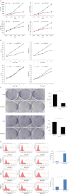
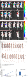
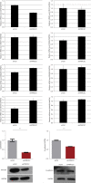
Results: Analysis of TCGA data sets revealed that patients with high PRRC2C expression had significantly shorter overall survival. PRRC2C was abundantly expressed in four human hepatocarcinoma cell lines. After knockdown PRRC2C, the proliferation of HCC cells were suppressed and the numbers of apoptotic cells increased. Migration and invasion ability of HCC cells were inhibited by PRRC2C knockdown. Meanwhile, PRRC2C silencing inhibited the tumor formation (indicated by reduced tumor volume and weight compared to the control group) in BALB/c (Bagg Albino Laboratory-bred strain) nude mice. The expressions of EMT-related genes N-cadherin and Vimentin were significantly lower in the PRRC2C knockdown group than in the control group.
Conclusions: PRRC2C promotes the proliferation and metastasis of liver cancer cells and inhibited apoptosis, potentially through upregulation of EMT related N-cadherin and Vimentin.
Keywords: Apoptosis; PRRC2C; hepatocellular; neoplasm metastasis; proliferation.
2023 Journal of Gastrointestinal Oncology. All rights reserved.
Conflict of interest statement
Conflicts of Interest: All authors have completed the ICMJE uniform disclosure form (available at https://jgo.amegroups.com/article/view/10.21037/jgo-23-10/coif). The authors have no conflicts of interest to declare.
Figures


Similar articles
-
Overexpression of the long non-coding RNA SPRY4-IT1 promotes tumor cell proliferation and invasion by activating EZH2 in hepatocellular carcinoma.Biomed Pharmacother. 2017 Jan;85:348-354. doi: 10.1016/j.biopha.2016.11.035. Epub 2016 Nov 28. Biomed Pharmacother. 2017. PMID: 27899259
-
Plumbagin promotes human hepatoma SMMC-7721 cell apoptosis via caspase-3/vimentin signal-mediated EMT.Drug Des Devel Ther. 2019 Jul 15;13:2343-2355. doi: 10.2147/DDDT.S204787. eCollection 2019. Drug Des Devel Ther. 2019. PMID: 31409969 Free PMC article.
-
TRIM66 promotes malignant progression of hepatocellular carcinoma by inhibiting E-cadherin expression through the EMT pathway.Eur Rev Med Pharmacol Sci. 2019 Mar;23(5):2003-2012. doi: 10.26355/eurrev_201903_17239. Eur Rev Med Pharmacol Sci. 2019. PMID: 30915743
-
Ventilagolin Suppresses Migration, Invasion and Epithelial-Mesenchymal Transition of Hepatocellular Carcinoma Cells by Downregulating Pim-1.Drug Des Devel Ther. 2021 Dec 1;15:4885-4899. doi: 10.2147/DDDT.S327270. eCollection 2021. Drug Des Devel Ther. 2021. PMID: 34880599 Free PMC article.
-
MAPKAPK5-AS1 drives the progression of hepatocellular carcinoma via regulating miR-429/ZEB1 axis.BMC Mol Cell Biol. 2022 Apr 25;23(1):21. doi: 10.1186/s12860-022-00420-x. BMC Mol Cell Biol. 2022. PMID: 35468721 Free PMC article.
References
LinkOut - more resources
Full Text Sources
Research Materials
