. 2023 May 8;51(8):3934-3949.
doi: 10.1093/nar/gkad140.

The ubiquitin-specific protease USP36 SUMOylates EXOSC10 and promotes the nucleolar RNA exosome function in rRNA processing

Affiliations

The ubiquitin-specific protease USP36 SUMOylates EXOSC10 and promotes the nucleolar RNA exosome function in rRNA processing

Yingxiao Chen et al. Nucleic Acids Res. .

Abstract

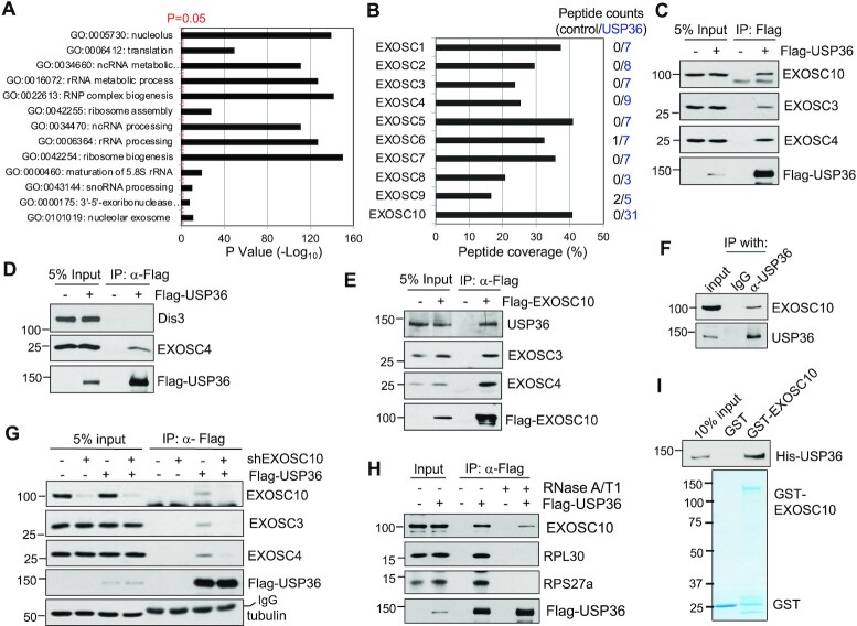
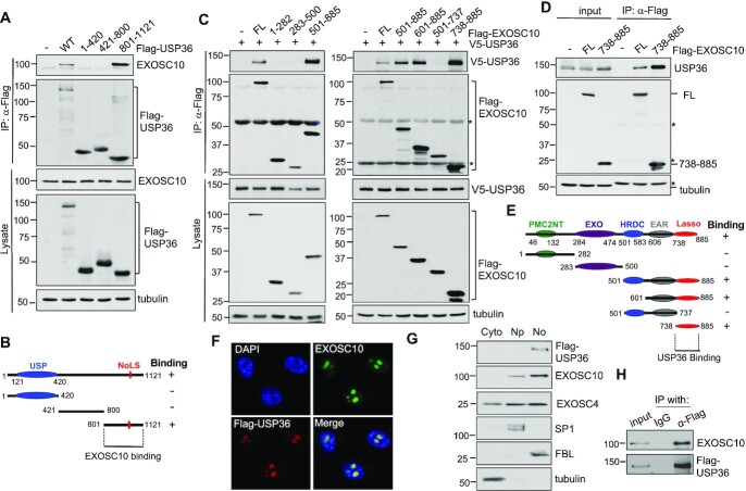
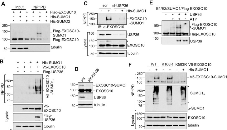
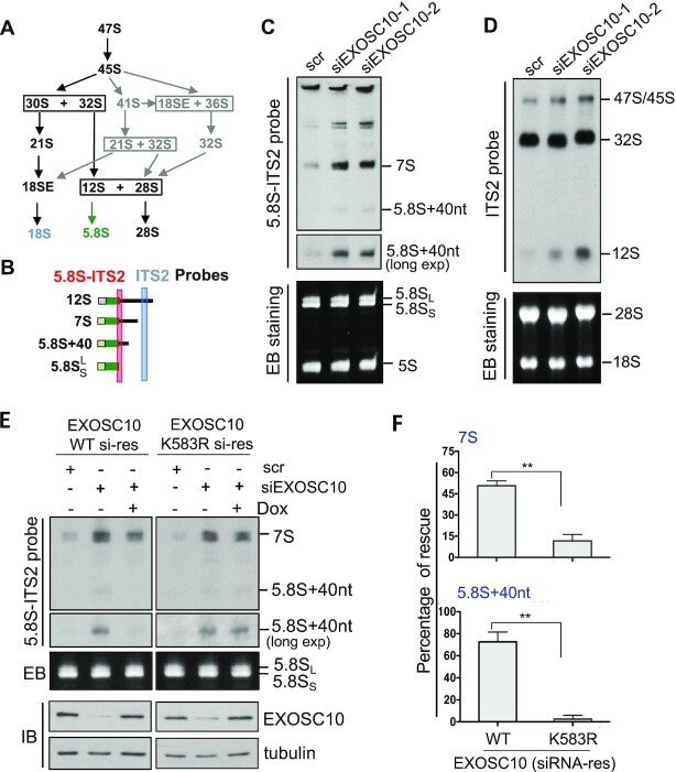
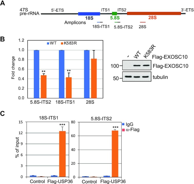
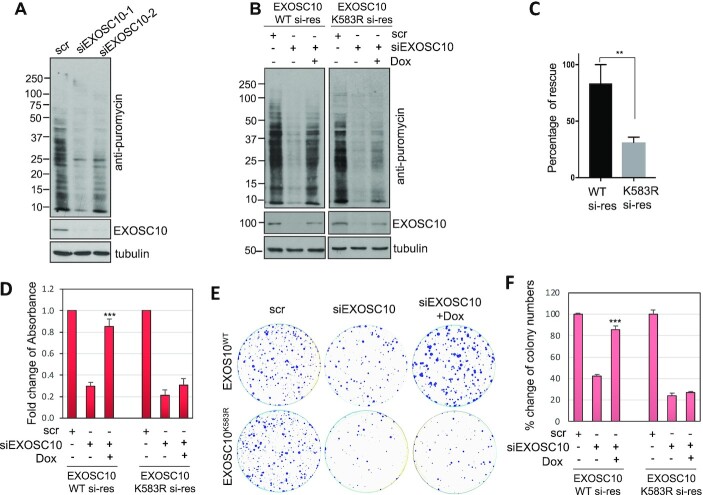
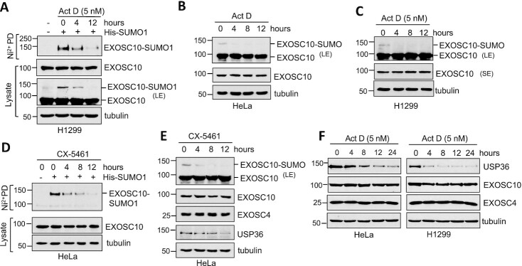
The RNA exosome is an essential 3' to 5' exoribonuclease complex that mediates degradation, processing and quality control of virtually all eukaryotic RNAs. The nucleolar RNA exosome, consisting of a nine-subunit core and a distributive 3' to 5' exonuclease EXOSC10, plays a critical role in processing and degrading nucleolar RNAs, including pre-rRNA. However, how the RNA exosome is regulated in the nucleolus is poorly understood. Here, we report that the nucleolar ubiquitin-specific protease USP36 is a novel regulator of the nucleolar RNA exosome. USP36 binds to the RNA exosome through direct interaction with EXOSC10 in the nucleolus. Interestingly, USP36 does not significantly regulate the levels of EXOSC10 and other tested exosome subunits. Instead, it mediates EXOSC10 SUMOylation at lysine (K) 583. Mutating K583 impaired the binding of EXOSC10 to pre-rRNAs, and the K583R mutant failed to rescue the defects in rRNA processing and cell growth inhibition caused by knockdown of endogenous EXOSC10. Furthermore, EXOSC10 SUMOylation is markedly reduced in cells in response to perturbation of ribosomal biogenesis. Together, these results suggest that USP36 acts as a SUMO ligase to promote EXOSC10 SUMOylation critical for the RNA exosome function in ribosome biogenesis.


Figures

Figure 1.
Figure 1.
USP36 interacts with the RNA exosome. (A) GO analysis of the USP36-interacting proteins identified by MS analysis of anti-Flag immunoprecipitates from Flag-USP36-expressing 293 cells. A vertical dashed line represents the significance score, P = 0.05. (B) RNA exosome components revealed by MS analysis of immunoprecipitates from 293-Flag-USP36 cells by anti-Flag antibody. Shown are the percentiles of peptide coverage of each RNA exosome component with the numbers (blue) of peptides indicated on the right from Flag-USP36-expressing cells. The peptide numbers from control 293 cells are shown in black. (C) USP36 interacts with the RNA exosome. 293 cells transfected with Flag-USP36 were subjected to co-IP with anti-Flag antibody followed by IB. (D) USP36 does not interact with Dis3. H1299 cells were transfected with Flag-USP36 and assayed by co-IP using anti-Flag antibody followed by IB. (E) EXOSC10 interacts with endogenous USP36. Cell lysates from H1299 cells transfected with Flag-EXOSC10 were immunoprecipitated with anti-Flag antibody followed by IB. (F) Endogenous USP36 interacts with endogenous EXOSC10. 293 cell lysates were immunoprecipitated with anti-USP36 antibody (Proteintech) followed by IB with anti-EXOSC10. (G) Flag-USP36 associates with the RNA exosome through EXOSC10. H1299 cells were transfected with empty vector or Flag-USP36 and infected with scrambled or EXOSC10 shRNA lentiviruses, followed by co-IP with anti-Flag and IB. (H) Interaction between USP36 and EXOSC10 is partially dependent on RNA. 293 cells transfected with Flag-USP36 were subjected to co-IP with anti-Flag antibody in the presence or absence of 100 μg/ml RNase A and 100 U/ml RNase T1 treatment followed by IB. (I) USP36 directly interacts with EXOSC10 in vitro. Purified GST or GST–EXOSC10 immobilized on glutathione beads was incubated with purified His-USP36. Bound proteins were assayed by IB with anti-USP36 (top). Coomassie staining of GST and GST–EXOSC10 proteins is shown in the bottom panel.
Figure 2.
Figure 2.
USP36 interacts with EXOSC10 via their C-terminal domains in the nucleolus. (A and B) EXOSC10 binds to the C-terminal domain of USP36. H1299 cells were transfected with Flag-USP36 or its deletion mutants as indicated, followed by co-IP with anti-Flag and IB analysis (A). The C-terminal EXOSC10-binding domain of USP36 is shown in (B). USP, ubiquitin-specific protease; NoLS, nucleolar localization signal. (C–E) USP36 binds to the Lasso region in the C-terminal domain (CTD) of EXOSC10. H1299 cells transfected with Flag-EXOSC10 or its deletion mutants together with (C) or without (D) V5-USP36 were subjected to co-IP with anti-Flag followed by IB. The diagram of EXOSC10 domains is shown in (E). PMC2NT, polycystin 2 N-terminal; EXO, exonuclease; HRDC, helicase and RNase D C-terminal; EAR, exosome-associated region. * indicates IgG. (F) Co-localization of Flag-USP36 and EXOSC10. HeLa cells transfected with Flag-USP36 were immunostained with anti-Flag (red) and anti-EXOSC10 (green). The nuclei were stained with DAPI (blue). (G) Subcellular distribution of Flag-USP36, EXOSC10 and EXOSC4 proteins. HeLa cells were fractionated into cytoplasmic (Cyto), nucleoplasmic (Np) and nucleolar fractions (No) followed by IB. Tubulin, SP1 and FBL were used as cytoplasmic, nucleoplasmic and nucleolar markers, respectively. (H) Co-IP of USP36 with EXOSC10 in the nucleolar fraction. Lysates from the nucleolar fraction shown in (G) were immunoprecipitated with anti-Flag antibody followed by IB with anti-EXOSC10 antibodies.
Figure 3.
Figure 3.
USP36 SUMOylates EXOSC10 in cells and in vitro. (A) EXOSC10 can be modified mainly by SUMO1. H1299 cells transfected with the indicated plasmids were subjected to Ni2+-NTA PD under denaturing conditions, followed by IB with anti-Flag antibody. (B) USP36 promotes SUMO1 modification of EXOSC10. H1299 cells transfected with the indicated plasmids were subjected to Ni2+-NTA PD followed by IB to detect EXOSC10 SUMOylation. The SUMO1-modified EXOSC10 is indicated. The protein expression is shown in the bottom panels. (C) Knockdown of USP36 attenuates SUMO1 modification of EXOSC10. HeLa cells transfected with His-SUMO1 and infected with scrambled (scr) or USP36 shRNA lentiviruses were subjected to Ni2+-NTA PD under denaturing conditions, followed by IB. * indicates SUMOylated EXOSC10 in cell lysates. (D) Knockdown of USP36 attenuates EXOSC10 modification by endogenous SUMO. HeLa cells infected with scr or USP36 shRNA lentiviruses were assayed by IB. (E) USP36 promotes SUMO1 modification of EXOSC10 in vitro. In vitro SUMOylation assays were performed with recombinant SUMO E1, Ubc9 (E2), SUMO1 and purified Flag-EXOSC10 with or without ATP and purified USP36 as indicated. (F) EXOSC10 is SUMOylated at K583. H1299 cells transfected with the indicated plasmids were subjected to Ni2+-NTA bead PD under denaturing conditions followed by IB.
Figure 4.
Figure 4.
SUMOylation of EXOSC10 plays a critical role in pre-rRNA processing. (A) Diagram showing the intermediate rRNA products of the major (black) and minor (gray) pre-rRNA processing pathways. (B) Diagram of the 12S rRNA processing. The positions of ITS2 and 5.8S–ITS2 junction probes for northern blot are indicated. (C and D) EXOSC10 is required for 12S rRNA processing. HeLa cells transfected with scr or two different EXOSC10 siRNAs were assayed for rRNA processing by northern blot (top panels) using the 5.8S–ITS2 junction (C) and ITS2 (D) probes as indicated. Ethidium bromide (EB) staining of the RNA gels is shown in the bottom panels. (E and F) WT EXOSC10, but not the K583R mutant, rescued EXOSC10 depletion-induced attenuation of 12S rRNA processing. HeLa cells stably expressing Dox-inducible siRNA-resistant EXOSC10 (WT or the K583R mutant) were transfected with scr or EXOSC10 siRNAs and treated with or without Dox. Total RNAs extracted from the cells were assayed by northern blot using the 5.8S–ITS2 junction probe and IB detection of the expression of EXOSC10 (E). The 7S and 5.8S + 40 nt species were quantified and the rescue efficiency is calculated from three independent experiments (F). **P<0.01, comparison between WT and the K583 mutant as determined by Student's t-test.
Figure 5.
Figure 5.
The SUMOylation of EXOSC10 is critical for its binding to pre-rRNA. (A) Diagram of 47S pre-rRNA showing the primers used for RT–qPCR assays indicated in the bottom. (B) Mutating K583 impairs the binding of EXOSC10 to rRNA precursors. 293 cells transfected with control or Flag-EXOSC10 (WT and the K583R mutant) plasmids were subjected to RNA-IP with anti-Flag followed by RT–qPCR. Shown is the fold reduction of relative rRNA binding of EXOSC10K583R compared with EXOSC10WT and normalized to empty vector-transfected cells from five independent experiments. The relative RNA enrichment was calculated by dividing RNAs in anti-Flag immunoprecipitates by that in control IgG immunoprecipitates. **P<0.01, compared with Flag-EXOSC10WT-transfected cells. P-values were calculated by Student's t-test. The expression of Flag-EXOSC10WT and Flag-EXOSC10K583R assayed by IB is shown on the right. (C) USP36 binds to pre-rRNA. HeLa cells transfected with control or Flag-USP36 were subjected to RNA-IP using anti-Flag or control mouse IgG, followed by RT–qPCR analysis. Shown are percentage enrichment relative to input from three independent experiments. P-values were calculated by Student's t-test. ***P<0.001.
Figure 6.
Figure 6.
SUMOylation of EXOSC10 plays a critical role in protein translation and cell growth. (A) Knockdown of EXOSC10 inhibits global protein translation. HeLa cells transfected with scr or one of the two EXOSC10 siRNAs were pulse-labeled with puromycin followed by IB with anti-puromycin to detect new protein synthesis. (B and C) The K583R mutant of EXOSC10 rescues the translation inhibition by endogenous EXOSC10 depletion less efficiently compared with WT EXOSC10. HeLa cells stably expressing Dox-inducible siRNA-resistant WT EXOSC10 or the K583R mutant were transfected with scr or EXOSC10 siRNAs and treated with or without Dox, followed by puromycin labeling and IB analysis (B). The percentage rescue is quantified from three independent experiments (C). Data were presented as mean ± standard deviation (SD). The P-value was determined by Student's t-test. **P<0.01. (DF) The K583R mutant rescues the growth inhibition by knockdown of endogenous EXOSC10 less efficiently compared with WT EXOSC10. HeLa cells stably expressing Dox-inducible siRNA-resistant WT EXOSC10 or the K583R mutant were transfected with scr or EXOSC10 siRNAs and treated with or without Dox, followed by MTT cell proliferation (D) and colony formation (E, F) assays. Shown are the fold changes of absorbance from four independent experiments (D), one representative colony formation (E) and the average percentage changes of the colony numbers (F) from three independent experiments. P-values shown were calculated by Student's t-test. ***P<0.001, compared with cells transfected with EXOSC10 siRNAs without induction of siRNA-resistant EXOSC10.
Figure 7.
Figure 7.
EXOSC10 SUMOylation and USP36 expression are reduced in cells following the perturbation of ribosome biogenesis. (A) EXOSC10 SUMOylation is reduced in cells in response to the treatment with a low dose of Act D. H1299 cells transfected with or without His-SUMO1 were treated with or without 5 nM Act D for different times, followed by Ni2+-NTA PD under denaturing conditions to detect SUMOylated EXOSC10 by IB. The protein expression is shown in the bottom panels. (B and C) EXOSC10 modified by endogenous SUMO is also reduced in cells in response to the low dose Act D treatment. HeLa (B) and H1299 (C) cells were treated with or without 5 nM Act D for different times, followed by IB. SUMOylated EXOSC10 is shown in IB with longer exposure (LE) in the top panels. (D) EXOSC10 SUMOylation is reduced in cells by the treatment with CX-5461. HeLa cells transfected with or without His-SUMO1 were treated with or without 1 μM CX-5461 for different times, followed by Ni2+-NTA PD under denaturing conditions to detect SUMOylated EXOSC10 by IB. The protein expression is shown in the bottom panels. (E) CX-5461 treatment reduces USP36 levels and attenuates EXOSC10 modification by endogenous SUMO. HeLa cells treated with 1 μM CX-5461 for different times were assayed by IB. (F) The level of USP36, but not of EXOSC10 and the core exosome component EXOSC4, is reduced by the low dose Act D treatment. HeLa and H1299 cells treated with 5 nM Act D for different times were analyzed by IB.

Similar articles

Cited by

References

    1. Kressler D., Hurt E., Bassler J.. A puzzle of life: crafting ribosomal subunits. Trends Biochem. Sci. 2017; 42:640–654. - PubMed
    1. Rodnina M.V., Wintermeyer W.. Recent mechanistic insights into eukaryotic ribosomes. Curr. Opin. Cell Biol. 2009; 21:435–443. - PubMed
    1. Januszyk K., Lima C.D.. The eukaryotic RNA exosome. Curr. Opin. Struct. Biol. 2014; 24:132–140. - PMC - PubMed
    1. Kilchert C., Wittmann S., Vasiljeva L.. The regulation and functions of the nuclear RNA exosome complex. Nat. Rev. Mol. Cell Biol. 2016; 17:227–239. - PubMed
    1. Zinder J.C., Lima C.D.. Targeting RNA for processing or destruction by the eukaryotic RNA exosome and its cofactors. Genes Dev. 2017; 31:88–100. - PMC - PubMed

Publication types

MeSH terms