. 2023 Feb;13(2):156-169.
doi: 10.1016/j.jpha.2022.12.002. Epub 2023 Jan 7.

Celastrol targeting Nedd4 reduces Nrf2-mediated oxidative stress in astrocytes after ischemic stroke

Affiliations

Celastrol targeting Nedd4 reduces Nrf2-mediated oxidative stress in astrocytes after ischemic stroke

Zexuan Hong et al. J Pharm Anal. 2023 Feb.

Abstract

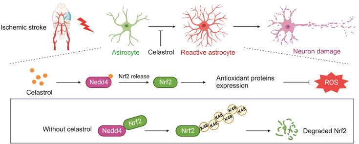
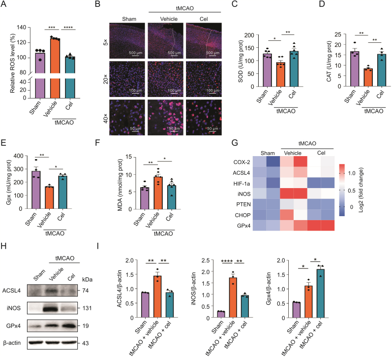
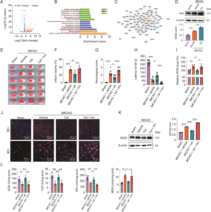
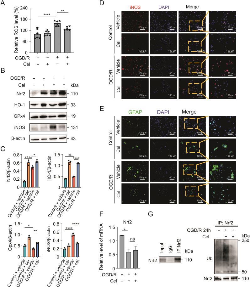
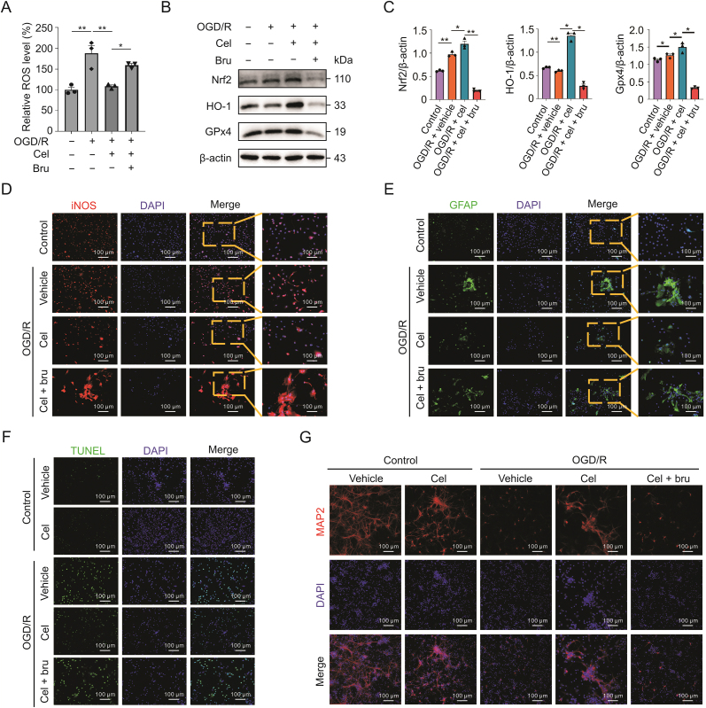
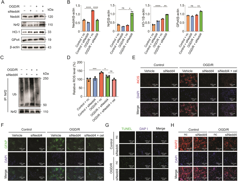
Stroke is the second leading cause of death worldwide, and oxidative stress plays a crucial role. Celastrol exhibits strong antioxidant properties in several diseases; however, whether it can affect oxidation in cerebral ischemic-reperfusion injury (CIRI) remains unclear. This study aimed to determine whether celastrol could reduce oxidative damage during CIRI and to elucidate the underlying mechanisms. Here, we found that celastrol attenuated oxidative injury in CIRI by upregulating nuclear factor E2-related factor 2 (Nrf2). Using alkynyl-tagged celastrol and liquid chromatography-tandem mass spectrometry, we showed that celastrol directly bound to neuronally expressed developmentally downregulated 4 (Nedd4) and then released Nrf2 from Nedd4 in astrocytes. Nedd4 promoted the degradation of Nrf2 through K48-linked ubiquitination and thus contributed to astrocytic reactive oxygen species production in CIRI, which was significantly blocked by celastrol. Furthermore, by inhibiting oxidative stress and astrocyte activation, celastrol effectively rescued neurons from axon damage and apoptosis. Our study uncovered Nedd4 as a direct target of celastrol, and that celastrol exerts an antioxidative effect on astrocytes by inhibiting the interaction between Nedd4 and Nrf2 and reducing Nrf2 degradation in CIRI.

Keywords: Celastrol; Cerebral ischemia; Nedd4; Nrf2; Oxidative stress; Reperfusion injury; Ubiquitylation.


Conflict of interest statement

The authors declare that there are no conflicts of interest.

Figures

Image 1
Graphical abstract
Fig. 1
Fig. 1
Celastrol significantly reduced cerebral infarction area in transient middle cerebral artery occlusion (tMCAO) mice. (A) Source and chemical structure of celastrol. (B) Overall scheme of the experiment design. (C and D) TTC staining of the brain slices and the percentage of the infarct volume. (E) hematoxylin and eosin (HE) and terminal deoxynucleotidyl transferase dUTP nick-end labeling (TUNEL) staining of brain sections. (F) Percentage of dead cells in the ischemic hemisphere. (G) Quantification of TUNEL-positive cells in the ischemic hemisphere. (H) Neurological deficit score of the mice. (I) Assessment of motor function by rotarod test. i.p.: intraperitoneal injection; cel: celastrol. All data shown are the means ± standard error of the mean (n = 7). P < 0.05, ∗∗P < 0.01, ∗∗∗P < 0.001, and ∗∗∗∗P < 0.0001.
Fig. 2
Fig. 2
Celastrol attenuated the oxidative stress after cerebral ischemic-reperfusion injury (CIRI). (A) Level of reactive oxygen species (ROS) in the brain tissues. (B) Immunostaining of inducible nitric oxide synthase (iNOS) in penumbra area. (C–F) Assessment of superoxide dismutase (SOD), catalase (CAT), glutathione peroxidase (GPx), and malondialdehyde (MDA) in brain tissues. (G) Heatmap presenting differentially expressed genes (DEGs) related to oxidative stress in different groups: each group involved two biological replicates containing three mice. (H and I) Protein expression of ACSL4, iNOS, and GPx4 in different groups measured by Western blotting analysis. All data shown are the means ± standard error of the mean (n = 3–6). cel: celastrol; tMCAO: transient middle cerebral artery occlusion; HIF-1α: hypoxia-inducible factor-1alpha; PTEN: phosphatase and tensin homolog; CHOP: CCAAT/enhancer-binding protein homologous protein. P < 0.05, ∗∗P < 0.01, ∗∗∗P < 0.001, and ∗∗∗∗P < 0.0001.
Fig. 3
Fig. 3
Celastrol alleviates oxidative damage via upregulating nuclear factor E2-related factor 2 (Nrf2). (A) Volcano plot analysis of the differentially expressed genes (DEGs) between celastrol-treated versus vehicle-treated mice after transient middle cerebral artery occlusion (tMCAO). Upregulated, downregulated, and non-differentially expressed genes are presented in red, blue, and gray dots, respectively. (B) Gene Ontology (GO) enrichment analysis of DEGs. BP: biological process; MF: molecular function; CC: cellular component. (C) Protein-protein interaction network among the DEGs. Nrf2 (gene name: Nfe2l2) is highlighted. (D) Immunoblotting showed protein expression of Nrf2 in vivo. (E and F) The 2,3,5-triphenyltetrazolium chloride (TTC) staining of the brain slices and the percentage of the infarct volume (n = 6). (G) Neurological scores of the mice (n = 7–9). (H) Assessment of motor function by the rotarod test (n = 7–9). (I) Effect of brusatol on the production of reactive oxygen species (ROS) after cerebral ischemic-reperfusion injury (CIRI). (J) Immunostaining showing the change of inducible nitric oxide synthase (iNOS) in penumbra area after inhibiting Nrf2. (K) Immunoblotting showing the protein level of iNOS in vivo. (L) Assessment of superoxide dismutase (SOD), catalase (CAT), glutathione peroxidase (GPx), and malondialdehyde (MDA) in brusatol-treated mice after CIRI, each group consisted of four to five mice. All data shown are the means ± standard error of the mean. cel: celastrol; bru: brusatol. P < 0.05, ∗∗P < 0.01, ∗∗∗P < 0.001, and ∗∗∗∗P < 0.0001.
Fig. 4
Fig. 4
Celasrol reduces the oxidative stress by decreasing the ubiquitylation of nuclear factor E2-related factor 2 (Nrf2) in astrocytes. (A) Effects of celastrol on reactive oxygen species (ROS) levels in astrocytes after oxygen-glucose deprivation/reoxygenation (OGD/R). (B and C) Protein levels of Nrf2, HO-1, glutathione peroxidase 4 (GPx4), and inducible nitric oxide synthase (iNOS) after celastrol treatment. (D) Immunostaining showing iNOS expression in astrocytes. (E) Activation of astrocytes after OGD/R following celastrol treatment. (F) The mRNA level of Nrf2 in astrocytes treated with celastrol. (G) Ubiquitinoylation of Nrf2 in astrocytes following celastrol treatment 24 h after reperfusion. Data shown are the means ± standard error of the mean. cel: celastrol; DAPI: 4',6-diamidino-2-phenylindole; GFAP: glial fibrillary acid protein. P < 0.05, ∗∗P < 0.01, and ∗∗∗∗P < 0.0001.
Fig. 5
Fig. 5
Inhibiting nuclear factor E2-related factor 2 (Nrf2) weakened the antioxidant effect of celastrol in astrocytes. (A) Effect of Nrf2 inhibition on the reactive oxygen species (ROS) in astrocytes after oxygen-glucose deprivation/reoxygenation (OGD/R). (B and C) Protein levels of Nrf2, HO-1, and glutathione peroxidase 4 (GPx4) after inhibition of Nrf2. (D) Expression of inducible nitric oxide synthase (iNOS) in astrocytes after inhibition of Nrf2. (E) Activation of astrocytes after OGD/R with inhibition of Nrf2. (F) Terminal deoxynucleotidyl transferase dUTP nick-end labeling (TUNEL) assays of neurons cocultured with astrocytes treated with celastrol treatment after OGD/R. (G) Microtubule associated protein 2 (MAP2) staining reflected damage to axons. Data are shown as the means ± standard error of the mean. cel: celastrol; bru: brusatol; DAPI: 4',6-diamidino-2-phenylindole; GFAP: glial fibrillary acid protein. P < 0.05 and ∗∗P < 0.01.
Fig. 6
Fig. 6
Celastrol bound to neuronally expressed developmentally downregulated 4 (Nedd4) and inhibited the ubiquitination of nuclear factor E2-related factor 2 (Nrf2) in astrocytes. (A) Structural formula of labeled celastrol. (B) Cellular thermal shift assay (CETSA) showing that celastrol bound to Nedd4 and reduced Nedd4 degradation with increasing temperatures. (C) Immunofluorescence showing the colocalization of celastrol-probe (cel-p) and Nedd4. (D and E) Western blot analysis of Nrf2, HO-1, and glutathione peroxidase 4 (GPx4) after Nedd4 overexpression (oe) in astrocytes. (F) Co-immunoprecipitation revealing the effect of celastrol on the binding between Nedd4 and Nrf2. (G) Immunofluorescence showing the binding of Nedd4 and Nrf2 in celastrol-treated astrocytes. Data shown are the means ± standard error of the mean. cel: celastrol; DAPI: 4',6-diamidino-2-phenylindole; nc: negative control. P < 0.05, ∗∗P < 0.01, and ∗∗∗P < 0.001.
Fig. 7
Fig. 7
Knockdown of neuronally expressed developmentally downregulated 4 (Nedd4) attenuated the oxidative stress by reducing the ubiquitinoylation of nuclear factor E2-related factor 2 (Nrf2). (A and B). Western blot analysis of the protein levels of Nrf2, HO-1, and glutathione peroxidase 4 (GPx4) after knockdown of Nedd4 (siNedd4). (C) Ubiquitinoylation of Nrf2 in astrocytes after knockdown of Nedd4 by transfecting siRNA. (D) Effect of inhibiting Nedd4 on the reactive oxygen species (ROS) of astrocytes after oxygen-glucose deprivation/reoxygenation (OGD/R). (E) Immunostaining revealing the expression of inducible nitric oxide synthase (iNOS) in astrocytes. (F) Glial fibrillary acid protein staining showing the activation of astrocytes. (G) Terminal deoxynucleotidyl transferase dUTP nick-end labeling (TUNEL) assay of neurons cocultured with astrocytes with Nedd4 knockdown after OGD/R. (H) Microtubule associated protein 2 (MAP2) staining reflecting the axonal damage. Data shown are the means ± standard error of the mean. cel: celastrol; IP: immunoprecipitation; Ub: ubiquitin; nc: negative control; DAPI: 4',6-diamidino-2-phenylindole; GFAP: glial fibrillary acid protein. P < 0.05, ∗∗P < 0.01, ∗∗∗P < 0.001, and ∗∗∗∗P < 0.0001.
Fig. 8
Fig. 8
Neuronally expressed developmentally downregulated 4 (Nedd4) catalyzed K48-linked ubiquitination of nuclear factor E2-related factor 2 (Nrf2). (A) Immunoblotting (IB) analysis of Nedd4 and Nrf2 respectively in human embryonic kidney (HEK) 293 T cells transfected with indicated DNA constructs (1, 3, and 5 μg). (B) Expression of Nrf2 in HEK 293 T cells co-transfected with Nrf2 (3 μg) and Nedd4 (1, 2, and 4 μg). (C) IB analysis of the level of Nrf2 in total protein and the ubiquitination of Nrf2 in HEK 293 T cells co-transfected with plasmids expressing Nrf2, Nedd4, and wild-type Ub, Ub-K48, or Ub-K63. IB analysis of ubiquitination of Nrf2 in (D) HEK 293 T and (E) C8-D1A cells when co-transfected with plasmids expressing Nrf2, Nedd4, and wild-type Ub or Ub-K48 upon celastrol treatment. Data shown are the means ± standard error of the mean. Ub: ubiquitin; cel: celastrol; IP: immunoprecipitation; WCL: whole cell lysate. ∗∗P < 0.01, ∗∗∗P < 0.001, and ∗∗∗∗P < 0.0001.

Similar articles

Cited by

References

    1. GBD 2019 Stroke Collaborators Global, regional, and national burden of stroke and its risk factors, 1990–2019: A systematic analysis for the Global Burden of Disease Study 2019. Lancet Neurol. 2021;20:795–820. - PMC - PubMed
    1. Kuriakose D., Xiao Z. Pathophysiology and treatment of stroke: Present status and future perspectives. Int. J. Mol. Sci. 2020;21:7609. - PMC - PubMed
    1. Sun M.-S., Jin H., Sun X., et al. Free radical damage in ischemia-reperfusion injury: An obstacle in acute ischemic stroke after revascularization therapy. Oxid. Med. Cell. Longev. 2018;2018 - PMC - PubMed
    1. Lin K., Zhang Z., Zhang Z., et al. Oleanolic acid alleviates cerebral ischemia/reperfusion injury via regulation of the gsk-3β/HO-1 signaling pathway. Pharmaceuticals (Basel) 2021;15:1. - PMC - PubMed
    1. Dringen R. Metabolism and functions of glutathione in brain. Prog. Neurobiol. 2000;62:649–671. - PubMed

LinkOut - more resources