. 2023 Mar 1;12(5):784.
doi: 10.3390/cells12050784.

Context-Dependent Role of Glucocorticoid Receptor Alpha and Beta in Breast Cancer Cell Behaviour

Affiliations

Context-Dependent Role of Glucocorticoid Receptor Alpha and Beta in Breast Cancer Cell Behaviour

Henriett Butz et al. Cells. .

Abstract

Background. The dual role of GCs has been observed in breast cancer; however, due to many concomitant factors, GR action in cancer biology is still ambiguous. In this study, we aimed to unravel the context-dependent action of GR in breast cancer. Methods. GR expression was characterized in multiple cohorts: (1) 24,256 breast cancer specimens on the RNA level, 220 samples on the protein level and correlated with clinicopathological data; (2) oestrogen receptor (ER)-positive and -negative cell lines were used to test for the presence of ER and ligand, and the effect of the GRβ isoform following GRα and GRβ overexpression on GR action, by in vitro functional assays. Results. We found that GR expression was higher in ER- breast cancer cells compared to ER+ ones, and GR-transactivated genes were implicated mainly in cell migration. Immunohistochemistry showed mostly cytoplasmic but heterogenous staining irrespective of ER status. GRα increased cell proliferation, viability, and the migration of ER- cells. GRβ had a similar effect on breast cancer cell viability, proliferation, and migration. However, the GRβ isoform had the opposite effect depending on the presence of ER: an increased dead cell ratio was found in ER+ breast cancer cells compared to ER- ones. Interestingly, GRα and GRβ action did not depend on the presence of the ligand, suggesting the role of the "intrinsic", ligand-independent action of GR in breast cancer. Conclusions. Staining differences using different GR antibodies may be the reason behind controversial findings in the literature regarding the expression of GR protein and clinicopathological data. Therefore, caution in the interpretation of immunohistochemistry should be applied. By dissecting the effects of GRα and GRβ, we found that the presence of the GR in the context of ER had a different effect on cancer cell behaviour, but independently of ligand availability. Additionally, GR-transactivated genes are mostly involved in cell migration, which raises GR's importance in disease progression.

Keywords: breast cancer; breast cancer progression; glucocorticoid receptor; glucocorticoid receptor alpha; glucocorticoid receptor beta; metastasis; migration; proliferation.


Conflict of interest statement

The authors declare no conflict of interest.

Figures

Figure 1
Figure 1
Mechanism of action of glucocorticoid receptor alpha (GRα) and beta (GRβ) in breast cancer cell. GR activity is strongly context-dependent, and determined by, among others, GR expression, splicing resulting in splice isoforms, posttranslational modifications and nuclear receptor crosstalk [8,9,10,23,24]. GRα activation can be ligand-dependent or -independent. By translocating into the nucleus, it binds to specific regulatory parts of the DNA (GR responsive elements, GREs) through which several genes’ expressions are induced or repressed in a cell type-specific manner. GRα and ER coactivation enhanced GRα binding to both GRE and oestrogen-responsive element (ERE), leading to an increased expression of pro-differentiating genes and negative regulators of pro-oncogenic Wnt signaling, and a decreased expression of epithelial–mesenchymal transition (EMT)-related genes. However, in the absence of ER, ligand-bound GRα binds to the GREs of several pro-tumourigenic genes, driving drug resistance and progression in TNBC (see details in the text, and in [6,23]). GRβ, due to its shorter sequence, cannot bind the ligand, but it is able to form a heterodimer with GRα. By binding to GREs, GRβ impairs GRα-mediated genomic actions, which is called the dominant–negative effect. While it has been described that GRβ is able to regulate proliferation and migration in other cell types, there is no clear evidence for its role in breast cancer development and progression (see details in the text, and in [22]).
Figure 2
Figure 2
Glucocorticoid receptor expression characterization in different normal tissue types (A) and normal breast tissue (B). ****: p < 0.0001.
Figure 3
Figure 3
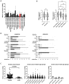
Glucocorticoid receptor expression in different cancer tissue types (A). (B) Immunostaining characteristics of GR using two commercially available antibodies (HPA004248: Cat#HPA004248, Atlas Antibodies; CAB010435: Cat#sc-8992, Santa Cruz Biotechnology). (C) Glucocorticoid receptor expression in female and male breast cancers; (D) GR encoding NR3C1 gene expression in primary vs. metastatic and in different subtypes of breast cancer cells. **: p < 0.01; ns: not significant.
Figure 4
Figure 4
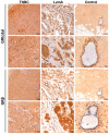
Representative images of glucocorticoid receptor protein staining in normal breast (control), triple-negative (TNBC) and oestrogen-positive (luminal A type, LumA) breast cancer, counterstained by hematoxylin. Tumour tissues show great variance in terms of the immunostaining pattern of GR. We show here the most intensively stained samples from both TNBC and LumA in each group.
Figure 5
Figure 5
Examples of the cytoplasmic (panel A) and mixed cytoplasmic+nuclear (panel B) staining patterns of the GRtotal and GRβ proteins. In Panel B, some nuclei are positive and some of them are negative for staining in the tumour tissue. The line indicates 50 μm. On this representative image, all tumours are of the triple-negative subtype.
Figure 6
Figure 6
(A) Discrimination of GRtotal and GRβ isoforms using Western blot. (B) Representative images of GRtotal and GRβ endogenous expression in triple-negative and ER+ breast cancer cells. (C) Densitometry of GRtotal and GRβ Western blot performed on triple-negative (S578T and MDA-MB231) and ER+ breast cancer cells (T47D and ZR751). Relative densities indicate GRtotal/actin and GRβ/actin values. *: p < 0.05; **: p < 0.01; ns: not significant.
Figure 7
Figure 7
Investigating viability (A), cell proliferation (B), and dead cell ratio (C) following GRα and GRβ transfection in the presence or absence of the ligand in triple-negative (HS578T and MDA-MB231) and ER+ breast cancer cells (T47D and ZR751). *: p < 0.05; **: p < 0.01; ***: p < 0.001; ****: p < 0.0001.
Figure 8
Figure 8
Representative cell migration images of triple-negative ((A) HS578T and (B) MDA-MB231) and ER+ breast cancer cells ((C) T47D) following GRα and GRβ overexpression. Photos were taken using 3.2× objective at 6–12 and 24–72 h following wounding.
Figure 9
Figure 9
Time-lapse (A,B) and comparative (C) results of cell migration of triple-negative ((A) HS578T and (B) MDA-MB231) and ER+ breast cancer cells ((C) T47D) following GRα and GRβ overexpression in the presence and the absence of the ligand. *: p < 0.05; **: p < 0.01; ***: p < 0.001; ****: p < 0.0001; ns: not significant.
Figure 10
Figure 10
Chord diagram of gene ontology biological process gene set enrichment of genes positively correlated with NR3C1 expression in breast cancer. The length of the element is proportional to the number of genes related to the GO term. The edges inside the chord diagram between the two elements denote the fact that there are common genes between them. Colours indicate p-values according to the scale. Red highlights the most significant biological process enhanced by GR action.

Similar articles

References

    1. Sung H., Ferlay J., Siegel R.L., Laversanne M., Soerjomataram I., Jemal A., Bray F. Global Cancer Statistics 2020: GLOBOCAN Estimates of Incidence and Mortality Worldwide for 36 Cancers in 185 Countries. CA A Cancer J. Clin. 2021;71:209–249. doi: 10.3322/caac.21660. - DOI - PubMed
    1. Nelson D.R., Brown J., Morikawa A., Method M. Breast Cancer-Specific Mortality in Early Breast Cancer as Defined by High-Risk Clinical and Pathologic Characteristics. PLoS ONE. 2022;17:e0264637. doi: 10.1371/journal.pone.0264637. - DOI - PMC - PubMed
    1. Cardoso F., Senkus E., Costa A., Papadopoulos E., Aapro M., André F., Harbeck N., Aguilar Lopez B., Barrios C.H., Bergh J., et al. 4th ESO–ESMO International Consensus Guidelines for Advanced Breast Cancer (ABC 4) Ann. Oncol. 2018;29:1634–1657. doi: 10.1093/annonc/mdy192. - DOI - PMC - PubMed
    1. Tsang J.Y.S., Tse G.M. Molecular Classification of Breast Cancer. Adv. Anat. Pathol. 2020;27:27–35. doi: 10.1097/PAP.0000000000000232. - DOI - PubMed
    1. Lamb C.A., Vanzulli S.I., Lanari C. Hormone Receptors in Breast Cancer: More than Estrogen Receptors. Medicina. 2019;79:540–545. - PubMed

Publication types

Grants and funding

This work was supported by a Hungarian Scientific Research Grant of the National Research, Development and Innovation Office NKFI FK 135065, the New National Excellence Program of the Ministry of Human Capacities (UNKP-22-5-SE-1) given to H.B., and by the Ministry of Innovation and Technology of Hungary from the National Research, Development and Innovation Fund, financed under the TKP2021-EGA/TKP2021-NVA/TKP2021-NKTA funding scheme, given to A.P. H.B. is a recipient of the Bolyai Research Fellowship of the Hungarian Academy of Sciences. H.B. and A.P. acknowledge financial support from the National Laboratories Excellence program (under the National Tumour Biology Laboratory project (NLP17)) and the Hungarian Thematic Excellence Programme (TKP2021-EGA-44).