Review
. 2023 Feb 17:11:1112319.
doi: 10.3389/fcell.2023.1112319. eCollection 2023.

Septins as membrane influencers: direct play or in association with other cytoskeleton partners

Affiliations
Review

Septins as membrane influencers: direct play or in association with other cytoskeleton partners

Béatrice Benoit et al. Front Cell Dev Biol. .

Abstract

The cytoskeleton comprises three polymerizing structures that have been studied for a long time, actin microfilaments, microtubules and intermediate filaments, plus more recently investigated dynamic assemblies like septins or the endocytic-sorting complex required for transport (ESCRT) complex. These filament-forming proteins control several cell functions through crosstalks with each other and with membranes. In this review, we report recent works that address how septins bind to membranes, and influence their shaping, organization, properties and functions, either by binding to them directly or indirectly through other cytoskeleton elements.

Keywords: ESCRT; actin; cytoskeleton; intermediate filament; membrane; microtubule; septin.


Conflict of interest statement

The authors declare that the research was conducted in the absence of any commercial or financial relationships that could be construed as a potential conflict of interest.

Figures
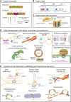
FIGURE 1
FIGURE 1
Septin structure, assembly and functions related to membranes. (A) Assembly of the septin cytoskeleton. In humans, the septin family comprises 13 members organized into 4 groups (2, 6, 7 and 3) according to their sequence homology. The organization of the four septin group into hexamers and octamers is depicted. Septin monomers either interact through their N- and C-terminal domains (NC/NC interfaces), or through their GTP-binding domain (G/G interfaces). These oligomers can then assemble into filaments or into rings. (B) Structural domains encountered in septin proteins. All the septins exhibit a GTP-binding domain (GBD), a septin unique element (SUE), and N- and C-terminal extensions (NTE, CTE) of variable length. Note that the CTE, which contains one or more coiled-coil regions is absent in group 3 septins. Membrane binding is mediated by two AH (amphipathic helix) and PB (polybasic domain) motifs. SEPT9 exhibits physical binding to F-actin through the first half of its NTE, and to microtubules through its N-terminal K/R-R/x-x-D/E repeat motifs. (C) Septin molecular functions. At the molecular level, septins can form diffusion barriers or act as scaffolds, as illustrated here in the context of binding to a positively curved membrane. As a barrier, septins can either restrict entry or concentrate molecules within a given domain. (D) Septins interact with lipids or proteins at membranes. Septins are enriched at positively curved membranes by binding to phosphoinositides. Through binding to other specific lipids (cardiolipin, Gb3) in bacteria, and through binding to very long chain fatty acid-bearing phosphoinositides in M. orizae fungus, septins ultimately regulate pathogen invasion. In yeast, the membrane recruitment of septins follows high concentrations of active Cdc42, directing further outgrowth of the bud. Through binding to specific proteins (Clathrin, Caveolin, FABP5, FSIT2, Anillin or GM130), septins are involved in a large array of cell processes in higher eukaryotes, including endocytosis, lipid storage, myelin sheet assembly, Golgi integrity and function in nucleating microtubules. (E) Septins can act as barriers or scaffolds at membranes. SEPT7 phosphorylation mediates PSD95 stability and restricts its mobility to favor dendritic maturation. At the base of primary cilia, they control protein entry and retain signaling receptors. Septins also organize endoplasmic reticulum-plasma membrane junctions for STIM1-Orai1 calcium signaling. Septins are regulators of autophagy, especially in yeast and neurons, where an interaction with Atg8 has been observed. SEPT2 can recruit Drp1 to mitochondrial constriction sites to favor Drp1-mediated fission. Septins also contribute to control vesicular trafficking machinery at several steps, including the SNARE-mediated membrane fusion.
FIGURE 2
FIGURE 2
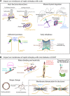
Impact on membranes of septin interplay with other cytoskeletons (A) Actin-related impacts. While facilitating cell invasion of specific bacteria, septins can also limit invasion either by enhancing host cell rigidity or by favoring autophagy-mediated degradation once bacteria have been internalized. During mesenchymal migration, septins contribute to the stabilization of nascent focal adhesions and to extracellular matrix degradation through metalloproteinase secretion. Septin-mediated enhancement of actomyosin contractility ultimately increase cell tension forces, thereby impacting the tumoral environment and modulating mechano-responsiveness. By linking the catenin complex to the actin cytoskeleton, septins favor cell-cell junctions required for the apico- basal polarity. During cytokinesis, membrane-associated septins and anillin are required to anchor the actomyosin contractile ring to the cell cortex. (B) Microtubule- and ESCRT-related impacts. Septins control the binding and motricity of molecular motors, thereby regulating the retrograde and anterograde transport on microtubules of proteins or vesicles, as illustrated with the clustering of lysosomes during cell adaptation to stress, the sorting of kinesin motor cargoes during entry into dendrites, or the control of cilia over-elongation. By guiding non-centrosomal Golgi-based microtubules, septins are also involved in focal adhesion disassembly in migrating cells. Delocalization of septins from microtubules impacts cell shape, as illustrated in platelets. During cytokinesis, membrane-bound septin rings demarcate the membrane sites for ESCRT III ring assembly prior to final abscission.

Similar articles

Cited by

References

    1. Addi C., Bai J., Echard A. (2018). Actin, microtubule, septin and ESCRT filament remodeling during late steps of cytokinesis. Curr. Opin. Cell Biol. 50, 27–34. 10.1016/j.ceb.2018.01.007 - DOI - PubMed
    1. Ageta-Ishihara N., Yamazaki M., Konno K., Nakayama H., Abe M., Hashimoto K., et al. (2015). A CDC42EP4/septin-based perisynaptic glial scaffold facilitates glutamate clearance. Nat. Commun. 6, 10090. 10.1038/ncomms10090 - DOI - PMC - PubMed
    1. Aigal S., Omidvar R., Stober K., Ziegelbauer J., Eierhoff T., Schampera J. N., et al. (2022). Septin barriers protect mammalian host cells against Pseudomonas aeruginosa invasion. Cell Rep. 41, 111510. 10.1016/j.celrep.2022.111510 - DOI - PubMed
    1. Arbizzani F., Mavrakis M., Hoya M., Ribas J. C., Brasselet S., Paoletti A., et al. (2022). Septin filament compaction into rings requires the anillin Mid2 and contractile ring constriction. Cell Rep. 39, 110722. 10.1016/j.celrep.2022.110722 - DOI - PubMed
    1. Au Yong J. Y. A., Wang Y.-M., Wang Y. (2016). The Nim1 kinase Gin4 has distinct domains crucial for septin assembly, phospholipid binding and mitotic exit. J. Cell Sci. 129, 2744–2756. 10.1242/jcs.183160 - DOI - PMC - PubMed

Grants and funding

This work was supported by the Ministère de l’Enseignement Supérieur et de la Recherche and by the Institut National de la Santé et de la Recherche Médicale.

LinkOut - more resources