. 2023 Feb 21:26:52-63.
doi: 10.1016/j.bioactmat.2023.02.009. eCollection 2023 Aug.

Targeted PERK inhibition with biomimetic nanoclusters confers preventative and interventional benefits to elastase-induced abdominal aortic aneurysms

Affiliations

Targeted PERK inhibition with biomimetic nanoclusters confers preventative and interventional benefits to elastase-induced abdominal aortic aneurysms

Nisakorn Yodsanit et al. Bioact Mater. .

Abstract

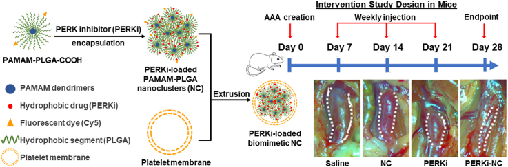
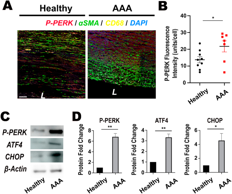
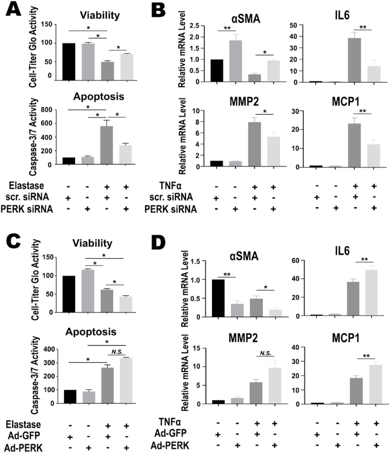
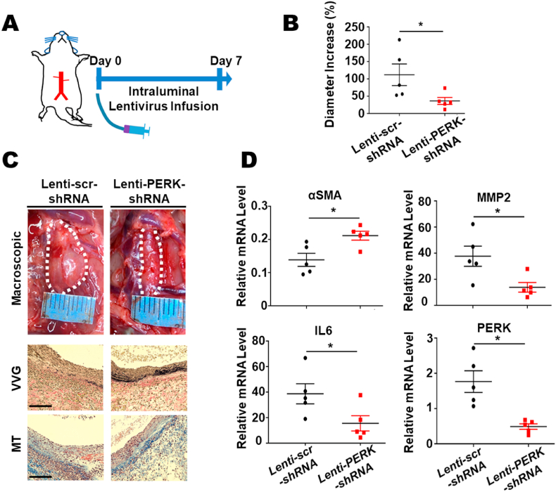
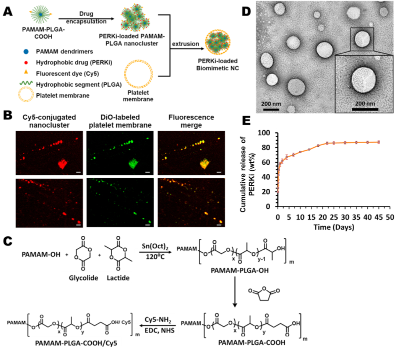
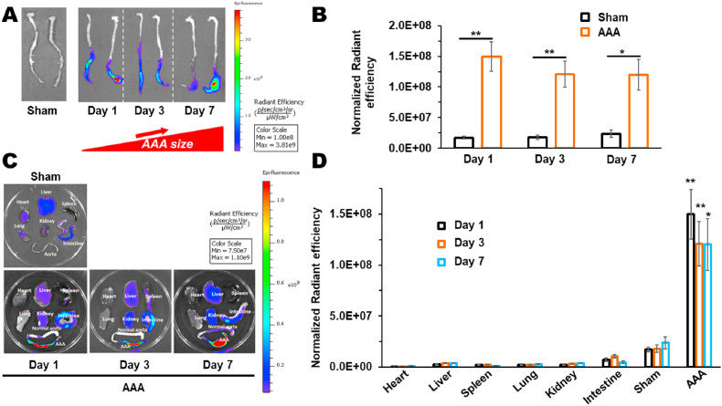
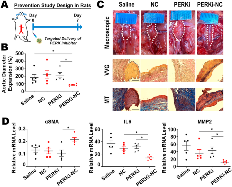
Abdominal aortic aneurysm (AAA) is a progressive aortic dilatation, causing ∼80% mortality upon rupture. Currently, there is no approved drug therapy for AAA. Surgical repairs are invasive and risky and thus not recommended to patients with small AAAs which, however, account for ∼90% of the newly diagnosed cases. It is therefore a compelling unmet clinical need to discover effective non-invasive strategies to prevent or slow down AAA progression. We contend that the first AAA drug therapy will only arise through discoveries of both effective drug targets and innovative delivery methods. There is substantial evidence that degenerative smooth muscle cells (SMCs) orchestrate AAA pathogenesis and progression. In this study, we made an exciting finding that PERK, the endoplasmic reticulum (ER) stress Protein Kinase R-like ER Kinase, is a potent driver of SMC degeneration and hence a potential therapeutic target. Indeed, local knockdown of PERK in elastase-challenged aorta significantly attenuated AAA lesions in vivo. In parallel, we also conceived a biomimetic nanocluster (NC) design uniquely tailored to AAA-targeting drug delivery. This NC demonstrated excellent AAA homing via a platelet-derived biomembrane coating; and when loaded with a selective PERK inhibitor (PERKi, GSK2656157), the NC therapy conferred remarkable benefits in both preventing aneurysm development and halting the progression of pre-existing aneurysmal lesions in two distinct rodent models of AAA. In summary, our current study not only establishes a new intervention target for mitigating SMC degeneration and aneurysmal pathogenesis, but also provides a powerful tool to facilitate the development of effective drug therapy of AAA.

Keywords: Abdominal aortic aneurysm; Biomimetic nanomedicine; ER stress; PERK; Targeted delivery.


Conflict of interest statement

The authors declare that they have no known competing financial interests or personal relationships that could have appeared to influence the work reported in this paper.

Figures

Image 1
Graphical abstract
Fig. 1
Fig. 1
The PERK Pathway is Activated in Clinical and Experimental Specimens of AAA. (A) Immunofluorescence co-staining of phospho-PERK (P-PERK, Thr980), SMC marker αSMA, and macrophage marker CD68 on sections of healthy aorta versus AAA lesions from de-identified human donors (note loss of SMC contents in AAA specimens). L: luminal side. (B) Quantification of P-PERK fluorescent intensity in human specimens. Mean ± SEM, n = 7-9. *P < 0.05. Unpaired Student's t-test. (C) Representative images of immunoblots for PERK pathway proteins (P-PERK, ATF4, CHOP) using tissue homogenates from healthy rat aorta versus elastase-induced AAA specimens. (D) Quantitation of P-PERK, ATF4, and CHOP levels based on immunoblot densitometry after normalization to β-Actin. Mean ± SEM, n = 4. *P < 0.05, **P < 0.01. Paired Student's t-test.
Fig. 2
Fig. 2
PERK Loss-of-function Reverses, its Gain-of-function Exacerbates SMC Degeneration In Vitro. To determine the specific role of PERK in driving the degenerative phenotype transition, human primary aortic SMCs were pretreated with either PERK siRNA (A, B) or overexpressing adenovirus (C, D) to achieve loss- and gain-of-function, respectively. SMCs were then subject to either transient exposure (5 min) to porcine elastase (∼1 unit/mL) to stimulate cell death (A, C), or TNFα (10 ng/mL) to induce the dedifferentiated/proteolytic/pro-inflammatory phenotypic transition. A–B: PERK silencing with siRNA. Starved SMCs were transfected with 50 nM PERK-specific siRNA for 48 h and then starved for 24 h prior to elastase or TNFα challenge. (A) Viability of SMCs was determined using CellTiter-Glo assay at 24 h post elastase challenge, and apoptotic index was determined using Caspase-Glo 3/7 assay at 4 h. (B) Quantitation of mRNA levels of inflammatory cytokine (IL6) and macrophage chemoattractant gene (MCP1), ECM proteolytic gene (MMP2), and SMC contractility gene (αSMA) in SMCs with PERK loss-of-function collected at 24 h post TNFα stimulation. (C–D) Adenovirus-mediated PERK gain-of-function. Cells were transduced with PERK-overexpressing adenovirus (Ad-PERK) or GFP control (Ad-GFP) for 6 h, recovered in complete medium for 24 h, followed by starvation for 24 h prior to elastase or TNFα challenge. Similar to A-B, SMC viability and apoptotic index (C), as well as quantitation of mRNA levels of representative genes of the proinflammatory, proteolytic, and SMC contractile genes (D), are presented. Mean ± SEM, n = 4-5; N.S. = no significance, *p < 0.05, **P < 0.01. One-way ANOVA with Tukey post-hoc analysis.
Fig. 3
Fig. 3
Local PERK Silencing Conferred Effective Control over Development of Elastase-induced AAA Lesions. Lentivirus expressing either scrambled control or PERK-specific shRNA was intraluminally infused immediately after the porcine elastase challenge to create AAA lesion. Aorta segments were collected at day 7 post the procedure for analysis. (A) Overview of experimental design. (B) Analysis of aortic diameter increase (fold change of maximal diameters at day 7 versus day 0 baseline). (C) Representative images of AAA lesions at macroscopic and microscopic levels (VVG: elastin staining shown as the black, wavy structure; MT: collagen staining shown in blue, and muscle content shown in red) at day 7 post procedure. Scale bar: 125 μm. (D) Tissue homogenate mRNA analysis of AAA-signature gene expression changes. Quantitation of mRNA levels of inflammatory cytokine genes (IL6), ECM proteolytic genes (MMP2), and SMC contractility gene (αSMA) in AAA segments from elastase-challenged aortic segments. Mean ± SEM, n = 5 rats; *p < 0.05. Mann-Whitney nonparametric test.
Fig. 4
Fig. 4
Design and Characterization of an AAA-targeting Biomimetic NC. (A) Overview/Illustration of experimental design. Drug-encapsulated biomimetic NC were prepared using extrusion method. (B) Confocal images demonstrating the successful coating process of biomimetic nanocluster. The merged fluorescence images show the colocalization of Cy5 and DiO which were labeled with PAMAM-PLGA-COOH and platelet membrane, respectively. Scale bars: 10 μm. (C) Synthesis schemes of PAMAM-PLGA-OH, PAMAM-PLGA-COOH and PAMAM-PLGA-COOH/Cy5. (D) TEM images of the platelet membrane-coated NC. Scale bars: 200 nm. (E) The in vitro release profile of PERKi from biomimetic NC in PBS (pH 7.4) with 2% tween80 at 37 °C. Mean ± SEM, n = 3.
Fig. 5
Fig. 5
Biodistribution of the biomimetic NC in a rat AAA model. Elastase-challenged rats or the sham control (perfused with heat-inactivated elastase) were intravenously injected with Cy5-loaded biomimetic NC. Aortas and other major organs from rats collected at day 1 (nascent AAAs), day 3 (small AAAs), and day 7 (fully established AAAs) were imaged using an IVIS spectrum luminescence system and were compared with those from the sham control. (A) Homing of the biomimetic NC to experimental AAA lesions of different stages. (B) Normalized radiant efficiency of AAA lesions (Fig. 5A) was determined based on Cy5 signal intensities captured (Ex/Em: 650/720 nm) after normalization to tissue weight. (C) Representative IVIS images of the major organs (heart, liver, spleen, lung, kidney, intestine, aorta). (D) Normalized radiant efficiency of major organs (Fig. 5C) was determined based on Cy5 signal intensities captured (Ex/Em: 650/720 nm) after normalization to tissue weight. Data are presented as mean ± SEM with n = 4 or 5 rats per group. Statistical analysis was performed using one-way ANOVA with Tukey's post hoc analysis. *P < 0.05, **P < 0.01 between the respective AAA and sham groups at each time point. Mean ± SEM, n = 4-5 rats; *P < 0.05, **P < 0.01. One-way ANOVA with Tukey post-hoc analysis.
Fig. 6
Fig. 6
Targeted Delivery of PERK Inhibitor Prevented AAA Formation. Targeted delivery of a selective PERK inhibitor prevented the elastase-induced aortic dilation and degeneration in a rat model. (A) Overview of experimental design. Immediately following the elastase-infusion procedure, rats were randomly assigned to a one-time intravenous injection with the following four treatments (all equivalent to 10 mg/kg payload): saline control (Saline), empty NC control (NC), free PERK inhibitor GSK2656157 (PERKi), and GSK2656157-encapsulated biomimetic NC (PERKi-NC). (B) Analysis of aortic diameter increase (fold change of maximal diameters at day 7 versus day 0 baseline). Mean ± SEM, n = 5-6 rats, *p < 0.05. One-way ANOVA with Tukey post-hoc analysis. (C) Representative images of AAA lesions at macroscopic and microscopic levels (VVG: elastin staining shown as the black, wavy structure; MT: collagen staining shown in blue, and muscle content shown in red) at day 7 post procedure. Scale bar: 125 μm. (D) Tissue homogenate mRNA analysis of AAA-signature gene expression changes. Quantitation of mRNA levels of inflammatory cytokine genes (IL6), ECM proteolytic genes (MMP2), and SMC contractility gene (αSMA) in AAA segments from elastase-challenged aortic segments. Mean ± SEM, n = 5 rats, *p < 0.05. One-way ANOVA with Tukey post-hoc analysis.
Fig. 7
Fig. 7
A Weekly Regimen of Targeted PERK Inhibition Therapy Halted the Growth of Pre-existing AAA lesions. (A) Overview of experimental design. A modified murine model was used to recapitulate the chronic disease progression of AAA. Ultrasound (abbreviated as US) imaging was performed at indicated time points as described in Supplementary Document. At day 7 post topical elastase application, mice with establish AAA lesions were randomly assigned to a weekly regimen with the following four treatments (all equivalent to 10 mg/kg payload): saline control (Saline), empty NC control (NC), free PERK inhibitor GSK2656157 (PERKi), and GSK2656157-encapsulated biomimetic NC (PERKi-NC). (B) Representative images of AAA lesions at macroscopic and microscopic levels (VVG: elastin staining shown as the black, wavy structure; MT: collagen staining shown in blue, and muscle content shown in red) at day 28 post procedure. Scale bar: 125 μm. (C) Analysis of aortic diameter increase (fold change of maximal diameters at day 28 versus day 0 baseline). Mean ± SEM, n = 10-12 mice. **P < 0.01. One-way ANOVA with Tukey post-hoc analysis. (D) Tissue homogenate mRNA analysis of AAA-signature gene expression changes. Quantitation of mRNA levels of inflammatory cytokine genes (IL6), ECM proteolytic genes (MMP2), and SMC contractility gene (αSMA) in AAA segments from elastase-challenged aortic segments. Mean ± SEM, n = 10 mice. *P < 0.05, **P < 0.01. One-way ANOVA with Tukey post-hoc analysis.

Similar articles

Cited by

References

    1. Sakalihasan N., Limet R., Defawe O.D. Abdominal aortic aneurysm. Lancet. Apr 30-May 6 2005;365(9470):1577–1589. doi: 10.1016/S0140-6736(05)66459-8. - DOI - PubMed
    1. Weintraub N.L. Understanding abdominal aortic aneurysm. N. Engl. J. Med. Sep 10 2009;361(11):1114–1116. doi: 10.1056/NEJMcibr0905244. - DOI - PMC - PubMed
    1. Lederle F.A., Kane R.L., MacDonald R., Wilt T.J. Systematic review: repair of unruptured abdominal aortic aneurysm. Ann. Intern. Med. May 15 2007;146(10):735–741. doi: 10.7326/0003-4819-146-10-200705150-00007. - DOI - PubMed
    1. Baxter B.T., Matsumura J., Curci J., et al. Non-invasive Treatment of Abdominal Aortic Aneurysm Clinical Trial (N-TA(3)CT): design of a Phase IIb, placebo-controlled, double-blind, randomized clinical trial of doxycycline for the reduction of growth of small abdominal aortic aneurysm. Contemp. Clin. Trials. May 2016;48:91–98. doi: 10.1016/j.cct.2016.03.008. - DOI - PMC - PubMed
    1. Baxter B.T., Matsumura J., Curci J.A., et al. Effect of doxycycline on aneurysm growth among patients with small infrarenal abdominal aortic aneurysms: a randomized clinical trial. JAMA. May 26 2020;323(20):2029–2038. doi: 10.1001/jama.2020.5230. - DOI - PMC - PubMed

LinkOut - more resources