CD19 CAR antigen engagement mechanisms and affinity tuning
- PMID: 36867678
- PMCID: PMC10228544
- DOI: 10.1126/sciimmunol.adf1426
CD19 CAR antigen engagement mechanisms and affinity tuning
Abstract
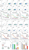
Chimeric antigen receptor (CAR) T cell therapy relies on T cells that are guided by synthetic receptors to target and lyse cancer cells. CARs bind to cell surface antigens through an scFv (binder), the affinity of which is central to determining CAR T cell function and therapeutic success. CAR T cells targeting CD19 were the first to achieve marked clinical responses in patients with relapsed/refractory B cell malignancies and to be approved by the U.S. Food and Drug Administration (FDA). We report cryo-EM structures of CD19 antigen with the binder FMC63, which is used in four FDA-approved CAR T cell therapies (Kymriah, Yescarta, Tecartus, and Breyanzi), and the binder SJ25C1, which has also been used extensively in multiple clinical trials. We used these structures for molecular dynamics simulations, which guided creation of lower- or higher-affinity binders, and ultimately produced CAR T cells endowed with distinct tumor recognition sensitivities. The CAR T cells exhibited different antigen density requirements to trigger cytolysis and differed in their propensity to prompt trogocytosis upon contacting tumor cells. Our work shows how structural information can be applied to tune CAR T cell performance to specific target antigen densities.
Conflict of interest statement
Competing interests:
The authors declare no competing interests.
Supplementary Materials
Materials and Methods
Figures


Similar articles
-
T-cells engineered with a novel VHH-based chimeric antigen receptor against CD19 exhibit comparable tumoricidal efficacy to their FMC63-based counterparts.Front Immunol. 2023 Feb 16;14:1063838. doi: 10.3389/fimmu.2023.1063838. eCollection 2023. Front Immunol. 2023. PMID: 36875091 Free PMC article.
-
Identification of Potent CD19 scFv for CAR T Cells through scFv Screening with NK/T-Cell Line.Int J Mol Sci. 2020 Dec 1;21(23):9163. doi: 10.3390/ijms21239163. Int J Mol Sci. 2020. PMID: 33271901 Free PMC article.
-
Complexities in comparing the impact of costimulatory domains on approved CD19 CAR functionality.J Transl Med. 2023 Jul 30;21(1):515. doi: 10.1186/s12967-023-04372-4. J Transl Med. 2023. PMID: 37518011 Free PMC article. Review.
-
Impact of Manufacturing Procedures on CAR T Cell Functionality.Front Immunol. 2022 Apr 13;13:876339. doi: 10.3389/fimmu.2022.876339. eCollection 2022. Front Immunol. 2022. PMID: 35493513 Free PMC article. Review.
-
Dual targeting of CD19 and CD22 against B-ALL using a novel high-sensitivity aCD22 CAR.Mol Ther. 2023 Jul 5;31(7):2089-2104. doi: 10.1016/j.ymthe.2023.03.020. Epub 2023 Mar 21. Mol Ther. 2023. PMID: 36945773 Free PMC article.
Cited by
-
Revolutionizing Cancer Treatment: Unleashing the Power of Combining Oncolytic Viruses with CAR-T Cells.Anticancer Agents Med Chem. 2024;24(19):1407-1418. doi: 10.2174/0118715206308253240723055019. Anticancer Agents Med Chem. 2024. PMID: 39051583 Review.
-
NK-92MI Cells Engineered with Anti-claudin-6 Chimeric Antigen Receptors in Immunotherapy for Ovarian Cancer.Int J Biol Sci. 2024 Feb 11;20(5):1578-1601. doi: 10.7150/ijbs.88539. eCollection 2024. Int J Biol Sci. 2024. PMID: 38481806 Free PMC article.
-
Programming CAR T Cell Tumor Recognition: Tuned Antigen Sensing and Logic Gating.Cancer Discov. 2023 Apr 3;13(4):829-843. doi: 10.1158/2159-8290.CD-23-0101. Cancer Discov. 2023. PMID: 36961206 Free PMC article.
-
Rational Protein Engineering to Enhance MHC-Independent T-cell Receptors.Cancer Discov. 2024 Nov 1;14(11):2109-2121. doi: 10.1158/2159-8290.CD-23-1393. Cancer Discov. 2024. PMID: 38980802
-
Trogocytosis of CAR molecule regulates CAR-T cell dysfunction and tumor antigen escape.Signal Transduct Target Ther. 2023 Dec 25;8(1):457. doi: 10.1038/s41392-023-01708-w. Signal Transduct Target Ther. 2023. PMID: 38143263 Free PMC article.
References
-
- Brentjens RJ, Latouche J-B, Santos E, Marti F, Gong MC, Lyddane C, King PD, Larson S, Weiss M, Rivière I, Sadelain M, Eradication of systemic B-cell tumors by genetically targeted human T lymphocytes co-stimulated by CD80 and interleukin-15. Nat. Med 9, 279–286 (2003). - PubMed
Publication types
MeSH terms
Substances
Grants and funding
LinkOut - more resources
Full Text Sources
Molecular Biology Databases

