. 2023 Feb 20:2023:2345279.
doi: 10.1155/2023/2345279. eCollection 2023.

Platelets Facilitate Wound Healing by Mitochondrial Transfer and Reducing Oxidative Stress in Endothelial Cells

Affiliations

Platelets Facilitate Wound Healing by Mitochondrial Transfer and Reducing Oxidative Stress in Endothelial Cells

Panshi Jin et al. Oxid Med Cell Longev. .

Abstract

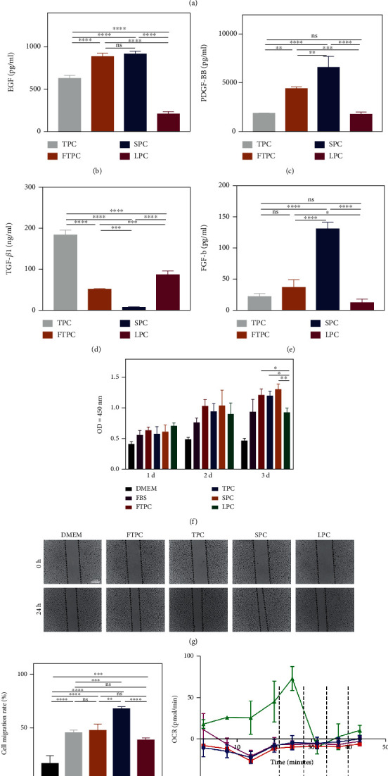
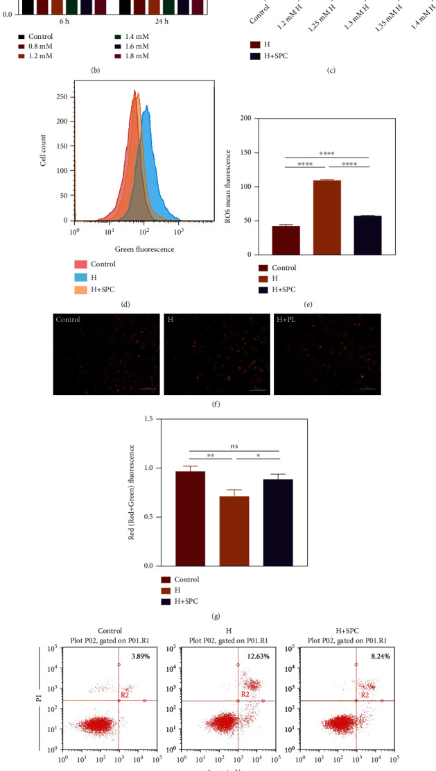
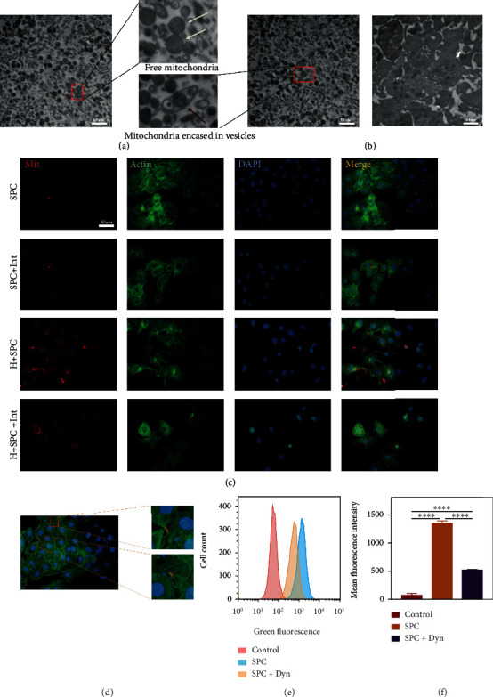
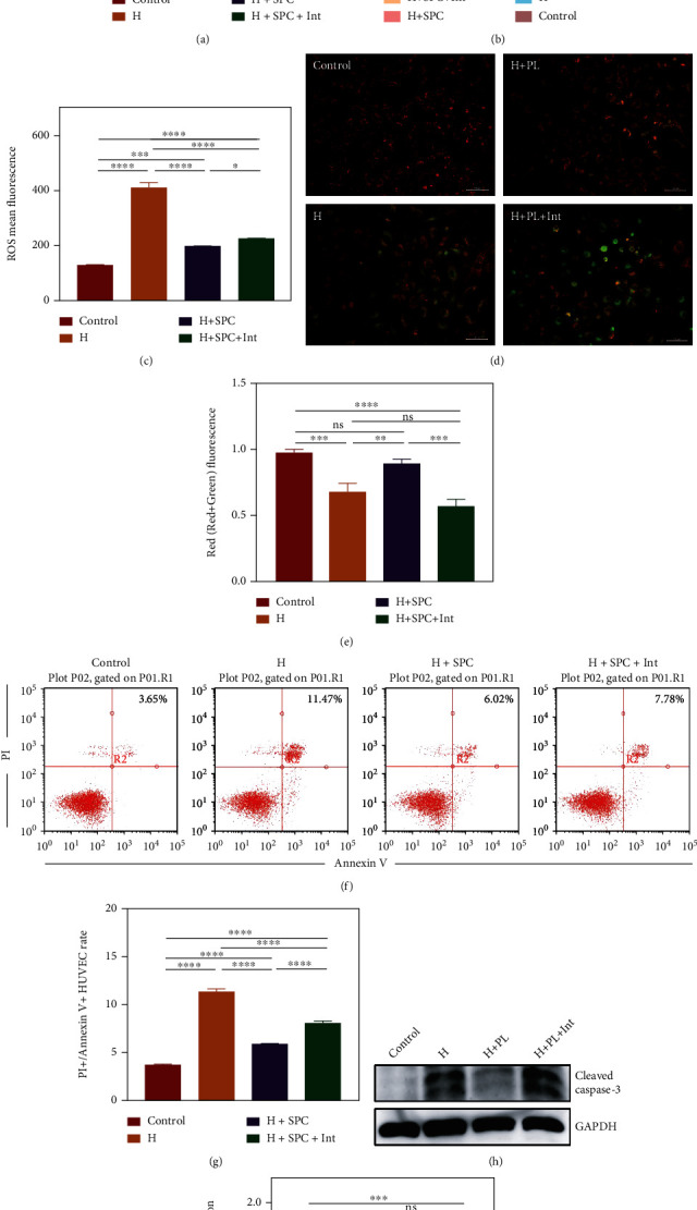
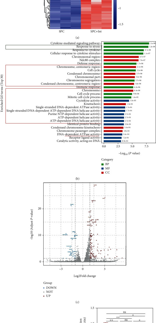
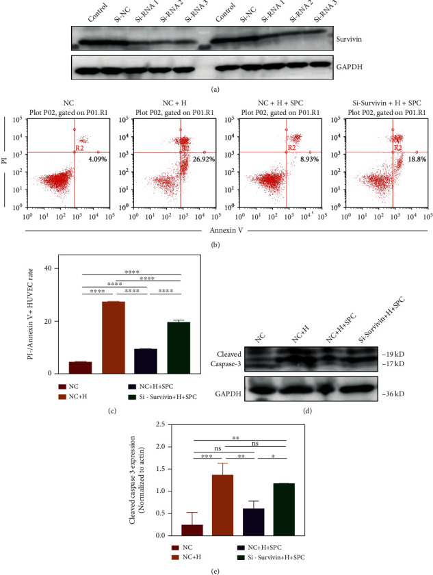
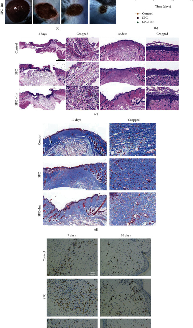
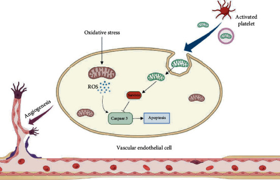
As a critical member in wound healing, vascular endothelial cells (ECs) impaired under high levels of reactive oxygen species (ROS) would hamper neovascularization. Mitochondria transfer can reduce intracellular ROS damage under pathological condition. Meanwhile, platelets can release mitochondria and alleviate oxidative stress. However, the mechanism by which platelets promote cell survival and reduce oxidative stress damage has not been clarified. Here, first, we selected ultrasound as the best method for subsequent experiments by detecting the growth factors and mitochondria released from manipulation platelet concentrates (PCs), as well as the effect of manipulation PCs on the proliferation and migration of HUVECs. Then, we found that sonicate platelet concentrates (SPC) decreased the level of ROS in HUVECs treated with hydrogen peroxide in advance, increased mitochondrial membrane potential, and reduced apoptosis. By transmission electron microscope, we saw that two kinds of mitochondria, free or wrapped in vesicles, were released by activated platelets. In addition, we explored that platelet-derived mitochondria were transferred to HUVECs partly by means of dynamin-dependent clathrin-mediated endocytosis. Consistently, we determined that platelet-derived mitochondria reduced apoptosis of HUVECs caused by oxidative stress. What is more, we screened survivin as the target of platelet-derived mitochondria via high-throughput sequencing. Finally, we demonstrated that platelet-derived mitochondria promoted wound healing in vivo. Overall, these findings revealed that platelets are important donors of mitochondria, and platelet-derived mitochondria can promote wound healing by reducing apoptosis caused by oxidative stress in vascular endothelial cells. And survivin is a potential target. These results further expand the knowledge of the platelet function and provide new insights into the role of platelet-derived mitochondria in wound healing.


Conflict of interest statement

The authors declare that they have no known competing financial interests or personal relationships that could have appeared to influence the work reported in this paper.

Figures

Figure 1
Figure 1
Comparison of manipulation platelet concentrates prepared by different methods. (a) The procedure of preparing platelet concentrates and manipulation platelet concentrates. (b–e) Enzyme-linked immunosorbent assay was used to detect the content of EGF, PDGF-BB, TGF-β1, and FGF-b in manipulation platelet concentrates. (f) CCK-8 assay was used to determine the cell viability of HUVECs treated with manipulation PCs. (g) Representative images of the wound closure of HUVECs treated with manipulation PCs at 0 h and 24 h and (h) statistical analysis of migration area (%) in scratch assay. (i) Oxygen consumption rate of mitochondria extracted from manipulation PCs (Adding ADP, oligomycin, FCCP, and rotenone in chronological order) and (j) Western blot showed the protein levels of COX IV and Tom 20. The data in the figures represent the mean ± SD. Significant differences between groups are indicated as p < 0.05, ∗∗p < 0.01, ∗∗∗p < 0.001, ∗∗∗∗p < 0.0001.
Figure 2
Figure 2
The effect of SPC on apoptosis in HUVECs. (a) Representative images of HUVECs treated with H2O2 for 6 h. (b) CCK-8 assay was used to determine the cell viability of HUVECs treated with H2O2 for 6 h and 24 h. (c) CCK-8 assay was used to determine the cell viability of HUVECs incubated with SPC after treated with H2O2 for 24 h. (d) Flow cytometric analysis of the ROS level in HUVECs incubated with SPC after treated with H2O2 for 24 h and (e) statistical analysis of ROS mean fluorescence. (f) Representative images of JC-1 staining of HUVECs incubated with SPC after treated with H2O2 for 24 h and (g) statistical analysis of Red/(Red+Green) fluorescence. (h) Flow cytometric analysis of apoptosis in HUVECs incubated with SPC after treated with H2O2 for 24 h and (i) the percentages of apoptotic cells (upper-right) based on total cell population. The data in the figures represent the mean ± SD. Significant differences between groups are indicated as p < 0.05, ∗∗p < 0.01, ∗∗∗p < 0.001, ∗∗∗∗p < 0.0001.
Figure 3
Figure 3
Platelet-derived mitochondria in SPC were taken up by HUVECs. (a) TEM was used to observe the mitochondria released by platelets treated with ultrasound. The white arrow pointed the exposed mitochondria, and the red arrow pointed to the membrane-wrapped mitochondria. (b) TEM was used to observe mitochondria within nonactivated platelets. (c, d) Representative images of HUVECs treated with H2O2 for 24 h after incubation with SPC or SPC following exposure to mitochondrial inhibitor and previously labeled with Mito Tracker Red. “Mit” labeled the mitochondria. “Actin” labeled the cytoskeleton. “DAPI” labeled the cell nucleus. (e) Flow cytometric analysis of the transfer of platelet-derived mitochondria in SPC to HUVECs previously treated with dynasore, an inhibitor of dynamin-dependent clathrin-mediated endocytosis, and (f) statistical analysis of mean fluorescence intensity. The data in the figures represent the mean ± SD. Significant differences between groups are indicated as p < 0.05, ∗∗p < 0.01, ∗∗∗p < 0.001, ∗∗∗∗p < 0.0001.
Figure 4
Figure 4
The effect of platelet-derived mitochondria in SPC on apoptosis in HUVECs. (a) CCK-8 assay was used to determine the cell viability of HUVECs incubated with SPC or SPC + Int after treated with H2O2 for 24 h. (b) Flow cytometric analysis of the ROS levels in HUVECs incubated with SPC or SPC + Int after treated with H2O2 for 24 h and (c) statistical analysis of ROS mean fluorescence. (d) Representative images of JC-1 staining of HUVECs incubated with SPC or SPC + Int after treated with H2O2 for 24 h and (e) statistical analysis of Red/(Red+Green) fluorescence. (f) Flow cytometric analysis of apoptosis in HUVECs incubated with SPC or SPC + Int after treated with H2O2 for 24 h and (g) the percentages of apoptotic cells (upper-right) based on total cell population. (h) Western blot showed the protein level of cleaved caspase 3 in HUVECs and (i) statistical analysis of the quantification of cleaved caspase 3 expression. The data in the figures represent the mean ± SD. Significant differences between groups are indicated as p < 0.05, ∗∗p < 0.01, ∗∗∗p < 0.001, ∗∗∗∗p < 0.0001.
Figure 5
Figure 5
Platelet-derived mitochondria in SPC increased HUVEC antiapoptosis gene survivin expression. (a) The heatmap of RNA-seq in the HUVECs treated with H2O2 for 24 h and then 24 h incubation with SPC or SPC + Int. (b) The GO enrichment analysis of the differential genes of HUVECs in two groups. (c) The volcano plot showed differential genes of HUVECs in two groups. (d) Western blot showed the protein level of survivin in HUVECs treated with H2O2 for 24 h and then 24 h incubation with SPC or SPC + Int and (e) statistical analysis of the quantification of survivin expression. The data in the figures represent the mean ± SD. Significant differences between groups are indicated as p < 0.05, ∗∗p < 0.01, ∗∗∗p < 0.001, ∗∗∗∗p < 0.0001.
Figure 6
Figure 6
Survivin mediated the protective effect of platelet-mitochondria in SPC on HUVECs. (a) Western blot showed the protein level of survivin in HUVECs treated with si-RNA or si-NC. (b) Flow cytometric analysis of apoptosis in HUVECs and (c) the percentages of apoptotic cells (bottom-right) based on total cell population. (d) Western blot showed the protein level of cleaved caspase 3 in HUVECs and (e) statistical analysis of the quantification of cleaved caspase 3 expression. The data in the figures represent the mean ± SD. Significant differences between groups are indicated as p < 0.05, ∗∗p < 0.01, ∗∗∗p < 0.001, ∗∗∗∗p < 0.0001.
Figure 7
Figure 7
Platelet-derived mitochondria in SPC promoted wound healing. (a) The gross view of wounds closure of mouse treated with PBS, SPC, and SPC + Int at days 0, 3, 7, and 10 and (b) statistical analysis of the wound healing rate (: control vs. SPC, #: control vs. SPC + Int, and &: SPC vs. SPC + Int). (c) HE staining of regenerated skin tissue in PBS, SPC, and SPC + Int groups at day 3 and day 12. (d) Masson staining of regenerated skin wounds in different groups at day 10. (e) IHC staining of CD31 in wound sections treated with PBS, SPC, and SPC + Int at day 7 and day 10. Significant differences between groups are indicated as p < 0.05, ∗∗p < 0.01, ∗∗∗p < 0.001, ∗∗∗∗p < 0.0001.
Figure 8
Figure 8
Diagram of our findings that platelet-derived mitochondria in SPC were taken up by HUVECs to reduce the apoptosis caused by oxidative stress. First, platelets were lysed or activated to release exposed or membrane-coated mitochondria. These mitochondria were then absorbed by HUVECs partly by means of dynamin-dependent clathrin-mediated endocytosis. Subsequently, platelet-derived mitochondria attenuated oxidative stress injury of HUVECs by upregulating the expression of survivin. Finally, platelet-derived mitochondria promoted vascularization and wound healing. Created with BioRender.com

Similar articles

Cited by

References

    1. Rodrigues M., Kosaric N., Bonham C. A., Gurtner G. C. Wound healing: a cellular perspective. Physiological Reviews . 2019;99(1):665–706. doi: 10.1152/physrev.00067.2017. - DOI - PMC - PubMed
    1. Gurtner G. C., Werner S., Barrandon Y., Longaker M. T. Wound repair and regeneration. Nature . 2008;453(7193):314–321. doi: 10.1038/nature07039. - DOI - PubMed
    1. Kim Y. W., Byzova T. V. Oxidative stress in angiogenesis and vascular disease. Blood . 2014;123(5):625–631. doi: 10.1182/blood-2013-09-512749. - DOI - PMC - PubMed
    1. Xian D., Song J., Yang L., Xiong X., Lai R., Zhong J. Emerging roles of redox-mediated angiogenesis and oxidative stress in dermatoses. Oxidative Medicine and Cellular Longevity . 2019;2019:14. doi: 10.1155/2019/2304018.2304018 - DOI - PMC - PubMed
    1. Pickles S., Vigié P., Youle R. J. Mitophagy and quality control mechanisms in mitochondrial maintenance. Current Biology . 2018;28(4):R170–R185. doi: 10.1016/j.cub.2018.01.004. - DOI - PMC - PubMed