Comparative Surface Electrostatics and Normal Mode Analysis of High and Low Pathogenic H7N7 Avian Influenza Viruses
- PMID: 36851517
- PMCID: PMC9960890
- DOI: 10.3390/v15020305
Comparative Surface Electrostatics and Normal Mode Analysis of High and Low Pathogenic H7N7 Avian Influenza Viruses
Abstract
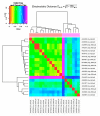
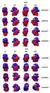
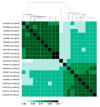
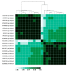
Influenza A viruses are rarely symptomatic in wild birds, while representing a higher threat to poultry and mammals, where they can cause a variety of symptoms, including death. H5 and H7 subtypes of influenza viruses are of particular interest because of their pathogenic potential and reported capacity to spread from poultry to mammals, including humans. The identification of molecular fingerprints for pathogenicity can help surveillance and early warning systems, which are crucial to prevention and protection from such potentially pandemic agents. In the past decade, comparative analysis of the surface features of hemagglutinin, the main protein antigen in influenza viruses, identified electrostatic fingerprints in the evolution and spreading of H5 and H9 subtypes. Electrostatic variation among viruses from avian or mammalian hosts was also associated with host jump. Recent findings of fingerprints associated with low and highly pathogenic H5N1 viruses, obtained by means of comparative electrostatics and normal modes analysis, prompted us to check whether such fingerprints can also be found in the H7 subtype. Indeed, evidence presented in this work showed that also in H7N7, hemagglutinin proteins from low and highly pathogenic strains present differences in surface electrostatics, while no meaningful variation was found in normal modes.
Keywords: HPAI; LPAI; SARS-CoV-2; electrostatic distance; fingerprint; influenza A virus; normal modes analysis; pandemic virus.
Conflict of interest statement
The authors declare no conflict of interest.
Figures


Similar articles
-
Normal modes analysis and surface electrostatics of haemagglutinin proteins as fingerprints for high pathogenic type A influenza viruses.BMC Bioinformatics. 2020 Aug 21;21(Suppl 10):354. doi: 10.1186/s12859-020-03563-w. BMC Bioinformatics. 2020. PMID: 32838732 Free PMC article.
-
An aptamer that binds efficiently to the hemagglutinins of highly pathogenic avian influenza viruses (H5N1 and H7N7) and inhibits hemagglutinin-glycan interactions.Acta Biomater. 2014 Mar;10(3):1314-23. doi: 10.1016/j.actbio.2013.12.034. Epub 2013 Dec 25. Acta Biomater. 2014. PMID: 24374323
-
Pandemic Avian Influenza and Intra/Interhaemagglutinin Subtype Electrostatic Variation among Viruses Isolated from Avian, Mammalian, and Human Hosts.Biomed Res Int. 2018 May 17;2018:3870508. doi: 10.1155/2018/3870508. eCollection 2018. Biomed Res Int. 2018. PMID: 29888260 Free PMC article.
-
[Highly pathogenic avian influenza in poultry (fowl plague); implications for human health].Bull Acad Natl Med. 2005 Nov;189(8):1817-26. Bull Acad Natl Med. 2005. PMID: 16737105 Review. French.
-
Avian influenza viruses in humans.Rev Sci Tech. 2009 Apr;28(1):161-73. doi: 10.20506/rst.28.1.1871. Rev Sci Tech. 2009. PMID: 19618624 Review.
Cited by
-
Phylogenetic Analysis and Codon Usage Bias Reveal the Base of Feline and Canine Chaphamaparvovirus for Cross-Species Transmission.Animals (Basel). 2023 Aug 14;13(16):2617. doi: 10.3390/ani13162617. Animals (Basel). 2023. PMID: 37627409 Free PMC article.
-
Host Membranes as Drivers of Virus Evolution.Viruses. 2023 Aug 31;15(9):1854. doi: 10.3390/v15091854. Viruses. 2023. PMID: 37766261 Free PMC article.
-
What Is life? Rethinking Biology in Light of Fundamental Parameters.Life (Basel). 2024 Feb 20;14(3):280. doi: 10.3390/life14030280. Life (Basel). 2024. PMID: 38541606 Free PMC article. Review.
-
Evolutionary changes in the number of dissociable amino acids on spike proteins and nucleoproteins of SARS-CoV-2 variants.Virus Evol. 2023 Jun 29;9(2):vead040. doi: 10.1093/ve/vead040. eCollection 2023. Virus Evol. 2023. PMID: 37583936 Free PMC article.
References
-
- Paget J., Spreeuwenberg P., Charu V., Taylor R.J., Iuliano A.D., Bresee J., Simonsen L., Viboud C., Global Seasonal Influenza-associated Mortality Collaborator Network Team. GLaMOR Collaborating Team Global mortality associated with seasonal influenza epidemics: New burden estimates and predictors from the GLaMOR Project. J. Glob. Health. 2019;9:020421. doi: 10.7189/jogh.09.020421. - DOI - PMC - PubMed
MeSH terms
Substances
Grants and funding
LinkOut - more resources
Full Text Sources
Medical
Miscellaneous

