. 2023 Feb 27;13(1):3354.
doi: 10.1038/s41598-023-29629-2.

Development of a novel ex vivo organ culture system to improve preservation methods of regenerative tissues

Affiliations

Development of a novel ex vivo organ culture system to improve preservation methods of regenerative tissues

Tomomi Yuta et al. Sci Rep. .

Abstract

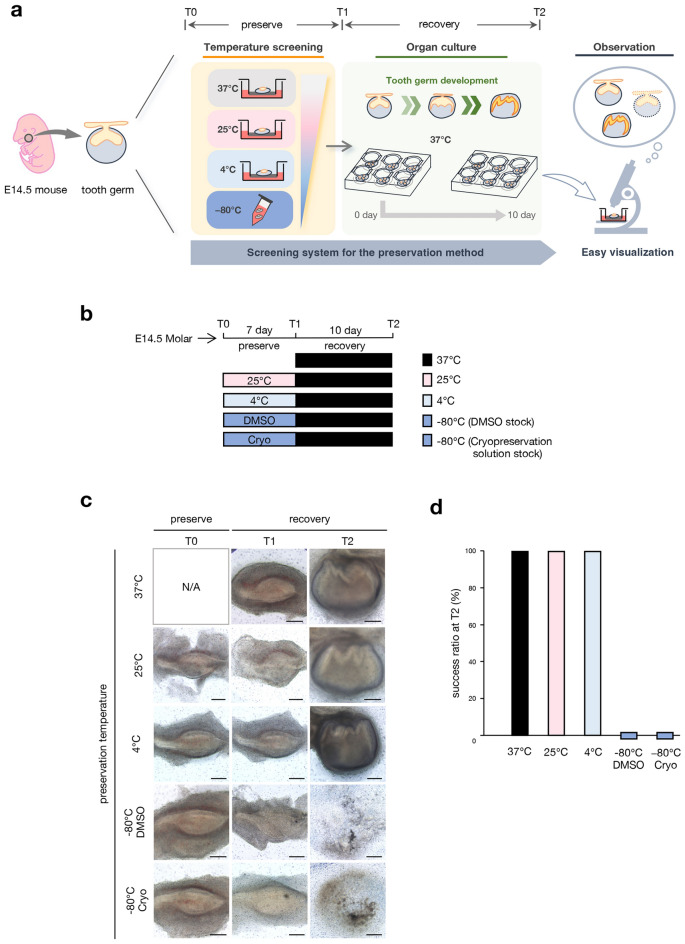
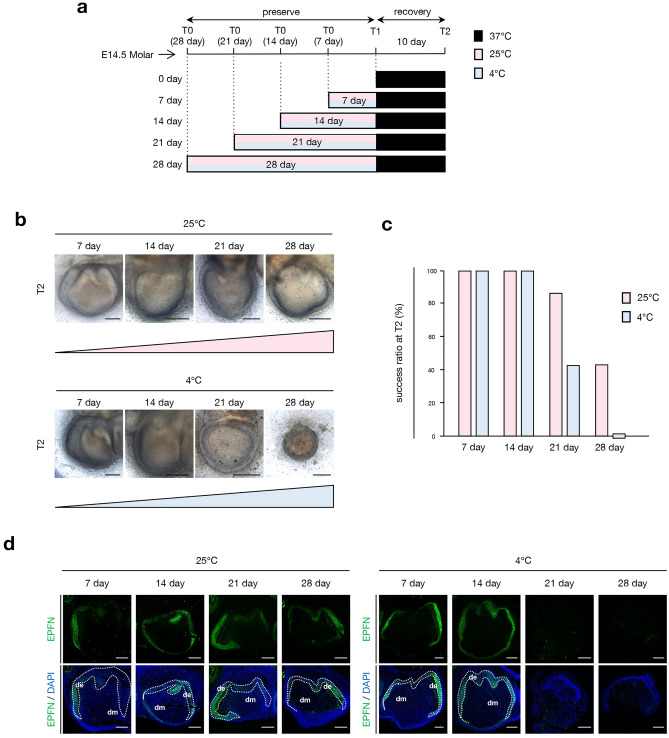
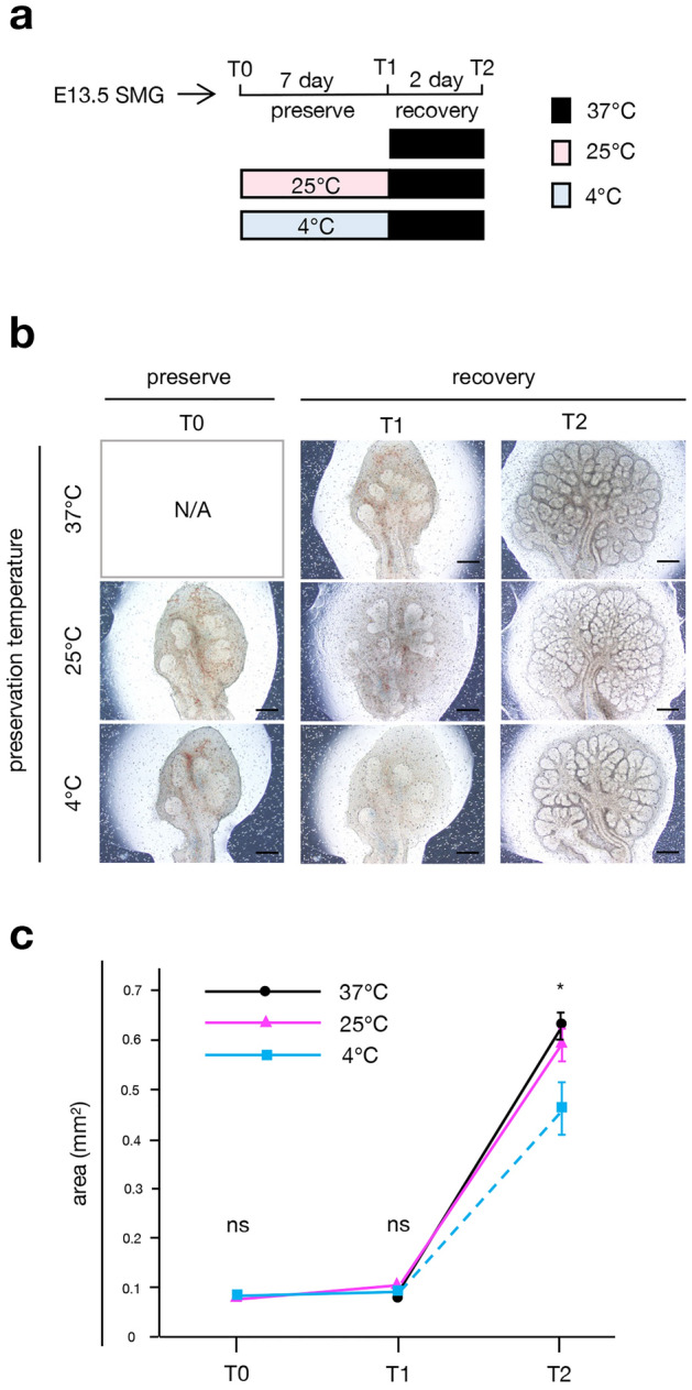
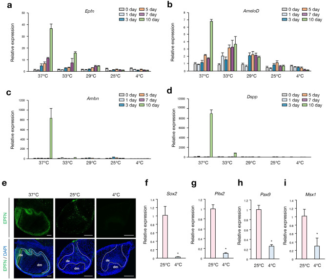
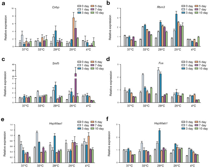
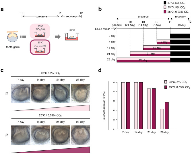
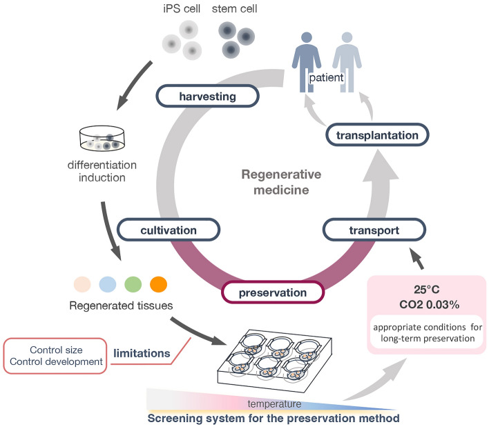
Recent advances in regenerative technology have made the regeneration of various organs using pluripotent stem cells possible. However, a simpler screening method for evaluating regenerated organs is required to apply this technology to clinical regenerative medicine in the future. We have developed a simple evaluation method using a mouse tooth germ culture model of organs formed by epithelial-mesenchymal interactions. In this study, we successfully established a simple method that controls tissue development in a temperature-dependent manner using a mouse tooth germ ex vivo culture model. We observed that the development of the cultured tooth germ could be delayed by low-temperature culture and resumed by the subsequent culture at 37 °C. Furthermore, the optimal temperature for the long-term preservation of tooth germ was 25 °C, a subnormothermic temperature that maintains the expression of stem cell markers. We also found that subnormothermic temperature induces the expression of cold shock proteins, such as cold-inducible RNA-binding protein, RNA-binding motif protein 3, and serine and arginine rich splicing factor 5. This study provides a simple screening method to help establish the development of regenerative tissue technology using a tooth organ culture model. Our findings may be potentially useful for making advances in the field of regenerative medicine.


Conflict of interest statement

The authors declare no competing interests.

Figures

Figure 1
Figure 1
Comparison of tissue preservation conditions using ex vivo developing mouse tooth germ culture. (a) Schematic of an experimental procedure for the screening of tissue preservation conditions using E14.5 tooth germ organ culture. T0: preservation temperature starting point, T1: preservation temperature endpoint, T2: recovery temperature endpoint. (b) Time course representation of E14.5 tooth germs preserved at 37 °C control, at 25 °C, at 4 °C, in DMSO at − 80 °C, and in cryopreservation solution at − 80 °C for 7 days, followed by a recovery period at 37 °C for 10 days. (c) Photographic analysis of cultured E14.5 tooth germs under different temperature conditions in (b). Scale bars, 200 µm. (d) Analysis of the development rate of cultured tooth germs under different temperature conditions in (c) (n = 6).
Figure 2
Figure 2
Comparison of low-temperature preservation conditions in extended-term preservation period. (a) Time course representation of tissue preservation for extended-term preservation at 37 °C control, low-temperature preservation (25 °C or 4 °C) for 7, 14, 21, and 28 days followed by recovery at 37 °C for 10 days. T0: preservation temperature starting point, T1: preservation temperature endpoint, T2: recovery temperature endpoint. (b) Photographic analysis of cultured E14.5 tooth germs in low-temperature preservation conditions for an extended term represented in (a). Scale bars, 200 µm. (c) Analysis of the development rate of cultured tooth germs under different temperature conditions and preservation terms in (b) (n = 7). (d) Immunofluorescence of cultured E14.5 tooth germs in low-temperature preservation conditions at T2 represented in (b). Green: EPFN, blue: DAPI. Dashed lines represent the border of the dental epithelium and mesenchyme. de dental epithelium, dm dental mesenchyme. Scale bars, 100 µm.
Figure 3
Figure 3
Comparison of tissue preservation condition using ex vivo developing submandibular gland culture. (a) Time course representation of tissue preservation at 37, 25, and 4 °C for 7 days, followed by a recovery period at 37 °C for 2 days. SMG: submandibular glands, T0: preservation temperature starting point, T1: preservation temperature endpoint, T2: recovery temperature endpoint. (b) Photographic analysis of cultured E13.5 submandibular glands in low-temperature preservation conditions represented in (a). Scale bars, 200 µm. (c) Changes in the area of cultured E13.5 submandibular gland buds at 37, 25, or 4 °C (n = 7). ns, p > 0.05: *p < 0.05. Error bars represent the mean ± SD.
Figure 4
Figure 4
Low-temperature preservation arrests the morphogenesis of cultured tooth germs. (a) Schematic representation of an experimental procedure for comparing preservation conditions with different temperatures using an E14.5 tooth germ organ culture. (b) Schematic diagram of the assessment of morphological changes during tooth germ organ culture. The development of cultured tooth germs was scored 0 to 5 according to their morphological changes. Score 0: no apparent change from starting point; score 1: epithelium thickening; score 2: epithelial invagination into the mesenchyme; score 3: multiple cusp formation; score 4: final morphogenesis; score 5: differentiation. (c) Photographic analysis of cultured E14.5 tooth germs in different temperature preservation conditions for 0, 1, 3, 5, 7, and 10 days. Scale bars, 200 µm. (d) Analysis of the developmental score of cultured tooth germs at different temperatures and under different preservation conditions in (c) (n = 20). *p < 0.05. Error bars represent mean ± SD.
Figure 5
Figure 5
Subnormothermic temperature preservation maintains the expression of differentiation marker genes and stem cell marker genes. (ad) RT-qPCR analysis of cultured E14.5 tooth germs at 37, 33, 29, 25, and 4 °C for 0, 1, 3, 5, 7, and 10 days (n = 3). The mRNA expressions of Epfn, AmeloD, Ambn, and Dspp were normalized to Gapdh. Error bars represent the mean ± SD. (e) Immunofluorescence of cultured E14.5 tooth germs at 37, 25, and 4 °C for 10 days. Green: EPFN, blue: DAPI. Dashed lines represent the border of the dental epithelium and mesenchyme. de dental epithelium, dm dental mesenchyme. Scale bars, 100 µm. (fi) RT-qPCR analysis of cultured E14.5 tooth germs at 25 and 4 °C for 10 days (n = 3). The mRNA expressions of Sox2, Pitx2, Pax9, and Msx1 were normalized to Gapdh. *p < 0.05. Error bars represent the mean ± SD.
Figure 6
Figure 6
Subnormothermic temperature preservation upregulated the expression of cold shock protein genes. (af) RT-qPCR analysis of cultured E14.5 tooth germs at 37, 33, 29, 25, and 4 °C for 0, 1, 3, 5, 7, and 10 days (n = 3). mRNA expression of Cirbp, Rbm3, Srsf5, Fus, Hsp90aa1, and Hsp90ab1 was normalized to Gapdh. Error bars represent the mean ± SD.
Figure 7
Figure 7
Concentration of CO2 does not affect the morphogenesis of cultured tooth germs. (a) A schematic of the experimental procedure for comparing conditions with different concentrations of CO2 using E14.5 tooth germ organ culture. T0: preservation temperature starting point, T1: preservation temperature endpoint, T2: recovery temperature endpoint. (b) Time course representation of tissue preservation in different concentrations of CO2 conditions (5% or 0.03%) for 7, 14, 21, and 28 days followed by recovery at 37 °C for 10 days. (c) A photographic analysis of cultured E14.5 tooth germs in conditions with different concentrations of CO2 is represented in (b). Scale bars, 200 µm. (d) Analysis of the development rate of cultured tooth germs under conditions with different concentrations of CO2 in (c) (n = 7).
Figure 8
Figure 8
Summary diagram of the results of tissue preservation conditions analyzed in this study.

Similar articles

Cited by

References

    1. Higashi H, et al. Transplantation of bioengineered liver capable of extended function in a preclinical liver failure model. Am. J. Transplant. 2022;22:731–744. doi: 10.1111/ajt.16928. - DOI - PMC - PubMed
    1. Takebe T, et al. Massive and reproducible production of liver buds entirely from human pluripotent stem cells. Cell Rep. 2017;21:2661–2670. doi: 10.1016/j.celrep.2017.11.005. - DOI - PubMed
    1. Lee JW, et al. Development of a 3D cell printed construct considering angiogenesis for liver tissue engineering. Biofabrication. 2016;8:015007. doi: 10.1088/1758-5090/8/1/015007. - DOI - PubMed
    1. Tohyama S, et al. Efficient large-scale 2D culture system for human induced pluripotent stem cells and differentiated cardiomyocytes. Stem Cell Rep. 2017;9:1406–1414. doi: 10.1016/j.stemcr.2017.08.025. - DOI - PMC - PubMed
    1. Nakanishi T, et al., editors. Etiology and Morphogenesis of Congenital Heart Disease: From Gene Function and Cellular Interaction to Morphology. Springer; 2016. - PubMed

Publication types