Review
. 2023 Jan 31;14(2):382.
doi: 10.3390/genes14020382.

Transfer RNA Modification Enzymes with a Thiouridine Synthetase, Methyltransferase and Pseudouridine Synthase (THUMP) Domain and the Nucleosides They Produce in tRNA

Affiliations
Review

Transfer RNA Modification Enzymes with a Thiouridine Synthetase, Methyltransferase and Pseudouridine Synthase (THUMP) Domain and the Nucleosides They Produce in tRNA

Hiroyuki Hori. Genes (Basel). .

Abstract

The existence of the thiouridine synthetase, methyltransferase and pseudouridine synthase (THUMP) domain was originally predicted by a bioinformatic study. Since the prediction of the THUMP domain more than two decades ago, many tRNA modification enzymes containing the THUMP domain have been identified. According to their enzymatic activity, THUMP-related tRNA modification enzymes can be classified into five types, namely 4-thiouridine synthetase, deaminase, methyltransferase, a partner protein of acetyltransferase and pseudouridine synthase. In this review, I focus on the functions and structures of these tRNA modification enzymes and the modified nucleosides they produce. Biochemical, biophysical and structural studies of tRNA 4-thiouridine synthetase, tRNA methyltransferases and tRNA deaminase have established the concept that the THUMP domain captures the 3'-end of RNA (in the case of tRNA, the CCA-terminus). However, in some cases, this concept is not simply applicable given the modification patterns observed in tRNA. Furthermore, THUMP-related proteins are involved in the maturation of other RNAs as well as tRNA. Moreover, the modified nucleosides, which are produced by the THUMP-related tRNA modification enzymes, are involved in numerous biological phenomena, and the defects of genes for human THUMP-related proteins are implicated in genetic diseases. In this review, these biological phenomena are also introduced.

Keywords: 4-thiouridine; C to U editing; N2-methylguanosine; N4-acetylcytidine; PUS10; deaminase; pseudouridine synthase; tRNA; tRNA methyltransferase; tRNA modification enzyme.


Conflict of interest statement

The author declares no conflict of interest.

Figures

Figure 1
Figure 1
Structures of modified nucleosides, which are produced by THUMP-related tRNA modification enzymes, and their positions in tRNA. (A) Structures of modified nucleosides, which are produced by THUMP-related tRNA modification enzymes. Modifications are indicated in red. Because uridine is produced by deamination of cytidine, the 4-O atom is colored in red. Because pseudouridine is synthesized by isomerization of uridine, the uracil base is enclosed in a red circle. (B) The typical tRNA structure is represented as a cloverleaf model. The numbers show the positions in tRNA. Conserved residues in tRNA are shown as letters: abbreviations, R, purine; Y, pyrimidine. Position 8 is conserved as U (red) in almost all tRNAs; however, in the case of M. kandleri, position 8 in precursor tRNA is C (orange). The colors correspond to the modified nucleosides in A: blue, m2G (and m22G); red, s4U; orange, U; cyan, ac4C; purple, Ψ. T. kodakarensis NAT10 homolog acetylates multiple positions in tRNA as described in the main text. (C) The modification positions are mapped on the L-shaped yeast tRNAPhe structure.
Figure 2
Figure 2
The modification pathways of THUMP-related tRNA methyltransferases. Eukaryotic, archaeal and bacterial enzymes are colored in red, blue and orange, respectively. The modification sites and modified nucleosides are enclosed by squares. (A) ArcTrm11 from P. abyssi and T. kodakarensis produces m2G10 and m22G10. The m22G10 modification is produced by the second methylation from m2G10. (B) S. cerevisiae Trm11 required a partner protein (Trm112) for the methylation and produces only m2G10. (C) ArcTrm11 from A. fulgidus and Halloferax volcanii requires a partner protein (arcTrm112) and produces both m2G10 and m22G10. (D) TrmN produces m2G6 from G6. (E) Trm14 produces m2G6 from G6. “?” means that T. kodakarensis Trm14 may produce m2G67 as well as m2G6; this modification has not been confirmed by purified protein. (F) Human THUMP3-TRMT112 complex produces m2G6 and m2G7 from G6 and G7, respectively.
Figure 3
Figure 3
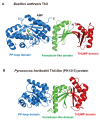
Structures of B. anthracis ThiI and P. horikoshii ThiI-like (PH1313) protein. (A) Structure of B. anthracis ThiI (PDB code: 2C5S) is represented by a cartoon model. Ferredoxin-like, THUMP and PP-loop domains are colored in green, red and blue, respectively. N and C show the N- and C-termini, respectively. Bound AMP is shown as a stick model. (B) Structure of P. horokoshii ThiI-like (PH1313) protein (PDB code: 1VBK) is shown by a cartoon model. Although this protein structure was solved as a dimer, only one subunit is shown. Ferredoxin-like, THUMP and PP-loop domains are colored in green, red and blue, respectively. The size of the PP-loop domain of this protein is smaller than that of B. anthracis ThiI due to the deletion of the C-terminal region.
Figure 4
Figure 4
(A) Secondary structure of minimum substrate RNA for ThiI. The modification position (U8) is colored in red. This RNA is a truncated RNA of E. coli tRNAPhe. The secondary structure is based on the complex of minimum substrate RNA and ThiI shown in panel B. (B) Crystal structure of the complex of the minimum substrate and T. maritima ThiI (PDB code: 4KR6). ThiI forms a dimer structure. To distinguish between the two subunits, one subunit is colored in pale green. The ferredoxin-like, THUMP and PP-loop domains in one subunit are colored in yellow, magenta and pale blue, respectively. The THUMP domain captures the CCA terminus of one minimum substrate RNA. The PP-loop domain in this subunit accesses U8 (red) in another minimum substrate RNA.
Figure 5
Figure 5
(A) Structure of mini-helix RNA. The modification position (C8) is colored in red. (B) Crystal structure of CDAT8. CDAT8 forms a dimer structure. To distinguish subunits, one subunit is colored in pale green. Deaminase, ferredoxin-like and THUMP domains in the other subunit are colored in pale blue, yellow and magenta, respectively.
Figure 6
Figure 6
Structures of P. abyssi Trm14 ((A): PDB code, 3TM4), T. thermophilus TrmN ((B): PDB code, 3TMA) and T. kodakarensis arcTrm11 ((C): PDB code, 5E71) are compared. The N-terminal ferredoxin-like domain, THUMP domain, Rossmann fold methyltransferase (methylase) domain and linker region are colored in yellow, red, blue and orange, respectively. Trm14 and TrmN modify G6 in tRNA while arcTrm11 modifies G10. The modification sites (G6 and G10) are mapped onto the L-shaped tRNA structure (D). G6, G10 and CCA terminus are highlighted as stick models. The distance between the THUMP and methylase domains of Trm14 and TrmN is shorter than that seen in arcTrm11. Because the THUMP domain captures the CCA terminus in tRNA, this short distance between the THUMP and methylase domains of Trm14 and TrmN enables the catalytic pocket in the methylase domain to access the modification site G6. In contrast, the longer distance between the THUMP and methylase domains of arcTrm11 is required for the positioning of the catalytic pocket with respect to the modification site G10. Thus, the N-terminal ferredoxin-like domain and linker region are important for the maintenance of the distance and angle between the THUMP and methylase domains, which decides the modification site in tRNA.
Figure 7
Figure 7
(A) Recognition sites of S. cerevisiae Trm11-Trm112 are marked on the L-shaped tRNA structure. The modification site (G10) and other recognition sites are colored in red and magenta, respectively. S. cerevisiae Trm11-Trm112 methylates standard tRNAs, which possess a regular size (5 nt) variable region, G10-C25 base pair and purine38 in addition to the CCA terminus. (B) Crystal structure of A. fulgidus arcTrm11-arcTrm112 (PDB code, 6ZXW) is represented by a cartoon model. The ferredoxin-like domain, THUMP domain, Rossmann fold methylase domain, and linker region are colored in yellow, red, blue and orange, respectively. Archaeal Trm112 is colored in green.
Figure 8
Figure 8
Structure of human PUS10 (PDB code, 2V9K) is represented by a cartoon model. N-terminal accessory and C-terminal pseudouridine synthase domains are colored in green and pale blue, respectively. The THUMP-related structure in the accessory domain is enclosed by a red circle. One Zn atom (magenta) is bound in the accessory domain.

Similar articles

Cited by

References

    1. Boccaletto P., Stefaniak F., Ray A., Cappannini A., Mukherjee S., Purta E., Kurkowska M., Shirvanizadeh N., Destefanis E., Groza P., et al. MODOMICS: A database of RNA modification pathways. 2021 update. Nucleic Acids Res. 2022;50:D231–D235. doi: 10.1093/nar/gkab1083. - DOI - PMC - PubMed
    1. Jühling F., Mörl M., Hartmann R.K., Sprinzl M., Stadler P.F., Pütz J. tRNAdb 2009: Compilation of tRNA sequences and tRNA genes. Nucleic Acids Res. 2009;37:D159–D162. doi: 10.1093/nar/gkn772. - DOI - PMC - PubMed
    1. Sajek M.P., Woźniak T., Sprinzl M., Jaruzelska J., Barciszewski J. T-psi-C: User friendly database of tRNA sequences and structures. Nucleic Acids Res. 2020;48:D256–D260. doi: 10.1093/nar/gkz922. - DOI - PMC - PubMed
    1. Lim K., Zhang H., Tempczyk A., Krajewski W., Bonander N., Toedt J., Howard A., Eisenstein E., Herzberg O. Structure of the YibK methyltransferase from Haemophilus influenzae (HI0766): A cofactor bound at a site formed by a knot. Proteins. 2003;51:56–67. doi: 10.1002/prot.10323. - DOI - PubMed
    1. Benítez-Páez A., Villarroya M., Douthwaite S., Gabaldón T., Armengod M.E. YibK is the 2’-O-methyltransferase TrmL that modifies the wobble nucleotide in Escherichia coli tRNA(Leu) isoacceptors. RNA. 2010;16:2131–2143. doi: 10.1261/rna.2245910. - DOI - PMC - PubMed

Publication types

Grants and funding

This work was supported by a Grant-in-Aid for Scientific Research (20H03211 to HH) from the Japan Society for the Promotion of Science (JSPS).

LinkOut - more resources