Review
. 2023 Jan 30;14(2):357.
doi: 10.3390/genes14020357.

Nonsense-Mediated mRNA Decay as a Mediator of Tumorigenesis

Affiliations
Review

Nonsense-Mediated mRNA Decay as a Mediator of Tumorigenesis

Preeti Nagar et al. Genes (Basel). .

Abstract

Nonsense-mediated mRNA decay (NMD) is an evolutionarily conserved and well-characterized biological mechanism that ensures the fidelity and regulation of gene expression. Initially, NMD was described as a cellular surveillance or quality control process to promote selective recognition and rapid degradation of erroneous transcripts harboring a premature translation-termination codon (PTC). As estimated, one-third of mutated and disease-causing mRNAs were reported to be targeted and degraded by NMD, suggesting the significance of this intricate mechanism in maintaining cellular integrity. It was later revealed that NMD also elicits down-regulation of many endogenous mRNAs without mutations (~10% of the human transcriptome). Therefore, NMD modulates gene expression to evade the generation of aberrant truncated proteins with detrimental functions, compromised activities, or dominant-negative effects, as well as by controlling the abundance of endogenous mRNAs. By regulating gene expression, NMD promotes diverse biological functions during development and differentiation, and facilitates cellular responses to adaptation, physiological changes, stresses, environmental insults, etc. Mutations or alterations (such as abnormal expression, degradation, post-translational modification, etc.) that impair the function or expression of proteins associated with the NMD pathway can be deleterious to cells and may cause pathological consequences, as implicated in developmental and intellectual disabilities, genetic defects, and cancer. Growing evidence in past decades has highlighted NMD as a critical driver of tumorigenesis. Advances in sequencing technologies provided the opportunity to identify many NMD substrate mRNAs in tumor samples compared to matched normal tissues. Interestingly, many of these changes are tumor-specific and are often fine-tuned in a tumor-specific manner, suggesting the complex regulation of NMD in cancer. Tumor cells differentially exploit NMD for survival benefits. Some tumors promote NMD to degrade a subset of mRNAs, such as those encoding tumor suppressors, stress response proteins, signaling proteins, RNA binding proteins, splicing factors, and immunogenic neoantigens. In contrast, some tumors suppress NMD to facilitate the expression of oncoproteins or other proteins beneficial for tumor growth and progression. In this review, we discuss how NMD is regulated as a critical mediator of oncogenesis to promote the development and progression of tumor cells. Understanding how NMD affects tumorigenesis differentially will pave the way for the development of more effective and less toxic, targeted therapeutic opportunities in the era of personalized medicine.

Keywords: cancer; gene expression; nonsense-mediated mRNA decay; splicing.


Conflict of interest statement

The authors declare no conflict of interest.

Figures

Figure 1
Figure 1
Regulation and dysregulation of nonsense-mediated mRNA decay (NMD) in physiology and pathology. NMD is an evolutionarily conserved pathway comprising a dual function to modulate gene expression: a surveillance or quality control mechanism to recognize and degrade erroneous mRNAs selectively; and a regulatory mechanism to control transcript abundance. By regulating gene expression, NMD promotes diverse physiological functions (shown at the top), including development, cell proliferation, differentiation, cellular stress, immune responses, etc. Mutations or alterations that impair the function or expression of proteins associated with the NMD pathway can be deleterious to cells and may cause pathological consequences (shown in the bottom), as implicated in developmental and neurological disability, genetic defect, cell signaling defect, hematopoietic defect, compromised immunity, and cancer. Therefore, NMD is strictly regulated in cells to safeguard the fidelity of gene expression.
Figure 2
Figure 2
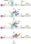
Schematics of the nonsense-mediated mRNA decay (NMD) pathway in mammalian cells. When a translating ribosome encounters a premature termination codon (PTC), the core NMD factor UPF1 interacts with the eukaryotic release factors eRF1 and eRF3, bound to the terminating ribosome, and induces premature translation termination. After this, the SURF complex is formed with SMG1 associated with SMG8 and SMG9, UPF1, eRF1, and eRF3. The SURF complex is then transformed into a decay-inducing complex (DECID), where UPF1 interacts with UPF2-UPF3B, either bound to the downstream EJC (the EJC-dependent NMD model) or disseminated in the cytoplasm (the EJC-independent NMD model). Subsequently, SMG1 induces the phosphorylation of UPF1. At this stage, translation is terminated with the separation of the ribosomal subunits, release factors, and developing peptide. Phosphorylated UPF1 triggers the mRNA decay by promoting the recruitment of different mRNA decay factors: SMG6 causes an endonucleolytic cleavage; SMG5-SMG7 heterodimer recruits the CCR4-NOT deadenylation complex, and/or PNRC2, and subsequently recruits the decapping complex (DCPC) to remove the cap-binding complex and the poly(A) tail. This facilitates 5′-to-3′ and 3′-to-5′ RNA degradation by XRN1 and the RNA exosome, respectively.
Figure 3
Figure 3
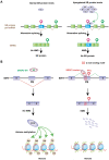
Regulation of NMD in cancer. (A) Schematics of different effectors of tumorigenesis, which are targeted by NMD. Tumor cells regulate NMD to target the mRNAs of these effectors to a favorable outcome that can help tumor growth and progression. (B) Differential regulation of NMD to promote tumorigenesis. Tumor cells acquire NMD-inducing nonsense mutations preferentially in tumor suppressor genes (e.g., TP53, WT1, RB, BRCA1/2) compared to oncogenes. Degradation of tumor suppressors via NMD is favorable for tumor development and growth. Tumor cells often induce NMD by upregulating NMD factors. Induction of NMD favors tumors by degrading proteins, which are toxic or detrimental to tumor cells. For example, in colorectal cancers with microsatellite instability (CRC MSI), NMD was induced by upregulation of several NMD factors (UPF1/2 and SMG1/6/7). Activated NMD helped the degradation of many premature termination codon (PTC)-containing mRNAs that are toxic against CRC MSI, such as a dominant negative (DN) mutant protein HSP110DE9. Tumor cells also often inhibit NMD by downregulating NMD factors or by deactivating mutations (mut) in NMD factors. Suppression of NMD favors tumors by upregulating oncoproteins or activating signaling pathways that favor tumorigenesis, metastasis, or adaptation to environmental stress. For example, disabling mutations in UPF1 in pancreatic adenosquamous carcinoma (ASC) suppressed NMD and upregulated the expression of a truncated TP53 isoform with dominant negative activity. In human adenocarcinoma (ADC), downregulated UPF1 inhibited NMD activity, which subsequently increased epithelial-mesenchymal transition (EMT) and metastatic events through upregulation of tumor growth factor β pathway (TGF-β). Therefore, tumor cells exploit complex regulation in NMD to favor their growth and progression.
Figure 4
Figure 4
Gene and protein structures of SR protein splicing factors. (A) Schematics of SR protein genes with coding exons, non-coding regions, and internal poison exons or 3′UTR poison sequences (not to scale). The presence of poison exons or 3′UTR poison sequences in each gene is shown on the right. (B) Schematics of protein structure and domain organization of SR proteins (not to scale). PE: poison exons; PS: poison sequences; RRM: RNA recognition motif; RS: serine-arginine rich domain; Zn: zinc knuckle.
Figure 5
Figure 5
Regulation of AS-NMD in cancer. (A) When SR proteins are overexpressed in normal cells, they autoregulate their expression via AS-NMD for homeostasis. SR protein genes harbor ultraconserved regions containing non-coding exons, also called poison exons (PEs) or 3′UTR poison sequences (PSs). When poison exons are included, they introduce a premature termination codon (PTC) and target the mRNAs for degradation. In contrast, splicing of the 3′UTR poison sequences introduces a new exon–exon junction, which marks the original stop codon as a PTC and elicits NMD. SR protein autoregulation via AS-NMD is inhibited in certain tumors. (B) SRSF2 is recurrently mutated in hematologic malignancy. One aberrant AS-NMD event promoted by the mutant SRSF2 is the inclusion of a poison exon in EZH2. EZH2 catalyzes histone methylation and functions in chromatin remodeling. Mutation in SRSF2 changes its binding preferences for a C-rich motif. This causes the inclusion of the EZH2 poison exon, which generates a PTC and is degraded by NMD. Therefore, the expression of EZH2 protein is downregulated, contributing to impaired hematopoietic differentiation.
Figure 6
Figure 6
Gene-specific NMD inhibition and targeted augmentation of protein synthesis. (A) A premature termination codon (PTC)-containing mRNA is degraded by nonsense-mediated mRNA decay (NMD), inhibiting the expression of the encoded protein. (B) Antisense oligonucleotides (ASO) targeting gene-specific exon junction complex (EJC)-binding sites can inhibit NMD, allowing the expression of a truncated protein from a PTC-containing mRNA. The truncated protein may or may not be functional depending on the presence or absence of the functional domain(s). (C) Gene-specific ASO-mediated NMD inhibition, along with the readthrough compound (RTC), can allow the expression of a full-length protein from a PTC-containing mRNA.

Similar articles

Cited by

References

    1. Popp M.W., Maquat L.E. Nonsense-Mediated mRNA Decay and Cancer. Curr. Opin. Genet. Dev. 2018;48:44–50. doi: 10.1016/j.gde.2017.10.007. - DOI - PMC - PubMed
    1. Lykke-Andersen S., Jensen T.H. Nonsense-Mediated mRNA Decay: An Intricate Machinery That Shapes Transcriptomes. Nat. Rev. Mol. Cell Biol. 2015;16:665–677. doi: 10.1038/nrm4063. - DOI - PubMed
    1. Miller J.N., Pearce D.A. Nonsense-Mediated Decay in Genetic Disease: Friend or Foe? Mutat. Res. Rev. Mutat. Res. 2014;762:52–64. doi: 10.1016/j.mrrev.2014.05.001. - DOI - PMC - PubMed
    1. Hwang J., Maquat L.E. Nonsense-Mediated mRNA Decay (NMD) in Animal Embryogenesis: To Die or Not to Die, That Is the Question. Curr. Opin. Genet. Dev. 2011;21:422–430. doi: 10.1016/j.gde.2011.03.008. - DOI - PMC - PubMed
    1. Brogna S., Wen J. Nonsense-Mediated mRNA Decay (NMD) Mechanisms. Nat. Struct. Mol. Biol. 2009;16:107–113. doi: 10.1038/nsmb.1550. - DOI - PubMed

Publication types

Substances

Grants and funding

This work was supported by the Edward P. Evans Foundation and the Winthrop P. Rockefeller Cancer Institute.