. 2023 Feb 11;12(4):590.
doi: 10.3390/cells12040590.

NbMLP43 Ubiquitination and Proteasomal Degradation via the Light Responsive Factor NbBBX24 to Promote Viral Infection

Affiliations

NbMLP43 Ubiquitination and Proteasomal Degradation via the Light Responsive Factor NbBBX24 to Promote Viral Infection

Liyun Song et al. Cells. .

Abstract

The ubiquitin-proteasome system (UPS) plays an important role in virus-host interactions. However, the mechanism by which the UPS is involved in innate immunity remains unclear. In this study, we identified a novel major latex protein-like protein 43 (NbMLP43) that conferred resistance to Nicotiana benthamiana against potato virus Y (PVY) infection. PVY infection strongly induced NbMLP43 transcription but decreased NbMLP43 at the protein level. We verified that B-box zinc finger protein 24 (NbBBX24) interacted directly with NbMLP43 and that NbBBX24, a light responsive factor, acted as an essential intermediate component targeting NbMLP43 for its ubiquitination and degradation via the UPS. PVY, tobacco mosaic virus, (TMV) and cucumber mosaic virus (CMV) infections could promote NbMLP43 ubiquitination and proteasomal degradation to enhance viral infection. Ubiquitination occurred at lysine 38 (K38) within NbMLP43, and non-ubiquitinated NbMLP43(K38R) conferred stronger resistance to RNA viruses. Overall, our results indicate that the novel NbMLP43 protein is a target of the UPS in the competition between defense and viral anti-defense and enriches existing theoretical studies on the use of UPS by viruses to promote infection.

Keywords: B-box zinc finger protein 24; MLP-like protein 43; potato virus Y; resistance; ubiquitination; ubiquitin–proteasome system.


Conflict of interest statement

The authors declare no conflict of interest.

Figures
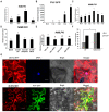
Figure 1
Figure 1
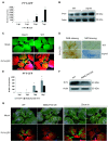
Expression pattern analysis of NbMLP43. (A) Trends in NbMLP43 expression in the root, stem, leaf, and flower. Data were analyzed with Duncan’s multiple range tests; different letters indicate that the values of the four treatments were significantly different (p < 0.05), which were analyzed the same as figures (BE). (B) Trends in PVY-GFP trend at 1, 3, 5, 7 dpi. (C) Trends in NbMLP43 expression after PVY inoculation. (D) NbMLP43 expression after spraying with 0.5 mM SA, 0.05 mM ethephon, or 0.1 mM Me-JA; water with 0.02% Tween 20 was used as a control. (E) NbMLP43 expression after silencing key signaling genes, namely NPR1, COI1, and EIN2. (F) SA content was detected before and after PVY infection in NbMLP43-OE plants, with wild-type as the control. Data were analyzed with independent sample t-test; * indicated that values of the two treatments were significantly different (p < 0.05). (G) Subcellular distribution of NbMLP43 in plant’s epidermis before and after PVY-infection.
Figure 2
Figure 2
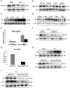
Functional validation of NbMLP43 based on mlp43 and NbMLP43-OE plants. (A) Detection of PVY CP expression in mlp43 and WT at 1, 3, 5, and 7 dpi at the RNA level. Data were analyzed with independent sample t-test; * indicated that values of the two treatments were significantly different (p < 0.05), which were analyzed the same way as figure E. (B) PVY CP protein levels in mlp43 and WT at 5 dpi. (C) PVY-GFP fluorescence in mlp43 and WT, with mock as a negative control. (D) DAB and NBT staining of WT and mlp43 plants inoculated with PVY-GFP. (E) Real-time PCR was used to detect the differences in virus expression at 1, 2, 3, and 4 dpi. NbMLP43-OE/PVY was the treatment group, whereas WT/PVY was the control group. (F) Differential expression of PVY CP protein was detected at 4 dpi. (G) GFP fluorescence at 12 dpi.
Figure 3
Figure 3
NbMLP43 was degraded through the UPS pathway. (A) PVY infection decreased NbMLP43 expression at the protein level. (B) NbMLP43 expression was detected after MG132 and 3MA treatment, with DMSO as a control. (C) Detection of PVY CP at 1, 3, 5, and 7 dpi. (D) Ubiquitination level in PVY infected plants at 1, 3, 5, and 7 dpi, with PBS treatment as mock. (E) PVY CP was detected at the RNA level after treatment with MG132 at 24 h and 48 h, and DMSO was treated as a control. Data were analyzed with independent sample t-test; * indicated that values of the two treatments were significantly different (p < 0.05), which were analyzed the same way as figure H. (F) PVY CP protein level after treatment with MG132 at 48 h. (G) Ubiquitination level of NbMLP43 after PVY infection using ubiquitin antibody and after K38 mutation. (H) Effect of NbMLP43 ubiquitination on PVY infection at the RNA level. (I) Effect of NbMLP43 ubiquitination on PVY infection at the protein level.
Figure 4
Figure 4
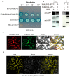
NbBBX24 interacted with NbMLP43. (A) Yeast point-to-point validation of the interaction between NbMLP43 and NbBBX24. (B) Interaction between NbMLP43 and NbBBX24 was verified with co-IP assay. (C) Subcellular distribution of NbMLP43 and NbBBX24. (D) YFP interaction signal of NbMLP43 and NbBBX24.
Figure 5
Figure 5
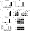
NbBBX24 acted as an intermediate factor that mediated the ubiquitinated degradation of NbMLP43. (A) Transcriptional levels of NbBBX24 were detected with light/dark photoperiods of 16/8 h and 8/16 h at 2 and 4 dpi under PVY infection. Data were analyzed with independent sample t-test; * indicated that values of the two treatments were significantly different (p < 0.05), the same as figure (B,C,F,H). (B) Transcriptional levels of PVY CP were detected with light/dark photoperiods of 16-h/8-h and 8-h/16-h at 2 and 4 dpi. (C) After silencing NbBBX24, transcriptional levels of PVY CP at light/dark photoperiods of 16/8 h and 8/16 h at 2 and 4 dpi under PVY infection. (D) Protein levels of PVY CP and ubiquitination levels of NbMLP43 at light/dark photoperiods of 16/8 h and 8/16 h at 4 dpi. (E) After silencing NbBBX24, protein levels of PVY CP at light/dark photoperiods of 16/8 h and 8/16 h at 4 dpi. (F) Expression of NbBBX24 under PVY infection at the transcriptional level. (H) Effects of silencing NbBBX24 on PVY infection at the transcriptional level. (G,I) Effects of silencing NbBBX24 on NbMLP43 ubiquitination, NbMLP43 itself, and PVY infection at the protein level.
Figure 6
Figure 6
Viral infection nonspecifically promoted ubiquitination of NbMLP43. (A) Yeast point-to-point validation of the interaction between NbBBX24 and PVY. (B) Effects of TMV (or CMV) infection on NbMLP43 ubiquitination and NbMLP43 itself. (C) Effects of TMV and CMV infection on the expression of NbBBX24 at the transcriptional level. Data were analyzed with independent sample t-test; * indicated that values of the two treatments were significantly different (p < 0.05), the same as figures (D,E). (D) Effect of NbMLP43 ubiquitination on TMV infection at the transcriptional level. (E) Effect of NbMLP43 ubiquitination on CMV infection at the transcriptional level. (F) Effects of NbMLP43 ubiquitination on TMV and CMV infection at the protein level. (G) Effects of NbMLP43 ubiquitination on TMV and CMV infection as identified from plant disease symptoms.
Figure 7
Figure 7
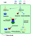
Article overview of virus hijacked UPS to degrade resistance protein NbMLP43. Viral infection induced NbMLP43 transcription but decreased NbMLP43 at the protein level. NbMLP43 conferred resistance to N. benthamiana against viral infection. The light response factor NbBBX24 specifically binded NbMLP43 for its ubiquitination and degradation via the UPS, enhancing viral infection. And non-ubiquitinated NbMLP43(K38R) conferred stronger resistance to RNA viruses.

Similar articles

Cited by

References

    1. Adriaenssens E.M., Cowan D.A. Using Signature Genes as Tools to Assess Environmental Viral Ecology and Diversity. Appl. Environ. Microbiol. 2014;80:4470–4480. doi: 10.1128/AEM.00878-14. - DOI - PMC - PubMed
    1. Nicaise V. Crop immunity against viruses: Outcomes and future challenges. Front. Plant Sci. 2014;5:660. doi: 10.3389/fpls.2014.00660. - DOI - PMC - PubMed
    1. Garcia-Ruiz H. Host factors against plant viruses. Mol. Plant Pathol. 2019;20:1588–1601. doi: 10.1111/mpp.12851. - DOI - PMC - PubMed
    1. Luo H. Interplay between the virus and the ubiquitin–proteasome system: Molecular mechanism of viral pathogenesis. Curr. Opin. Virol. 2015;17:1–10. doi: 10.1016/j.coviro.2015.09.005. - DOI - PMC - PubMed
    1. Wang Y., Yang N., Zheng Y., Yue J., Bhadauria V., Peng Y.-L., Chen Q. Ubiquitination in the rice blast fungus Magnaporthe oryzae: From development and pathogenicity to stress responses. Phytopathol. Res. 2022;4:1. doi: 10.1186/s42483-021-00106-w. - DOI

Publication types

MeSH terms

Grants and funding

This research was supported by grants from the Shandong Provincial Natural Science Foundation Project (ZR2021MC058); the Major Green Prevention and Control projects (110202101045(LS-05), 110202101050(LS-10)).