Review
. 2023 Feb 4;13(2):294.
doi: 10.3390/biom13020294.

The Actin Network Interfacing Diverse Integrin-Mediated Adhesions

Affiliations
Review

The Actin Network Interfacing Diverse Integrin-Mediated Adhesions

Benjamin Geiger et al. Biomolecules. .

Abstract

The interface between the cellular actin network and diverse forms of integrin-mediated cell adhesions displays a unique capacity to serve as accurate chemical and mechanical sensors of the cell's microenvironment. Focal adhesion-like structures of diverse cell types, podosomes in osteoclasts, and invadopodia of invading cancer cells display distinct morphologies and apparent functions. Yet, all three share a similar composition and mode of coupling between a protrusive structure (the lamellipodium, the core actin bundle of the podosome, and the invadopodia protrusion, respectively), and a nearby adhesion site. Cytoskeletal or external forces, applied to the adhesion sites, trigger a cascade of unfolding and activation of key adhesome components (e.g., talin, vinculin, integrin), which in turn, trigger the assembly of adhesion sites and generation of adhesion-mediated signals that affect cell behavior and fate. The structural and molecular mechanisms underlying the dynamic crosstalk between the actin cytoskeleton and the adhesome network are discussed.

Keywords: actin; cell–matrix adhesions; focal adhesions; integrins; invadopodia; podosomes; vinculin.


Conflict of interest statement

All authors declare that there are no conflicts of interest associated with this article.

Figures

Figure 5
Figure 5
The effect of matrix composition and mechanical properties on focal adhesion and actin network organization. (A) Human foreskin fibroblasts (HFF) were plated in serum-free medium on glass coverslips coated with fibronectin or vitronectin and integrin-specific blocking antibodies, and were added as described. The cells were allowed to spread for 3 h, and then fixed and immunostained for vinculin or paxillin. Notice the extensive spreading of fibronectin, mediated by integrin α5β1, and broad distribution of focal adhesion throughout the entire ventral membrane compared to the reduced spreading and preferential formation of peripheral focal adhesions by cells adhering to vitronectin via integrin αvβ3. Modified from the Ph.D. thesis of B. Zimerman [70]. (B) HFF cells stably expressing paxillin-YFP (green) were plated on fibronectin-coated PDMS substrates with different rigidities, and fixed at 6 h following seeding. Cells were stained with TRITC–phalloidin (red) and DAPI (blue). (See also Ref. [65]). Notice the marked polarization of the cell plated on the rigid elastomer compared to the largely radial spreading on the softer matrix.
Figure 6
Figure 6
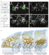
Activated vinculin remodels branched actin network into a bundle. (A) Activated vinculin interacts with surface-attached talin VBS1 to initiate actin bundles. A talin-VBS1 was patterned onto a glass. The patterned surface was mounted into a flow chamber, allowing a mix of inactive vinculin, actin monomers, Arp2/3, and WAVE WA (2). Bundling of Arp2/3-branched actin networks by vinculin was observed only at the sites of patterned VBS1. Arrowheads indicate bundles formed by the recruitment of actin branches labeled with stars. The circle shows the position of the VBS1-coated pattern decorated by vinculin (green in the upper left panel). Scale bar, 3 µm. (B) A model describing the bundling of branched actin networks by membrane-bound talin-activated vinculin at nascent adhesions. A schematic view of a nascent integrin-based adhesion site, localized in proximity to the leading edge of a cell (left). (1) The Arp2/3 branched actin network treadmills and flows centripetally. Vinculin is activated by interacting with talin [89,90]. (2) At nascent adhesion sites, talin-activated vinculin stably binds and bundles mobile branched (this work) networks, likely affecting actin dynamics [87]. (3) Actin retrograde flow generates tension that further activates talin and vinculin, reinforcing the link to integrins. Adhesion-anchored vinculin interacting with the flowing and branched actin networks initiate bundles at the nascent adhesion site. Vinculin engages the retrograde actin flow, applying tension to the mechanoresponsive components of the adhesion, enabling adhesion self-sustained assembly dynamics and the recruitment of additional focal adhesion components such as myosin II, α-actinin, and zyxin [79,87,92]. The figure was modified from [45].
Figure 1
Figure 1
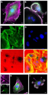
Association of F-actin with the main forms of integrin-mediated adhesions. (A) Vinculin-rich focal adhesions (FA, green), associated with the termini of actin-based stress fibers (magenta) in REF52 cells. Small ’focal complexes’ (FX) the precursors of focal adhesions are seen along the cell’s edge, and elongated ‘fibrillar adhesions’ (FB), associated with the ventral cell membrane, are found in the perinuclear region. (B) Podosomes (P) consisting of an F-actin core (magenta), and vinculin-rich adhesion ring (green) in developing (upper-right) and mature (lower-left) murine osteoclast; (CF) cultured melanoma cell line (WM793) seeded on fluorescently labeled gelatin matrix ((E), red) and incubated for 5 h. The cells were then fixed and stained for actin ((C), green) and DAPI ((D), blue) for the detection of invadopodia and nuclei, respectively. The merged image is shown in (F). Note the prominence of actin-rich invadopodia “under” the nuclei and their close association with the dark spots, corresponding to degraded regions in the underlying gelatin matrix. (G) A magnified image of a WM793 cell depicting the relative distributions of actin-rich invadopodia in magenta (G) and vinculin in green (G’). The arrows point to a close association (yet, often with variable intensities) between the cytoskeletal-protrusive and the adhesive domains of the invadopodia. (H,I) Untreated MEF and MEF expressing constitutively active pp60Src (Y527F) were stained for vinculin (green) and actin (magenta). Notice the apparent transition of the adhesive cytoskeletal complex from the typical focal adhesion–stress fiber phenotype in the untreated cell (H) to podosome- or invadopodia-like phenotype in the cells expressing the deregulated pp60Src (I).
Figure 2
Figure 2
Molecular and functional networking. An illustration depicting the basic interplay between cells and the surrounding soluble and insoluble microenvironments. The integrin adhesome consists of multiple molecular components that concertedly form a “scaffolding domain” that physically links the actin cytoskeleton to the extracellular matrix and “regulatory domain” that drives the assembly and reorganization of the scaffolding components. The outermost components of the scaffolding domain are diverse heterodimeric integrin receptors that bind directly to the external surfaces. Via their transmembrane cytoplasmic “tails”, integrins interact with diverse adaptor proteins (e.g., talin, vinculin, and paxillin) that directly and indirectly connect the adhesion complex to the actin cytoskeleton. The regulatory domain consists of different signaling pathways, involving different serine and threonine (S/T)- and tyrosine (Y)-specific kinases (K) and phosphatases (P). The assembly and remodeling of the adhesion sites are affected by external signals, some of which are chemical (e.g., matrix composition, adhesive ligands, and soluble and immobilized growth factors) and others are physical (e.g., matrix rigidity, pressure, shear stress, cytoskeletal polymerization, and contraction mechanics). For further details, see “Integrin adhesions as chemical and mechanical sensors of the pericellular environment” below.
Figure 3
Figure 3
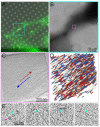
A mixed polarity of actin filaments and multiprotein complexes associated with focal adhesions. (A) Fluorescence microscopy image of a MEF cell expressing vinculin/venus-fluorescent protein, plated on a fibronectin-coated EM grid. The position indicated in the image, marked turquoise and magenta, was identified after vitrification using cryo-electron microscopy. (B) A low magnification cryo-EM image of a cell protrusion, in which the focal adhesion site was identified (turquoise) and a tomogram was acquired (Magenta). (C) A 9 nm thick section through a tomogram is framed in magenta (A,B). The direction of the cell edge is marked by the blue arrow and the cell interior by the red arrow. (D) The 3D isosurface rendering view of the tomogram is overlayed by the polarity analysis of the filaments (filaments with the barbed end toward the distal end of the cell are marked in blue, and those oriented toward the cell body are marked red (colored arrows in (C)). (E) A 9 nm slice image of the tomograms, acquired at focal adhesion, reveals macromolecular structures similar to those previously identified [34], indicated by the turquoise arrowheads. The magenta arrowheads point to ribosomes.
Figure 4
Figure 4
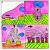
A schematic “artist’s depiction” of the protrusion–adhesion coupling model in the “classical forms” of integrin adhesions. The model shows a proposed mechanical coupling between flowing or treadmilling actin filaments and nearby integrin adhesion in four “prototypic” systems. The adhesion complexes in (AC) are highlighted yellow, and a rough “zoom-in” into the yellow highlighted box is shown enlarged in (D). (A) Spreading of a cell (e.g., fibroblast) that forms focal complexes (left highlighted box), interacting with the centripetally flowing actin filaments in the lamellipodia, or lamellae (flow direction indicated by the thick arrow). F-actin and its polarity are represented by the black lines (arrows indicate the pointed end of the actin filaments). The right highlighted box refers to mature focal adhesions, located in more central regions of the cell. Actin-mediated forces that are applied to the nascent and mature adhesions are generated by the lamellipodial flow and the stress fiber, respectively. (B) A schematic view of an invadopodium, with an actin core bundle that polymerizes, thereby pushing on the ventral cell membrane and producing an invasive protrusion (down-pointing arrow). The elongating actin bundle can also apply force to the nearby nucleus (up-pointing arrow), leading to its indentation. It is proposed that in its upward treadmilling, the core actin bundle applies shear forces to the nearby adhesion site located at the “neck” of the protrusion, thereby reinforcing the adhesion. (C) A schematic view of a podosome located at the sealing zone of a multinuclear osteoclast, displaying a “central actin bundle” that treadmills “upwards” while pushing the ventral membrane toward the underlying bone surface, thereby contributing to the sealing of the “resorption lacuna” of the osteoclast. It is further suggested that the central actin bundle is cross-linked to “lateral actin fibers” that, in turn, interact with the nearby (ring-like) adhesion site and pull on it. (D) A close-up schematic depiction of the key molecular events induced by a directional flow of actin on the assembly of the integrin (colored green and red) with talin (purple) and vinculin (at different stages of unfolding; colored red). Actin filaments with different polarities are represented by arrowed black lines, with the arrow indicating the pointed end of the filaments, (for further details, see discussion of the mechanisms underlying the mechanosensing capacity of integrin adhesions below).

Similar articles

Cited by

References

    1. Chastney M.R., Conway J.R.W., Ivaska J. Integrin adhesion complexes. Curr. Biol. 2021;31:R536–R542. doi: 10.1016/j.cub.2021.01.038. - DOI - PubMed
    1. Humphries J.D., Chastney M.R., Askari J.A., Humphries M.J. Signal transduction via integrin adhesion complexes. Curr. Opin. Cell Biol. 2019;56:14–21. doi: 10.1016/j.ceb.2018.08.004. - DOI - PubMed
    1. Bouvard D., Brakebusch C., Gustafsson E., Aszodi A., Bengtsson T., Berna A., Fassler R. Functional consequences of integrin gene mutations in mice. Circ. Res. 2001;89:211–223. doi: 10.1161/hh1501.094874. - DOI - PubMed
    1. Danen E.H.J., Sonnenberg A. Integrins in regulation of tissue development and function. J. Pathol. 2003;200:471–480. doi: 10.1002/path.1416. - DOI - PubMed
    1. Zaidel-Bar R., Ballestrem C., Kam Z., Geiger B. Early molecular events in the assembly of matrix adhesions at the leading edge of migrating cells. J. Cell Sci. 2003;116:4605–4613. doi: 10.1242/jcs.00792. - DOI - PubMed

Publication types

MeSH terms

LinkOut - more resources