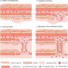
Angiogenesis after ischemic stroke
- PMID: 36829053
- PMCID: PMC10310733
- DOI: 10.1038/s41401-023-01061-2
Angiogenesis after ischemic stroke
Abstract
Owing to its high disability and mortality rates, stroke has been the second leading cause of death worldwide. Since the pathological mechanisms of stroke are not fully understood, there are few clinical treatment strategies available with an exception of tissue plasminogen activator (tPA), the only FDA-approved drug for the treatment of ischemic stroke. Angiogenesis is an important protective mechanism that promotes neural regeneration and functional recovery during the pathophysiological process of stroke. Thus, inducing angiogenesis in the peri-infarct area could effectively improve hemodynamics, and promote vascular remodeling and recovery of neurovascular function after ischemic stroke. In this review, we summarize the cellular and molecular mechanisms affecting angiogenesis after cerebral ischemia registered in PubMed, and provide pro-angiogenic strategies for exploring the treatment of ischemic stroke, including endothelial progenitor cells, mesenchymal stem cells, growth factors, cytokines, non-coding RNAs, etc.
Keywords: angiogenesis; endothelial progenitor cells; ischemic stroke; secreted proteins; stem cells.
© 2023. The Author(s), under exclusive licence to Shanghai Institute of Materia Medica, Chinese Academy of Sciences and Chinese Pharmacological Society.
Conflict of interest statement
The authors declare no competing interests.
Figures


Similar articles
-
Neural Stem Cells for Early Ischemic Stroke.Int J Mol Sci. 2021 Jul 19;22(14):7703. doi: 10.3390/ijms22147703. Int J Mol Sci. 2021. PMID: 34299322 Free PMC article. Review.
-
Neural stem cell therapy for subacute and chronic ischemic stroke.Stem Cell Res Ther. 2018 Jun 13;9(1):154. doi: 10.1186/s13287-018-0913-2. Stem Cell Res Ther. 2018. PMID: 29895321 Free PMC article. Review.
-
Angiogenic Actions of Paeoniflorin on Endothelial Progenitor Cells and in Ischemic Stroke Rat Model.Am J Chin Med. 2021;49(4):863-881. doi: 10.1142/S0192415X21500415. Epub 2021 Apr 7. Am J Chin Med. 2021. PMID: 33829966
-
Effects of ML351 and tissue plasminogen activator combination therapy in a rat model of focal embolic stroke.J Neurochem. 2021 May;157(3):586-598. doi: 10.1111/jnc.15308. Epub 2021 Feb 5. J Neurochem. 2021. PMID: 33481248
-
Tissue plasminogen activator (tPA) and matrix metalloproteinases in the pathogenesis of stroke: therapeutic strategies.CNS Neurol Disord Drug Targets. 2008 Jun;7(3):243-53. doi: 10.2174/187152708784936608. CNS Neurol Disord Drug Targets. 2008. PMID: 18673209 Free PMC article. Review.
Cited by
-
Il1r2 and Tnfrsf12a in transcranial magnetic stimulation effect of ischemic stroke via bioinformatics analysis.Medicine (Baltimore). 2024 Jan 26;103(4):e36109. doi: 10.1097/MD.0000000000036109. Medicine (Baltimore). 2024. PMID: 38277520 Free PMC article.
-
Lactylation and Ischemic Stroke: Research Progress and Potential Relationship.Mol Neurobiol. 2024 Nov 14. doi: 10.1007/s12035-024-04624-4. Online ahead of print. Mol Neurobiol. 2024. PMID: 39541071 Review.
-
β-asarone induces viability and angiogenesis and suppresses apoptosis of human vascular endothelial cells after ischemic stroke by upregulating vascular endothelial growth factor A.PeerJ. 2024 Jun 27;12:e17534. doi: 10.7717/peerj.17534. eCollection 2024. PeerJ. 2024. PMID: 38948219 Free PMC article.
-
Out of the core: the impact of focal ischemia in regions beyond the penumbra.Front Cell Neurosci. 2024 Mar 5;18:1336886. doi: 10.3389/fncel.2024.1336886. eCollection 2024. Front Cell Neurosci. 2024. PMID: 38504666 Free PMC article. Review.
-
Metabolomic discoveries for early diagnosis and traditional Chinese medicine efficacy in ischemic stroke.Biomark Res. 2024 Jun 20;12(1):63. doi: 10.1186/s40364-024-00608-7. Biomark Res. 2024. PMID: 38902829 Free PMC article. Review.
References
-
- Chen YC, Wu JS, Yang ST, Huang CY, Chang C, Sun GY, et al. Stroke, angiogenesis and phytochemicals. Front Biosci (Sch Ed). 2012;4:599–610. - PubMed
Publication types
MeSH terms
Substances
LinkOut - more resources
Full Text Sources
Medical

