

A Novel Dressing Composed of Adipose Stem Cells and Decellularized Wharton's Jelly Facilitated Wound Healing and Relieved Lymphedema by Enhancing Angiogenesis and Lymphangiogenesis in a Rat Model
- PMID: 36826903
- PMCID: PMC9960849
- DOI: 10.3390/jfb14020104
A Novel Dressing Composed of Adipose Stem Cells and Decellularized Wharton's Jelly Facilitated Wound Healing and Relieved Lymphedema by Enhancing Angiogenesis and Lymphangiogenesis in a Rat Model
Abstract
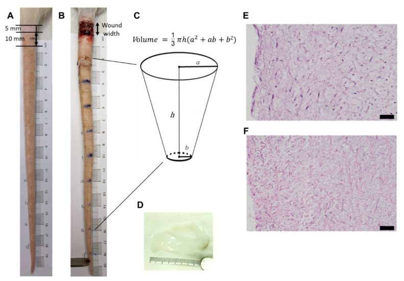
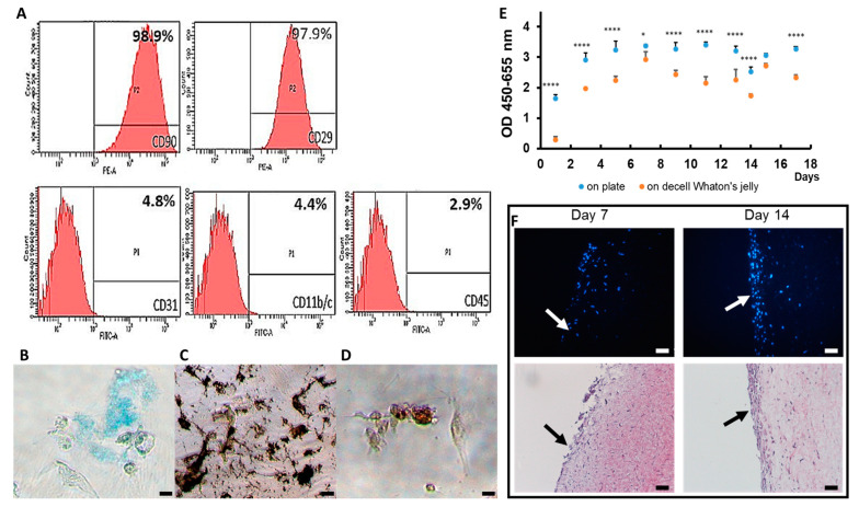
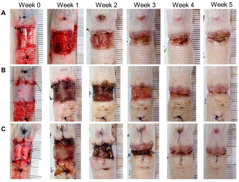
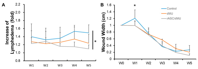
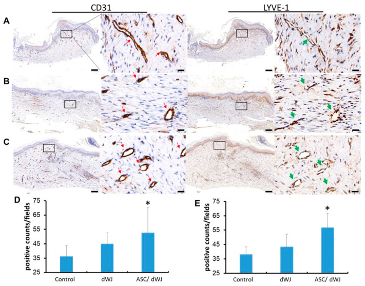
Lymphedema causes tissue swelling due to the accumulation of lymphatic fluid in the tissue, which delays the process of wound-healing. Developing effective treatment options of lymphedema is still an urgent issue. In this study, we aim to fabricate tissue-engineered moist wound dressings with adipose stem cells (ASCs) and decellularized Wharton's jelly (dWJ) from the human umbilical cord in order to ameliorate lymphedema. Rat ASCs were proliferated and an apparent layer was observed on dWJ at day 7 and 14. A rat tail lymphedema model was developed to evaluate the efficacy of the treatment. Approximately 1 cm of skin near the base of the rat tail was circularly excised. The wounds were treated by secondary healing (control) (n = 5), decellularized Wharton's jelly (n = 5) and ASC-seeded dWJ (n = 5). The wound-healing rate and the tail volume were recorded once a week from week one to week five. Angiogenesis and lymphangiogenesis were assessed by immunochemistry staining with anti-CD31 and anti-LYVE1. The results showed that the wound-healing rate was faster and the tail volume was lesser in the ASC-seeded dWJ group than in the control group. More CD31+ and LYVE-1+ cells were observed at the wound-healing area in the ASC-seeded dWJ group than in the control group. This proves that tissue-engineered moist wound dressings can accelerate wound-healing and reduce lymphedema by promoting angiogenesis and lymphangiogenesis.
Keywords: angiogenesis; decellularized Wharton’s jelly; lymphangiogenesis; lymphedema; rat adipose stem cell; wound-healing.
Conflict of interest statement
The authors declare no conflict of interest.
Figures
Similar articles
-
Injectable decellularized Wharton's jelly hydrogel containing CD56+ umbilical cord mesenchymal stem cell-derived exosomes for meniscus tear healing and cartilage protection.Mater Today Bio. 2024 Sep 19;29:101258. doi: 10.1016/j.mtbio.2024.101258. eCollection 2024 Dec. Mater Today Bio. 2024. PMID: 39347017 Free PMC article.
-
The effect of allogenic human Wharton's jelly stem cells seeded onto acellular dermal matrix in healing of rat burn wounds.J Cosmet Dermatol. 2020 Apr;19(4):995-1001. doi: 10.1111/jocd.13109. Epub 2019 Sep 25. J Cosmet Dermatol. 2020. PMID: 31556227
-
The healing effect of Wharton's jelly stem cells seeded on biological scaffold in chronic skin ulcers: A randomized clinical trial.J Cosmet Dermatol. 2019 Dec;18(6):1961-1967. doi: 10.1111/jocd.12931. Epub 2019 May 24. J Cosmet Dermatol. 2019. PMID: 31127705 Clinical Trial.
-
Lymphangiogenesis: novel strategies to promote cutaneous wound healing.Burns Trauma. 2024 Sep 26;12:tkae040. doi: 10.1093/burnst/tkae040. eCollection 2024. Burns Trauma. 2024. PMID: 39328366 Free PMC article. Review.
-
Absence of Wharton's Jelly at the Abdominal Site of the Umbilical Cord Insertion. Rare Case Report and Review of the Literature.Medicina (Kaunas). 2021 Nov 18;57(11):1268. doi: 10.3390/medicina57111268. Medicina (Kaunas). 2021. PMID: 34833486 Free PMC article. Review.
Cited by
-
[Research advances on stem cell-based treatments in animal studies and clinical trials of lymphedema].Zhongguo Xiu Fu Chong Jian Wai Ke Za Zhi. 2024 Jan 15;38(1):99-106. doi: 10.7507/1002-1892.202309045. Zhongguo Xiu Fu Chong Jian Wai Ke Za Zhi. 2024. PMID: 38225848 Free PMC article. Chinese.
-
Decellularized Umbilical Cord as a Scaffold to Support Healing of Full-Thickness Wounds.Biomimetics (Basel). 2024 Jul 3;9(7):405. doi: 10.3390/biomimetics9070405. Biomimetics (Basel). 2024. PMID: 39056846 Free PMC article.
References
-
- Executive Committee of the International Society of Lymphology The diagnosis and treatment of peripheral lymphedema. Lymphology. 2003;36:84–91. - PubMed
-
- Cheng M.-H., Chang D.W., Patel K.M. Principles and Practice of Lymphedema Surgery. Elsevier Health Sciences; Mumbai, India: 2015.
Grants and funding
LinkOut - more resources
Full Text Sources
Miscellaneous
