. 2023 Feb 7:10:1097955.
doi: 10.3389/fmolb.2023.1097955. eCollection 2023.

Mead acid inhibits retinol-induced irritant contact dermatitis via peroxisome proliferator-activated receptor alpha

Affiliations

Mead acid inhibits retinol-induced irritant contact dermatitis via peroxisome proliferator-activated receptor alpha

Azusa Saika et al. Front Mol Biosci. .

Abstract

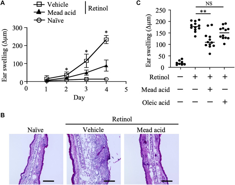
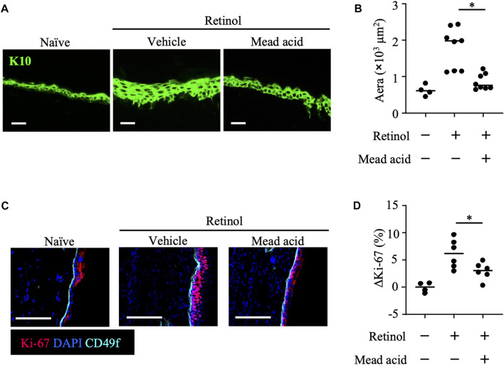
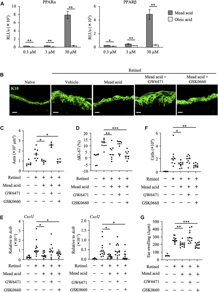
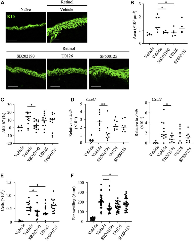
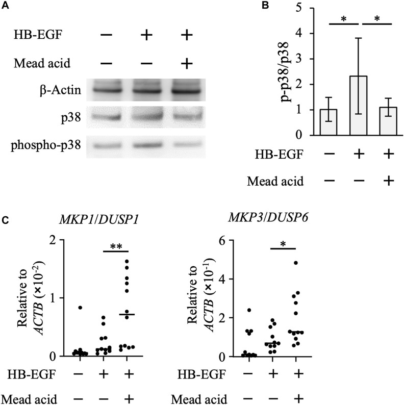
Retinol is widely used in topical skincare products to ameliorate skin aging and treat acne and wrinkles; however, retinol and its derivatives occasionally have adverse side effects, including the induction of irritant contact dermatitis. Previously, we reported that mead acid (5,8,11-eicosatrienoic acid), an oleic acid metabolite, ameliorated skin inflammation in dinitrofluorobenzene-induced allergic contact hypersensitivity by inhibiting neutrophil infiltration and leukotriene B4 production by neutrophils. Here, we showed that mead acid also suppresses retinol-induced irritant contact dermatitis. In a murine model, we revealed that mead acid inhibited keratinocyte abnormalities such as keratinocyte hyperproliferation. Consistently, mead acid inhibited p38 MAPK (mitogen-activated protein kinase) phosphorylation, which is an essential signaling pathway in the keratinocyte hyperplasia induced by retinol. These inhibitory effects of mead acid were associated with the prevention of both keratinocyte hyperproliferation and the gene expression of neutrophil chemoattractants, including Cxcl1 and Cxcl2, and they were mediated by a PPAR (peroxisome proliferator-activated receptor)-α pathway. Our findings identified the anti-inflammatory effects of mead acid, the use of which can be expected to minimize the risk of adverse side effects associated with topical retinoid application.

Keywords: hyperproliferation; inflammation; irritant contact dermatitis (ICD); keratinocyte; lipid metabolite; mead acid; oleic acid; retinol.


Conflict of interest statement

The authors declare that the research was conducted in the absence of any commercial or financial relationships that could be construed as a potential conflict of interest.

Figures

FIGURE 1
FIGURE 1
Retinol-induced irritant contact dermatitis (ICD) is inhibited by mead acid treatment. Retinol-induced ICD was induced by topical application of retinol for 4 consecutive days. Mice were treated topically with vehicle or mead acid 45 min after each retinol application. (A) Ear thickness was measured daily. Ear swelling (Δµm) was calculated as (Ear thickness [µm] on the indicated day)—(Ear thickness [µm] before the first retinol application). n = 8/group. Data were combined from two independent experiments (mean ± SEM values). Significant differences (p < 0.05) shown are between the vehicle and mead-acid-treatment groups. (B) For histological analysis, frozen ear sections were stained on day 5 with hematoxylin and eosin. Representative images from three independent experiments are shown. Scale bars, 100 μm. (C) Ear thickness was measured on day 4. Ear swelling (Δµm) was calculated as (Ear thickness [µm] on day 4)—(Ear thickness [µm] before the first retinol application). Horizontal bars indicate median values. Data are representative of three independent experiments. NS, not significant; **p < 0.01.
FIGURE 2
FIGURE 2
Mead acid inhibits neutrophil infiltration in retinol-induced irritant contact dermatitis (ICD). ICD was induced by topical application of retinol for 4 consecutive days. Mice were treated topically with vehicle or mead acid after each retinol treatment. (A) Cells were isolated from murine ears and used for flow cytometric analysis. Numbers indicate the percentages of Ly6G+ CD11b+ neutrophils. Representative images are shown. (B) The number of Ly6G+ CD11b+ neutrophils was determined on the basis of total cell numbers and flow cytometric data. (C) Frozen ear sections obtained on day 4 were stained with the indicated antibodies for immunohistologic analysis. Representative images from two independent experiments are shown. Bars, 100 μm. (D) Ear tissues were homogenized to isolate mRNA, and Cxcl1 and Cxcl2 expression was measured by quantitative RT-PCR analysis and normalized to that of Actb. Data are combined from four independent experiments. Horizontal bars indicate median values. *p < 0.05.
FIGURE 3
FIGURE 3
Mead acid inhibits keratinocyte abnormalities induced by retinol. Retinol was applied topically for 4 consecutive days to induce ICD. Mice were treated topically with vehicle or mead acid after retinol treatment. (A) Frozen ear sections obtained on day 4 were stained with anti-K10 antibody. Scale bars, 20 μm. Representative images from three independent experiments are shown. (B) The area stained for K10 was calculated by using BZ-X700 Analyzer software. (C) Frozen ear sections were stained with the indicated antibodies. Representative images from two independent experiments are shown. Bars, 100 μm. (D) The percentage of Ki-67-expressing keratinocytes gated as CD45 CD31 CD34 CD49f+ was measured by flow cytometry. The rate of increase of Ki-67+ cells [ΔKi-67 (%)] was calculated as (Ki-67 [%])—(Ki-67 [%] average in retinol-untreated mice) on day 4. Data are combined from four independent experiments. Horizontal bars indicate median values. *p < 0.05.
FIGURE 4
FIGURE 4
Mead acid reduces retinol-induced irritant contact dermatitis (ICD) via a PPARα-mediated pathway. (A) Activation levels of PPARα and PPARβ/δ were measured by using a reporter assay system following a 24 h exposure to mead acid or oleic acid (0.3, 3, or 30 µM) or vehicle. Data are combined from two independent experiments (average ±SD values). Statistical significance was determined by using Welch’s t-test. (B–G) ICD was induced by topical application of retinol. Mice were co-treated with mead acid and a receptor antagonist (GW6471 or GSK0660). (B) Frozen ear sections were stained with anti-K10 antibody. Representative images from two independent experiments are shown. Scale bars, 20 μm. (C) The area stained for K10 was calculated by using BZ-X700 Analyzer software. (D) The percentage of Ki-67-expressing keratinocytes gated as CD45 CD31 CD34 CD49f+ was measured by flow cytometry. The rate of increase of Ki-67-positive cells [ΔKi-67 (%)] was calculated as (Ki-67 [%])—(Ki-67 [%] average in retinol-untreated mice) on day 4. Data are combined from four independent experiments. (E) Ear tissues were homogenized to isolate mRNA, and Cxcl1 and Cxcl2 expression was measured by quantitative RT-PCR analysis and normalized to that of Actb. Data are combined from four independent experiments. (F) The number of Ly6G+ CD11b+ neutrophils was determined on the basis of total cell numbers and flow cytometric data. (G) Ear thickness was measured on day 4. Ear swelling (Δµm) was calculated as (Ear thickness [µm] on day 4)—(Ear thickness [µm] before the first retinol application). Horizontal bars indicate median values. RLUs, relative light units. *p < 0.05; **p < 0.01; ***p < 0.001.
FIGURE 5
FIGURE 5
Inhibition of p38 MAPK decreases keratinocyte abnormalities induced by retinol. After topical application of retinol, the p38MAPK inhibitor SB202190, the MAPK/ERK inhibitor U1026, the JNK inhibitor SP600125, or vehicle was applied topically to both sides of mouse ears. (A) Frozen ear sections obtained on day 4 were stained with anti-K10 antibody. Representative images from three independent experiments are shown. Scale bars, 20 μm. (B) The area stained for K10 was calculated by using BZ-X700 Analyzer software. (C) The percentage of Ki-67-expressing keratinocytes gated as CD45 CD31 CD34 CD49f+ was measured by flow cytometry. The rate of increase of Ki-67-positive cells [ΔKi-67 (%)] was calculated as (Ki-67 [%] on day 4)—(Ki-67 [%] average in retinol-untreated mice on day 4). Data are combined from four independent experiments. (D) Ear tissues were homogenized to isolate mRNA, and Cxcl1 and Cxcl2 expression was measured by quantitative RT-PCR analysis and normalized to that of Actb. Data are combined from four independent experiments. (E) The number of Ly6G+ CD11b+ neutrophils was determined on the basis of total cell numbers and flow cytometric data. (F) Ear thickness was measured on day 4. Ear swelling (Δµm) was calculated as (Ear thickness [µm] on day 4)—(Ear thickness [µm] before the first retinol application). Horizontal bars indicate median values. Data are combined from five independent experiments. *p < 0.05; **p < 0.01; ***p < 0.001.
FIGURE 6
FIGURE 6
Mead acid attenuates p38 MAPK activation. HaCaT cells were pretreated with mead acid or vehicle before stimulation with HB-EGF (1 ng/mL). (A) The expression levels of phospho-p38, p38, and β-actin were measured by western blotting. Representative images from three independent experiments are shown. (B) The ratio of phospho-p38 to p38 was calculated by dividing the intensity of expression of phospho-p38 by the intensity of that of p38. (C) The expression levels of MKP1/DUSP1 and MKP3/DUSP6 were measured by quantitative RT-PCR analysis and normalized to that of ACTB. Data are combined from four independent experiments. *p < 0.05; **p < 0.01.

Similar articles

Cited by

References

    1. Al Tanoury Z., Piskunov A., Rochette-Egly C. (2013). Vitamin A and retinoid signaling: Genomic and nongenomic effects. J. Lipid Res. 54, 1761–1775. 10.1194/jlr.R030833 - DOI - PMC - PubMed
    1. Beckenbach L., Baron J. M., Merk H. F., Loffler H., Amann P. M. (2015). Retinoid treatment of skin diseases. Eur. J. Dermatol. 25, 384–391. 10.1684/ejd.2015.2544 - DOI - PubMed
    1. Boukamp P., Petrussevska R. T., Breitkreutz D., Hornung J., Markham A., Fusenig N. E. (1988). Normal keratinization in a spontaneously immortalized aneuploid human keratinocyte cell line. J. Cell. Biol. 106, 761–771. 10.1083/jcb.106.3.761 - DOI - PMC - PubMed
    1. Buchanan P. J., Gilman R. H. (2016). Retinoids: Literature review and suggested algorithm for use prior to facial resurfacing procedures. J. Cutan. Aesthet. Surg. 9, 139–144. 10.4103/0974-2077.191653 - DOI - PMC - PubMed
    1. Cattani F., Gallese A., Mosca M., Buanne P., Biordi L., Francavilla S., et al. (2006). The role of CXCR2 activity in the contact hypersensitivity response in mice. Eur. Cytokine Netw. 17, 42–48. - PubMed

Grants and funding

This work was supported by the Ministry of Education, Culture, Sports, Science and Technology of Japan (MEXT)/Japan Society for the Promotion of Science KAKENHI (grant numbers 21K20769 to AS; 19K07617 to TN; 22K15004 to KH; and 21H02757 to JK); the Japan Agency for Medical Research and Development (AMED; grant number 22ae0121035s012 to KH and grant numbers 22fk0108145h0003, 22ae0121042h0002, and 223fa727001h0001 to JK); the Ministry of Health and Welfare of Japan and Public/Private R&D Investment Strategic Expansion Program: PRISM (grant number 20AC5004 to JK); the Cross-Ministerial Strategic Innovation Promotion Program (SIP) (grant number 18087292 to JK); the Grant for the Joint Research Project of the Institute of Medical Science, the University of Tokyo (to JK); the Ono Medical Research Foundation (to JK); and the Canon Foundation (to JK).

LinkOut - more resources