Two differentially stable rDNA loci coexist on the same chromosome and form a single nucleolus
- PMID: 36821584
- PMCID: PMC9992848
- DOI: 10.1073/pnas.2219126120
Two differentially stable rDNA loci coexist on the same chromosome and form a single nucleolus
Abstract
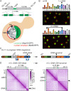
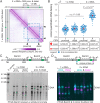
The nucleolus is the most prominent membraneless compartment within the nucleus-dedicated to the metabolism of ribosomal RNA. Nucleoli are composed of hundreds of ribosomal DNA (rDNA) repeated genes that form large chromosomal clusters, whose high recombination rates can cause nucleolar dysfunction and promote genome instability. Intriguingly, the evolving architecture of eukaryotic genomes appears to have favored two strategic rDNA locations-where a single locus per chromosome is situated either near the centromere (CEN) or the telomere. Here, we deployed an innovative genome engineering approach to cut and paste to an ectopic chromosomal location-the ~1.5 mega-base rDNA locus in a single step using CRISPR technology. This "megablock" rDNA engineering was performed in a fused-karyotype strain of Saccharomyces cerevisiae. The strategic repositioning of this locus within the megachromosome allowed experimentally mimicking and monitoring the outcome of an rDNA migratory event, in which twin rDNA loci coexist on the same chromosomal arm. We showed that the twin-rDNA yeast readily adapts, exhibiting wild-type growth and maintaining rRNA homeostasis, and that the twin loci form a single nucleolus throughout the cell cycle. Unexpectedly, the size of each rDNA array appears to depend on its position relative to the CEN, in that the locus that is CEN-distal undergoes size reduction at a higher frequency compared to the CEN-proximal counterpart. Finally, we provided molecular evidence supporting a mechanism called paralogous cis-rDNA interference, which potentially explains why placing two identical repeated arrays on the same chromosome may negatively affect their function and structural stability.
Keywords: Hi-C maps; cis duplicated rDNA loci; megablock chromosome engineering; nucleolus.
Conflict of interest statement
The authors have organizational affiliations to disclose: J.D.B. is a Founder and Director of CDI Labs, Inc., a Founder of Neochromosome, Inc, a Founder of and Consultant to ReOpen Diagnostics, and serves or served on the Scientific Advisory Board of the following: Logomix, Inc., Modern Meadow, Inc., Rome Therapeutics, Inc., Sample6, Inc., Sangamo, Inc., Tessera Therapeutics, inc., and the Wyss Institute. The remaining authors declare no competing interests. The authors have stock ownership to disclose: J.D.B. has substantive interest in CDI Labs, Neochromosome, and ReOpen Diagnostics.
Figures


Similar articles
-
Quantification of the dynamic behaviour of ribosomal DNA genes and nucleolus during yeast Saccharomyces cerevisiae cell cycle.J Struct Biol. 2019 Nov 1;208(2):152-164. doi: 10.1016/j.jsb.2019.08.010. Epub 2019 Aug 23. J Struct Biol. 2019. PMID: 31449968
-
Links between nucleolar activity, rDNA stability, aneuploidy and chronological aging in the yeast Saccharomyces cerevisiae.Biogerontology. 2014 Jun;15(3):289-316. doi: 10.1007/s10522-014-9499-y. Epub 2014 Apr 8. Biogerontology. 2014. PMID: 24711086 Free PMC article.
-
Expression of rRNA genes and nucleolus formation at ectopic chromosomal sites in the yeast Saccharomyces cerevisiae.Mol Cell Biol. 2006 Aug;26(16):6223-38. doi: 10.1128/MCB.02324-05. Mol Cell Biol. 2006. PMID: 16880531 Free PMC article.
-
Ribosomal DNA and the nucleolus at the heart of aging.Trends Biochem Sci. 2022 Apr;47(4):328-341. doi: 10.1016/j.tibs.2021.12.007. Epub 2022 Jan 18. Trends Biochem Sci. 2022. PMID: 35063340 Review.
-
Ribosomal DNA and the Nucleolus as Keystones of Nuclear Architecture, Organization, and Function.Trends Genet. 2019 Oct;35(10):710-723. doi: 10.1016/j.tig.2019.07.011. Epub 2019 Aug 22. Trends Genet. 2019. PMID: 31447250 Free PMC article. Review.
Cited by
-
Context-dependent neocentromere activity in synthetic yeast chromosome VIII.Cell Genom. 2023 Nov 9;3(11):100437. doi: 10.1016/j.xgen.2023.100437. eCollection 2023 Nov 8. Cell Genom. 2023. PMID: 38020969 Free PMC article.
References
-
- Long E. O., Dawid I. B., Repeated genes in eukaryotes. Annu. Rev. Biochem. 49, 727–764 (1980). - PubMed
-
- Sylvester J. E., et al. , The human ribosomal RNA genes: Structure and organization of the complete repeating unit. Hum. Genet. 73, 193–198 (1986). - PubMed
-
- Warner J. R., The economics of ribosome biosynthesis in yeast. Trends Biochem. Sci. 24, 437–440 (1999). - PubMed
-
- Lafontaine D. L. J., Riback J. A., Bascetin R., Brangwynne C. P., The nucleolus as a multiphase liquid condensate. Nat. Rev. Mol. Cell Biol. 22, 165–182 (2021). - PubMed
Publication types
MeSH terms
Substances
Grants and funding
LinkOut - more resources
Full Text Sources
Molecular Biology Databases

